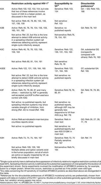
The past decade has seen an explosion of knowledge on the ability of human innate restriction factors, including the APOBEC3 family of proteins, to counteract infection by human immunodeficiency virus type 1 (HIV-1). All seven APOBEC3 family members have been found to have varying levels of activity against HIV-1 under different experimental conditions (Table 1). This antiretroviral activity is due primarily to the ability of APOBEC3 proteins to deaminate viral cDNA cytidines to uridines and inhibit viral reverse transcription and integration (Refs Reference Chiu and Greene1, Reference Harris and Liddament2).
Studies reporting restriction activity, susceptibility to Vif and mutational context preferences of APOBEC3 proteins with respect to HIV-1

aSingle-cycle activity here is defined as the suppression of infectivity relative to negative restriction controls of at least 50%. Spreading infection activity is defined as the substantial suppression of viral spread for the duration of the experiment shown. Some experiments involved the modification of APOBEC3 proteins by, for example, fusion to a helper protein. Where this occurs, restriction is counted as if the protein were wild type.
bAny data concerning the Vif sensitivity of modified APOBEC3 proteins as described above are disregarded in this table.
cReferences for the dinucleotide preferences of APOBEC3 mutational activity are preferentially chosen from papers with data on mutation of the HIV-1 genome. Studies on other substrates generally agree with those listed here, but variation among papers may occur.
Abbreviations: A3A–H, APOBEC3A–H; HIV-1, human immunodeficiency virus type 1; Vif, virion infectivity factor.
HIV-1 averts restriction by APOBEC3 proteins through the action of its accessory protein virion infectivity factor (Vif), which decreases the encapsidation of certain APOBEC3 proteins (Ref. Reference Malim and Emerman3) (Table 1). It is not entirely clear, however, to what extent Vif is successful in protecting the integrity of the viral genome. Multiple lines of evidence suggest that the neutralisation of APOBEC3 proteins by Vif is not absolute. Thus, APOBEC3 proteins may be partially effective at restricting HIV-1 infectivity in vivo despite the presence of Vif. Supporting this view, the effects of hypermutation on the viral genome are readily apparent in patient viral sequences. Recent evidence additionally suggests this mutational activity may have the further undesired effect of providing HIV-1 with a reservoir of genetic diversity that contributes to viral immune evasion and drug resistance.
Here, we briefly review what is known about the basic biology of APOBEC3–Vif interactions. We then summarise current clinical data on correlations between APOBEC3 function and the natural history of HIV-1 infection to ascertain what may occur when HIV-1 and APOBEC3 proteins encounter each other in vivo. Finally, we discuss strategies to harness the restrictive capacity of APOBEC3 proteins to successfully treat HIV-1 infection.
HIV-1 Vif is a viral counterdefence against restriction by APOBEC3 proteins
HIV-1 Vif is required for growth on certain cell types and in vivo
Lentiviruses are a genus within the broader family of retroviruses. In addition to the ubiquitous gag, pol and env genes, lentiviruses possess several accessory genes that have integral roles in viral pathogenesis (recently reviewed by Refs Reference Malim and Emerman3, Reference Gramberg, Sunseri and Landau4). The prototypical lentivirus, HIV-1, contains two accessory genes involved in the regulation of viral gene expression and four additional accessory genes more directly involved in pathogenesis. One of these encodes the virion infectivity factor (Vif).
The vif gene is located in the centre of the HIV-1 genome, overlapping the 3′ end of pol and the 5′ end of the accessory gene vpr. Expressed from a partially spliced, Rev-dependent subgenomic mRNA (Refs Reference Garrett, Tiley and Cullen5, Reference Schwartz, Felber and Pavlakis6), the protein product is 192 residues, highly basic and 23 kDa in size (Refs Reference Kan7, Reference Lee8, Reference Sodroski9). Vif is required for productive infection in vivo and for infection of primary CD4+ T cells, monocytes and macrophages ex vivo (Refs Reference Desrosiers10, Reference Strebel11, Reference Gabuzda12, Reference Gabuzda13). This requirement, however, is variable in some human T cell lines (Ref. Reference Fisher14), allowing these lines to be grouped as ‘permissive’ for the replication of Vif-deficient HIV-1 (e.g. CEM-SS, SupT1) or ‘nonpermissive’ (e.g. CEM, H9).
When produced in permissive cells, virions from Vif-deficient HIV-1 have no quantitative or qualitative defects relative to wild-type virus except for the absence of the Vif protein itself. (Refs Reference Strebel11, Reference Fisher14). When produced in nonpermissive cells, however, Vif-deficient HIV-1 virions rarely complete reverse transcription (Refs Reference Simon and Malim15, Reference Courcoul16, Reference Goncalves17, Reference Sova and Volsky18).
Given the above, two scenarios were considered likely to explain the phenotype of Vif-deficient viruses on nonpermissive cells: (1) permissive cells contain an endogenous factor that complements the function lacking in a Vif-deficient virus or (2) nonpermissive cells contain a Vif-suppressible factor with antiviral restriction activity. To determine which hypothesis was correct, two groups tested the infectivity of virions produced from heterokaryons formed by the fusion of permissive and nonpermissive cells (Refs Reference Madani and Kabat19, Reference Simon20). Under these conditions, the production of infectious virions would support the former hypothesis, while the production of noninfectious virions would support the latter. In fact, such heterokaryons formed from the fusion of permissive and nonpermissive cells produce virions with diminished infectivity, indicating that nonpermissive cells contain a Vif-sensitive dominant restriction factor.
APOBEC3G is a Vif-sensitive restrictor of HIV-1 infectivity with DNA cytidine deaminase activity
To identify the cellular factor suggested by cell fusion experiments, Sheehy et al. sought to isolate genes uniquely expressed in a nonpermissive cell line called CEM (Ref. Reference Sheehy21) by suppression subtractive hybridisation, which simultaneously suppresses the recovery of transcripts common to CEM and its permissive derivative CEM-SS and enriches the recovery of transcripts that are more abundant in CEM. Among many sequences, a cDNA encoding APOBEC3G (A3G) was isolated, and engineering naturally permissive CEM-SS cells to express A3G rendered these cells nonpermissive for the replication of Vif-deficient but not wild-type virus. A3G therefore accounts for the nonpermissive phenotype of CEM, although a number of studies since then suggest that other APOBEC3 proteins may also contribute to the nonpermissive phenotype (Table 1). Of particular note, passage of Vif-deficient HIV-1 on permissive T cells stably expressing A3G yields viral variants that grow efficiently in the presence of A3G but not in CEM cells (Ref. Reference Haché22), demonstrating that other factors beyond A3G are also capable of restricting HIV-1.
At approximately the same time, two other groups independently identified A3G, not for its antiviral activity but rather for its enzymatic activity, which provided a crucial clue about the mechanism by which A3G might restrict HIV-1. Jarmuz et al. identified A3G as part of a family of polynucleotide cytidine deaminases related to the mRNA-editing enzyme APOBEC1, which edits the apolipoprotein B mRNA (Ref. Reference Jarmuz23). (The acronym APOBEC derives from APOBEC1 and stands for ‘apolipoprotein B mRNA editing, catalytic polypeptide’; human APOBEC3 proteins are designated ‘apolipoprotein B mRNA editing, catalytic polypeptide-like 3′ followed by the letter, A–H, identifying the specific protein.) Meanwhile, Harris and colleagues hypothesised, based on homology between APOBEC1 and the DNA mutator activation-induced deaminase (AID/AICDA), which is involved in antibody diversification, that APOBEC1 and its homologues can act on DNA substrates. Indeed, APOBEC1, A3G, A3C and AID were all shown to mutate DNA (Refs Reference Petersen-Mahrt, Harris and Neuberger24, Reference Harris, Petersen-Mahrt and Neuberger25).
HIV-1 restriction by A3G is mediated by deamination of viral cDNA cytidine during reverse transcription
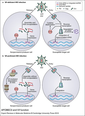
Soon after the fortuitous convergence of data showing A3G's capacity to restrict HIV-1 and deaminate DNA cytidines, several groups demonstrated that the major basis for retroviral restriction is the deamination of minus-strand viral cDNA during reverse transcription (Refs Reference Harris26, Reference Mangeat27, Reference Zhang28). The proviral DNA of a Vif-deficient virus grown in the presence of A3G becomes heavily mutated, with characteristic genomic plus-strand G-to-A transitions. This strand bias is only explained by deamination of the minus-strand reverse transcript, which after deamination from C-to-U templates A rather than G on the viral plus-strand (Refs Reference Harris26, Reference Mangeat27, Reference Zhang28, Reference Lecossier29) (Fig. 1). Furthermore, some catalytically defective A3G mutants are unable to as effectively reproduce the nonpermissive phenotype, which represents important additional evidence for the dependence of restriction on deaminase activity (Refs Reference Mangeat27, Reference Zhang28, Reference Browne, Allers and Landau30, Reference Holmes31, Reference Miyagi32, Reference Schumacher33).
APOBEC3 and Vif function. (a) Vif-deficient HIV infection. In a ‘nonpermissive producer cell’ that expresses the DNA cytidine deaminase APOBEC3G (A3G), gene expression of Vif-deficient HIV-1 proceeds normally, but during assembly A3G (pink circles) is encapsidated along with normal virion components. Virions with encapsidated A3G bud from a producer cell and enter a target cell normally. Reverse transcription, however, is blocked by the presence of A3G, either by the direct inhibition of cDNA synthesis or the degradation of uridine-containing cDNA, arising from cytidine deamination. Uridine-containing cDNA templates A on the viral plus-strand, resulting in G-to-A hypermutation. A direct A3G-mediated block to integration has also been proposed (not shown). (b) Vif-proficient HIV infection. To prevent encapsidation, Vif expressed from a Vif-proficient virus links A3G to an E3 ligase complex, which results in the ubiquitination of A3G and its eventual degradation in the proteasome. Alternatively, Vif may directly inhibit encapsidation, lower A3G expression and/or inhibit A3G catalytic activity (not shown). Subsequent release, entry, reverse transcription and integration are shown proceeding normally. Abbreviations: dsDNA, double-stranded DNA; HIV, human immunodeficiency virus.

Although hypermutation is clearly a crucial part of the retrovirus-restriction mechanism, evidence also exists that the catalytic activity of A3G may not wholly explain its ability to restrict HIV-1 (Refs Reference Holmes31, Reference Shindo34, Reference Newman35, Reference Bishop, Holmes and Malim36, Reference Bishop37, Reference Iwatani38, Reference Guo39, Reference Yang40, Reference Li41, Reference Luo42). These studies suggest deaminase-independent effects on various specific steps in reverse transcription or integration (Fig. 1), although at least one report finds some of these same effects to be dependent on catalytic activity (Ref. Reference Mbisa43). While a substantial quantity of evidence suggests deaminase-independent restriction by A3G, the sheer diversity of the steps at which A3G is proposed to exert its deaminase-independent effects makes it highly improbable that all of these phenomena are physiological. In fact, several groups have suggested that the deaminase-independent effects of A3G may represent overexpression artifacts, as stable expression of physiological levels of catalytically inactive A3G has little if any effect on HIV-1 (Refs Reference Browne, Allers and Landau30, Reference Miyagi32, Reference Schumacher, Nissley and Harris44). Thus, it appears that deaminase activity is largely required for the inhibitory activity of A3G under physiological conditions. Whether this will also be the case for other APOBEC3 proteins, which may have more pronounced deaminase-independent effects (Ref. Reference Holmes31), remains to be seen. Moreover, it is important to note that deaminase-independent mechanisms may be more central to the restriction of many other retroelements including hepatitis B virus and LINE-1 (Refs Reference Turelli45, Reference Bogerd46, Reference Muckenfuss47, Reference Stenglein and Harris48, and reviewed recently by Refs Reference Holmes, Malim and Bishop49, Reference Bonvin and Greeve50).
It was proposed early on that the effect of A3G-mediated deamination might be to shunt mutated viral cDNA into a base-excision repair pathway that would ultimately result in the degradation of the viral genome (Refs Reference Harris26, Reference Harris51). Under this model, a uracil DNA glycosylase such as UNG would recognise and excise cDNA uracils, leaving an abasic site at which an endonuclease such as APEX (APEX1) would cleave the cDNA backbone. While one report supports such a scenario (Ref. Reference Yang52), different data sets focusing on uracil DNA glycosylase functions imply either reparative (Refs Reference Chen53, Reference Mansky54, Reference Priet55) or degradative (Ref. Reference Schröfelbauer56) effects on the viral genome. Alternatively, uracil DNA glycosylase activity may simply be irrelevant to the viral lifecycle (Refs Reference Schumacher33, Reference Kaiser and Emerman57). Further work will be required to elucidate the potential mechanisms by which viral cDNA is processed following APOBEC3-mediated deamination.
HIV-1 Vif preserves viral infectivity by preventing A3G encapsidation
Many mechanisms are plausible to explain how HIV-1 Vif counteracts the restrictive capacity of A3G. Most authors agree that this occurs by inhibiting A3G packaging, although the details of the mechanism(s) by which this inhibition occurs are debatable. At least part of the mechanism by which Vif overcomes restriction by A3G involves the Vif-mediated degradation of A3G through a proteasome-dependent pathway (Refs Reference Conticello, Harris and Neuberger58, Reference Marin59, Reference Sheehy, Gaddis and Malim60, Reference Stopak61, Reference Yu62, Reference Mehle63). Specifically, Vif binds both to A3G and to an E3 ubiquitin ligase complex consisting of cullin-5 (CUL5), ring-box protein 2 (RBX2/RNF7) and elongins B (TCEB2) and C (TCEB1). By bringing A3G into proximity with the ubiquitin ligase complex, Vif facilitates the ubiquitination and eventual degradation of A3G through the proteasome (Refs Reference Yu62, Reference Mehle64, Reference Yu65) (Fig. 1).
Evidence also exists suggesting that Vif can directly inhibit A3G encapsidation (Refs Reference Sheehy, Gaddis and Malim60, Reference Kao66, Reference Mariani67). Such a mechanism need not require degradation as an end point. Other reports indicate a Vif-dependent reduction in A3G expression (Refs Reference Stopak61, Reference Kao66), and it is also possible that Vif may function by directly inhibiting the catalytic activity of A3G (Ref. Reference Santa-Marta68).
The mechanisms proposed above are not necessarily mutually exclusive. While details may differ, an overarching message is readily apparent in the current Vif literature: preventing the encapsidation of A3G is crucial for the ability of Vif to preserve the infectivity of HIV-1. In fact, reducing the encapsidation of A3G by simply increasing particle production to titrate out the restriction factor is at least one mechanism by which HIV-1 can resist restriction in the absence of Vif function (Ref. Reference Haché22).
Evidence for APOBEC3 function in vivo, and clinical implications
Multiple APOBEC3 family proteins have activity against HIV-1
While discussion to this point has focused on a basic description of the interactions between Vif and A3G, there are six additional proteins in the APOBEC3 family – APOBEC3A (A3A), APOBEC3B (A3B), APOBEC3C (A3C), APOBEC3DE (A3DE), APOBEC3F (A3F) and APOBEC3H (A3H) (Refs Reference Harris and Liddament2, Reference Jarmuz23, Reference Conticello69, Reference Wedekind70) – all of which have been implicated in the mutation and restriction of HIV-1 under certain conditions. A summary of references with data concerning the restriction activity and Vif sensitivity of each APOBEC3 protein is provided in Table 1; current in-depth reviews of the differential properties of these APOBEC3 proteins may be found elsewhere (e.g. Refs Reference Chiu and Greene1, Reference Malim71).
While the results apparent in Table 1 do reflect a lack of consensus on the ability of certain APOBEC3 proteins to function against HIV-1, there are areas of agreement. Most notably, it is well accepted that A3F and, more prominently, A3G have strong activity against HIV-1. Moreover, there is a second tier consisting of A3B and A3DE where antiviral activity is generally reproducible in overexpression single-cycle infectivity assays but may not be apparent in a more physiological spreading infection system. Further study of these proteins, particularly in spreading infections, will be required to determine definitively their ability to act as dominant restriction factors. It is generally accepted, however, that A3G, A3F, A3DE and perhaps A3H (see below) are Vif-sensitive, while A3B is not, which implies that these Vif-sensitive factors are likely the most relevant for the restriction of HIV-1 in vivo. That said, the fact that A3B expression increases significantly in lymphoid tissue from acutely infected patients along with A3F and A3G (Ref. Reference Li72) may also suggest a role for this Vif-insensitive protein.
A3H represents a special case since only one known allele appears to encode a protein that is stable in human cells, and stability is required for its ability to effectively restrict HIV-1 (Refs Reference OhAinle73, Reference Harari74, Reference Tan75). It is also presently unclear to what extent this APOBEC3 protein is Vif-sensitive (Table 1), although the fact that some have found it sensitive to Vif suggests that it may be important for the restriction of HIV-1. Perhaps even more muddled is the case of A3C. A3C may be sensitive to Vif, but its restriction activity against HIV-1 is relatively weak, arguing against a role for this APOBEC3 protein in vivo. It should be noted, however, that A3C is quite active against simian immunodeficiency virus (Ref. Reference Yu76). While some case can thus be made for the ability of six of seven human APOBEC3 proteins to dominantly restrict HIV-1, there are few findings to support such a role for A3A, as the references showing the strongest effects require the fusion of A3A to a helper protein that shuttles it into the viral core (see discussion below).
Beyond restriction and Vif-sensitivity phenotypes, it is important to emphasise one key characteristic that helps to distinguish among APOBEC3 proteins: the sequence context in which they typically mutate DNA cytidines to uridines (Table 1). A3G preferentially mutates cytidine residues that are preceded by another cytidine, with an even stronger preference for the context 5′-CCCA-3′ in minus-strand viral cDNA, where the underlined deoxycytidine is the nucleotide preferentially targeted (Refs Reference Harris26, Reference Mangeat27, Reference Zhang28, Reference Yu77). In plus-strand reverse-transcribed cDNA, this manifests as 5′-TGGG-3′ to 5′-TAGG-3′ mutations, or 5′-GG-3′ to 5′-AG-3′ in the less stringent dinucleotide context. Other APOBEC3 proteins, however, generally display a preference for the mutation of cytidines preceded by another deoxyribonucleotide, most often thymidine (e.g. (Refs Reference Bishop78, Reference Liddament79, Reference Wiegand80, Reference Zheng81). This results in 5′-TC-3′ to 5′-TU-3′ transitions, which manifest on plus-strand viral cDNA as 5′-GA-3′ to 5′-AA-3′ transitions.
The identification of these different sequence contexts is important for assessing which APOBEC3 proteins act on a given viral substrate in a context where multiple APOBEC3 proteins are expressed such as in vivo. Furthermore, assessing these contexts helps one to appreciate how hypermutation can be detrimental to a virus. Aside from deleterious amino acid or other mutations that may occur in unique sequence contexts, any UGG codon encoding tryptophan is susceptible to the creation of a UAG or perhaps even a UAA stop, particularly in the presence of A3G. Similarly, the AUG start codon may place the wobble base in an APOBEC3-susceptible hotspot depending on the identity of the next base in the sequence, thus making the ablation of initiating methionines another distinct possibility. Extensive mutation may also disrupt HIV-1 genomic RNA secondary structures, which are important for the regulation of HIV-1 gene expression (Ref. Reference Watts82).
APOBEC3 proteins mutate the HIV-1 genome
Even prior to the discovery of APOBEC3 proteins, the tendency for HIV-1 to acquire G-to-A mutations was evident (Refs Reference Janini83, Reference Vartanian84, Reference Vartanian85) – a phenomenon analogous to the original observation of hypermutation in spleen necrosis virus (Ref. Reference Pathak and Temin86). These early reports generally noted a preferential sequence context for hypermutation of 5′-GA-3′ and, to a lesser extent, 5′-GG-3′, suggesting that, given what is now known, multiple APOBEC3 proteins account for these early descriptions of hypermutation (Ref. Reference Liddament79).
While more-recent studies at times note stronger preferences for one context or another under a given condition (Refs Reference Kieffer87, Reference Pace88, Reference Piantadosi89), a theme uniting recent literature on the in vivo hypermutation of HIV-1 is that it frequently occurs in both major dinucleotide contexts. One study indicates, for example, that hypermutated sequences separate roughly into thirds: sequences with hypermutation predominantly in a 5′-GA-3′ context, sequences with hypermutation predominantly in a 5′-GG-3′ context, and sequences with hypermutation occurring in roughly equal proportions in each context (Ref. Reference Land90). Another reports the occurrence of 5′-GG-3′ context mutations in approximately 95% of hypermutated sequences versus the occurrence of 5′-GA-3′ context mutations in approximately 60% of hypermutated sequences (Ref. Reference Gandhi91). Furthermore, mutations in both contexts frequently occur in the same cloned sequence. For example, 5′-GG-3′ hypermutations occur within 95% of predominantly 5′-GA-3′ context clones (Ref. Reference Gandhi91), while another data set shows a very slight preference for hypermutation in a 5′-GA-3′ context with a strong 5′-GG-3′ component (Ref. Reference Ulenga92).
It is reasonable to conclude, therefore, that multiple APOBEC3 proteins are active in vivo and that at least one of these is A3G with its strong 5′-GG-3′ context preference. The identities of the APOBEC3 proteins responsible for the reported 5′-GA-3′ hypermutations, however, remain unknown. While A3F is certainly a likely candidate based on its expression, Vif sensitivity and restriction profiles (see below; Table 1), it is entirely possible that other APOBEC3 proteins account for at least a portion of this hypermutation. A more careful elucidation of the expression and restriction capacities of APOBEC3 proteins in vivo as well as the interactions of these proteins with HIV-1 in vitro will be required to define the true nature of the in vivo APOBEC3 repertoire, which may itself vary from person to person (e.g. Ref. Reference Kidd93).
Regardless of which APOBEC3 proteins are actively hypermutating HIV-1, it is clear that their overall effect is significant. Estimates of the frequency of hypermutated sequences in vivo range from approximately 7% to 20% with wide variation in the proportions seen in any one individual (Refs Reference Kieffer87, Reference Pace88, Reference Piantadosi89, Reference Land90, Reference Gandhi91). In fact, these figures almost certainly underestimate the total proportion of proviral sequences mutated by APOBEC3 proteins. Since a sequence must typically reach a given threshold of G-to-A mutations to be defined as ‘hypermutated’, methods of quantifying hypermutants fail to account for sequences that have been mutated by APOBEC3 proteins at a lower level. Furthermore, extremely hypermutated sequences may fail to replicate and amplify by PCR while still potentially contributing to the overall mutational load of a viral population through recombination (Ref. Reference Mulder, Harari and Simon94). As briefly discussed above, the deamination of cDNA cytidines may also create a substrate for the downstream degradation of the viral genome, which would further diminish the apparent proportion of viral genomes affected by APOBEC3 proteins.
APOBEC3 proteins may contribute to viral adaptation
Several authors have previously discussed the theoretical possibility that drug-resistance mutations could be attributable to APOBEC3-mediated hypermutation, citing the frequent occurrence of drug-resistance mutations in sequence contexts consistent with the action of APOBEC3 proteins (Refs Reference Berkhout and de Ronde95, Reference Haché, Mansky and Harris96, Reference Pillai, Wong and Barbour97). Combined with the common occurrence of hypermutation in vivo, it would seem that a role for APOBEC3 proteins in aiding the evolution of HIV-1 drug resistance is likely. Nevertheless, the actual data in the literature on this topic are still limited. For example, one study demonstrates that, in the presence of a vif allele defective for the neutralisation of A3G, HIV-1 can readily acquire a particular drug-resistance mutation by recombination with a lethally hypermutated genome residing in the same cell (Ref. Reference Mulder, Harari and Simon94). Computer modelling to assess the likelihood of drug-resistance mutation acquisition from A3G action further suggests that HIV-1 does acquire drug-resistance mutations from A3G activity at a low rate (Ref. Reference Jern98). Also of interest, a recent paper on HIV-1 sequence evolution proposes that APOBEC3 proteins may be involved in the acquisition of immune-escape mutations early in HIV-1 infection (Ref. Reference Wood99).
While the consensus of the data to date is that APOBEC3 proteins are likely to contribute to HIV-1 evolution, many questions remain unanswered. For example, how often does this actually occur in vivo, and what is the relative contribution of APOBEC3 proteins versus other sources of mutation such as reverse transcriptase? Which APOBEC3 proteins are primarily responsible? To what extent does selective pressure contribute to the amplification of mutations caused by APOBEC3 proteins? Much work remains to be done in determining the impact of APOBEC3 proteins on the adaptability of HIV-1. The field would particularly benefit from studies that expand beyond A3G to consider the APOBEC3 proteins likely to cause 5′-GA-3′ context mutations.
Correlation of hypermutation levels, APOBEC3 expression and clinical indicators
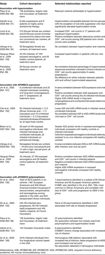
While hypermutation clearly occurs in vivo, the data concerning its correlation with clinical parameters of infection are less clear; a summary of pertinent research on this topic is provided in Table 2. For example, one study shows a significant correlation between hypermutation in both 5′-GA-3′ and 5′-GG-3′ contexts and reduced viral load (Ref. Reference Pace88), consistent with the higher levels of hypermutation found in another group of patients with low viral loads (Ref. Reference Vázquez-Pérez100). Strikingly, the authors of the first study conclude that the drop in viral load attributable to hypermutation is notably greater than that attributable to other known protective alleles such as CCR5Δ32. Corroborating this effect, 17 Kenyan women with significant hypermutation levels have a statistically significant increase in CD4+ cell counts (Ref. Reference Land90). Furthermore, a positive correlation is found between adenosine content and CD4+ cell counts throughout this entire cohort of over 200 Kenyan women when the total adenosine content of patient proviruses is taken as a surrogate for hypermutation.
Summary of linkage studies between clinical indicators and hypermutation, APOBEC3 expression or APOBEC3 polymorphism

Abbreviations: A3B, APOBEC3B; A3F, APOBEC3F; A3G, APOBEC3G; AIDS, acquired immune deficiency syndrome; HIV, human immunodeficiency virus.
By contrast, hypermutation does not significantly correlate with viral load in a cohort of Senegalese female sex workers, although levels of hypermutation do generally correlate with A3G mRNA expression (Ref. Reference Ulenga92). Additional evidence suggesting no correlation between hypermutation and improved clinical status has been found by comparing patients on highly active antiretroviral therapy (HAART) with elite suppressors (patients who maintain high CD4+ cell counts and undetectable viral loads) (Ref. Reference Gandhi91). In contrast with the Senegalese cohort, however, no correlation between A3G expression and the fraction of hypermutated sequences existed in these groups. A third cohort, consisting of 28 Kenyan women, also shows no correlation between hypermutation and viral load or CD4+ cell counts (Ref. Reference Piantadosi89).
Others have more directly assessed the impact of specific APOBEC3 proteins by correlating their mRNA expression levels with clinical indicators; results of these studies are also summarised in Table 2. Using this approach, strong negative correlations are found between A3G mRNA levels and viraemia, concordant with positive correlations between A3G mRNA levels and CD4+ cell counts (Refs Reference Vázquez-Pérez100, Reference Jin101), although one study does not reproduce these associations (Ref. Reference Cho102). Nevertheless, other data indicate that patients with a low viral set point have significantly higher levels of A3G mRNA both preinfection and postinfection than patients with a high viral set point; analysis for A3F yields the same correlation with postinfection A3F mRNA levels but not preinfection mRNA levels (Ref. Reference Ulenga103). Furthermore, the peripheral blood mononuclear cells of HIV-exposed seronegative patients show higher A3G expression and decreased susceptibility to ex vivo infection (Ref. Reference Biasin104), and HIV-exposed seronegative individuals have higher A3G expression levels than healthy controls (Ref. Reference Vázquez-Pérez100).
Thus, the preponderance of evidence indicates a correlation between higher A3G expression and improved clinical status. This correlation may be less apparent or even absent, however, when hypermutation is taken as a surrogate for APOBEC3 action. Failing to find a correlation when substituting hypermutation for APOBEC3 function may not be that surprising, though, given that reducing the probability of successful reverse transcription and integration appears to be an important part of the mechanism by which APOBEC3 proteins restrict HIV-1. Interpretations of data correlating hypermutation with clinical indicators are somewhat confounded because it is not clear what has occurred when no correlation is found. If APOBEC3 proteins are highly effective, the viral genome may simply not persist long enough to integrate. Furthermore, it is possible that some APOBEC3 repertoires and/or individual APOBEC3 alleles may be protective, leading some patients to be selectively excluded from studies due to a lack of apparent HIV-1 infection. Regardless, the positive data in these studies, particularly those focusing on APOBEC3 expression, do indicate promise for therapies aimed at the APOBEC3–Vif axis. The field would benefit from further research correlating clinical indicators with both hypermutation and the expression of more APOBEC3 family members in larger patient cohorts. Individuals known to be relatively resistant to HIV-1 should also be closely examined for APOBEC3 sequence and copy number variations.
APOBEC3 polymorphisms may be important for restriction activity against HIV-1
Relatively little is known about the functional relevance of human APOBEC3 polymorphisms, but several attempts have been made to correlate clinical status with APOBEC3 variations, as summarised in Table 2. One survey of the APOBEC3G alleles in a French cohort divided into rapid and slow progressors reports 29 polymorphisms, two of which affect the amino acid sequence, but none of these correlates with the rate at which these groups progress to disease (Ref. Reference Do105). Others find that the H186R allele identified in this cohort does, however, correlate with CD4+ cell depletion and rapid progression to conditions defining AIDS (acquired immune deficiency syndrome) in African American patients (Ref. Reference An106), although there is no apparent association with risk of transmission. Another allelic variant, C40693T in intron 4 of APOBEC3G, is associated with an increased risk of infection in a cohort of Caucasian patients (Ref. Reference Valcke107).
While these associations are suggestive, their functional basis is far from clear. For example, there is no apparent difference between the wild-type and H186R alleles of APOBEC3G in an in vitro single-cycle infectivity assay (Ref. Reference Do105), and no experimental data are available for the C40693T variant. As the authors of the latter study rightly point out, however, the close association of APOBEC3G with other APOBEC3 genes on chromosome 22 raises the distinct possibility that a given association may actually reflect linkage with an undetected variation elsewhere in the locus rather than a direct effect of the identified polymorphism (Ref. Reference Valcke107).
In the case of APOBEC3H, activity against HIV-1 is strongly influenced by polymorphisms. Four major haplotypes of human APOBEC3H exist, but only one of these appears to be expressed stably and able to inhibit HIV-1 (Refs Reference OhAinle73, Reference Harari74, Reference Tan75). This active variant, APOBEC3H haplotype II, is present in roughly 50% of individuals of African descent, 10–18% of individuals of European descent and 3–4% of individuals of Asian descent (Ref. Reference OhAinle73). Another common APOBEC3 polymorphism is an APOBEC3B deletion, which occurs in approximately 20% of the world's population (Ref. Reference Kidd93). In contrast with APOBEC3H haplotype II, however, deletion of APOBEC3B is relatively rare in African and European populations but common in East Asian (36.9%), Amerindian (57.7%) and Oceanic (92.9%) populations.
Whether these or other unidentified allelic variants are of significance for susceptibility to HIV-1 infection or disease progression is unknown, although at least one recent report has found a statistically significant increased risk of HIV-1 infection and progression in individuals with homozygous APOBEC3B deletions (Ref. Reference An108). It is important to point out, however, that most APOBEC3 proteins have evolved under positive selection, implying that the challenges posed by ancient and presumably divergent pathogens have resulted in the rapid evolution of these restriction factors (Refs Reference OhAinle109, Reference Sawyer, Emerman and Malik110). While this selective pressure may not have been applied by HIV-1 given its relatively recent arrival in the human population, selection for APOBEC3 proteins better able to restrict other pathogens may have inadvertently selected cross-reactivity for the restriction of HIV-1. Thus, it is reasonable to predict that, despite the relative dearth of data presently available, variation at the genetic level will account for some effect on HIV-1 in vivo. Furthermore, it is possible that ongoing selection by the HIV-1 epidemic itself may result in the expansion of protective APOBEC3 alleles not yet identified.
Studies of APOBEC3 effects in primary cell types
CD4+ T cells are the primary reservoir of HIV-1 infection in vivo, but relatively little is known about the APOBEC3 repertoire found in these cells. One report suggests that A3G forms the basis of a largely uncharacterised, deaminase-independent target-cell restriction of incoming HIV-1 virions in resting CD4+ T cells (Ref. Reference Chiu111), an effect enhanced by treatment with interferon α (IFN-α/IFNA1), which upregulates A3G expression (Ref. Reference Chen112). The reproducibility of this phenomenon, however, has been questioned (Refs Reference Santoni de Sio and Trono113, Reference Kamata, Nagaoka and Chen114). Regardless, evidence indicates that among activated CD4+ T cells, the Th1 subset of helper T cells expresses greater levels of A3G and A3F, which are further enhanced by the autocrine action of IFN-γ (IFNG) (Ref. Reference Vetter115). Greater expression, in turn, results in greater encapsidation of APOBEC3 proteins in HIV-1 produced from these cells and consequently lower viral infectivity than in the Th2 subset of helper T cells, even for Vif-proficient viruses. In general, most APOBEC3 family proteins with the exception of A3A and, to a lesser extent, A3B appear to be expressed in primary CD4+ T cells; among T cell subsets, however, A3G is expressed at somewhat higher levels than A3F (Ref. Reference Koning116).
Studies with primary myeloid lineage cells have also noted a prominent effect of IFN treatment on the ability of APOBEC3 proteins to restrict HIV-1, linking higher expression of A3G and A3A to the relative resistance of monocytes versus differentiated macrophages to infection (Ref. Reference Peng117). Similarly, A3G and A3F are responsible for preventing HIV-1 from infecting immature dendritic cells, an effect that augments as dendritic cells mature and increase their A3G expression (Ref. Reference Pion118). Expression of IFN-α and, to a lesser extent, IFN-γ have also been linked to increased APOBEC3 levels and increased resistance to HIV-1 infection; this effect is so dramatic that the upregulation of APOBEC3 proteins may effectively overwhelm normal Vif function and enable restriction of wild-type HIV-1 in macrophages (Refs Reference Peng117, Reference Peng119). Of note, IFN-α clearly upregulates all APOBEC3 proteins in macrophages and dendritic cells with the possible exceptions of A3B and A3C, indicating that these are part of the normal IFN response (Ref. Reference Koning116). This upregulation is particularly astounding for A3A (Refs Reference Koning116, Reference Peng117). Given the common expression of many APOBEC3 proteins in primary cell types and their IFN inducibility, it is tempting to speculate that the combination of different APOBEC3 proteins may yield emergent properties not readily apparent in the majority of in vitro studies to date, which almost exclusively focus on the effects of single APOBEC3 proteins.
Strategies for the clinical application of APOBEC3–Vif biology: increasing the encapsidation of APOBEC3 proteins
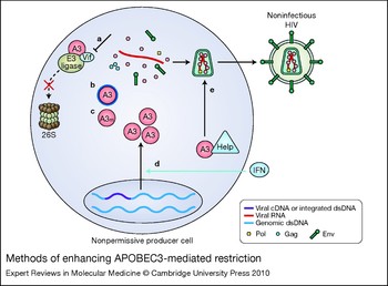
Strategies aimed at the therapeutic use of APOBEC3 proteins to restrict HIV-1, despite their diversity, are united by one goal: to increase the amount of APOBEC3 proteins encapsidated in HIV-1 virions (Fig. 2). If substantial quantities of APOBEC3 proteins are packaged in a particular virion, even in a Vif-proficient virus (see below), the genomic RNA of that virion is not likely to become a replication-competent provirus. Moreover, while the mutagenic capacity of APOBEC3 proteins carries with it at least some risk for contributing to viral adaptation (Refs Reference Mulder, Harari and Simon94, Reference Jern98), particularly when amplified, the risk associated with A3G mutagenic activity specifically may be tolerable relative to the potential benefits of unleashing its antiviral activity (Ref. Reference Jern98). To increase the packaging of APOBEC3 proteins, two general strategies are tenable: (1) indirectly increasing encapsidation by preventing the degradation of APOBEC3 proteins; or (2) directly increasing encapsidation by enhancing APOBEC3 expression or access to the viral core.
Methods of enhancing APOBEC3-mediated restriction. Methods of enhancing restriction by APOBEC3 proteins are united in the common goal of delivering more APOBEC3 proteins to the viral core. One strategy is to indirectly increase APOBEC3 encapsidation by protecting APOBEC3 proteins from degradation (dashed, blocked arrow). This may occur by (a) targeting inhibitors to a Vif surface that interacts with APOBEC3 proteins or with cellular proteins involved in Vif-mediated degradation, (b) chemically shielding APOBEC3 proteins, or (c) employing a degradation-resistant variant of an APOBEC3 protein (A3m) such as A3G D128K. Alternatively, encapsidation of APOBEC3 proteins could be directly increased by (d) boosting APOBEC3 expression, particularly through interferon (IFN) upregulation, or (e) fusion to a privileged molecule with core access (triangle labelled ‘Help’), which may shuttle APOBEC3 proteins to the viral core with great enough efficiency to overcome Vif-mediated degradation. All methods will theoretically result in greater APOBEC3 encapsidation (pink circles) and restriction of HIV-1, even in the presence of Vif. Abbreviations: dsDNA, double-stranded DNA; HIV, human immunodeficiency virus.

Indirectly increasing encapsidation by preventing the degradation of APOBEC3 proteins
A pharmaceutical approach
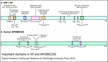
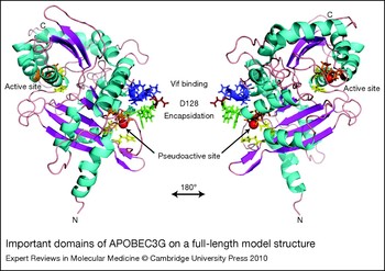
Prevention of the degradation of APOBEC3 proteins would be most simply accomplished by blocking the interaction of APOBEC3 proteins with Vif (Fig. 2a and 2b). To that end, a great deal of effort has been expended attempting to elucidate the important interacting regions on Vif and on APOBEC3 proteins (Refs Reference Russell and Pathak120, Reference Simon121, Reference Zhang122, Reference He123, Reference Tian124, Reference Yamashita125, Reference Huthoff126, Reference Huthoff and Malim127, Reference Russell128, Reference Zhang129, Reference Pery130, Reference Chen131, Reference Dang132 and others cited elsewhere in this article). While a thorough discussion of the many details involved in these investigations is beyond the scope of this review, a schematic summary of important regions on Vif and on A3G with particular attention to their putative interaction domains is provided in Figure 3, and biologically relevant regions of A3G are highlighted on a recently published full-length structural model of this restriction factor (Ref. Reference Harjes133) in Figure 4.
Important domains in Vif and APOBEC3G. While many papers have identified residues critical to Vif function, only putative continuous interaction domains in Vif (a) and APOBEC3G (A3G) (b) are depicted here for the sake of clarity. Exceptions are made for two important lysines in Vif (K22 and K26) as well as the C-terminal zinc-binding domain of Vif. Vif and A3G are internally to scale, but Vif is depicted at twice its actual size relative to A3G. HIV-1 Vif residues shown are those found in the HXB2 reference sequence (accession number K03455). A3G corresponds to reference sequence NP_068594. Note that K26 and 23SLV25 of Vif are required for neutralisation of A3G and A3F, respectively, but these residues may not mediate direct interaction with either APOBEC3 protein. Similarly, one group finds that while A3G D128 is important in permitting the degradation of A3G, it does not mediate direct binding to Vif. See main text for references and discussion. HIV, human immunodeficiency virus.

Important domains of APOBEC3G on a full-length model structure. Important domains of APOBEC3G (A3G) identified by genetic analysis are highlighted on a full-length model structure of A3G (Ref. Reference Harjes133) with colouration carried out in Pymol. See main text for references and discussion. Side chains are shown for key regions, and the right panel represents a rotation of 180º about the y-axis. Red spheres, zinc; green side chains (124YYFW127), encapsidation determinant (overlaps 126–127 start of Vif-binding region 126FWDPDYQ132); red side chain (D128), a key residue for Vif sensitivity; blue side chains (129PDYQ132), the remainder of the putative Vif-binding site; yellow side chains (H65 & E67 and H257 & E259), the HxE of the pseudocatalytic and catalytic N- and C-terminal domains, respectively; orange side chains (96PC97 & C100 and 287PC288 & C291), the PCxxC of the active and pseudoactive N- and C-terminal domains, respectively.

In surveying this literature, it is worthwhile to note that several discontinuous surfaces on Vif have been implicated in the binding of specific APOBEC3 proteins (Fig. 3a). This indicates that the APOBEC3-interaction surfaces on HIV-1 Vif are structurally complex and that three-dimensional structures of Vif bound to APOBEC3 proteins will likely be required both to understand and to effectively target this interaction. In contrast, what is known of the Vif-interaction surfaces on APOBEC3 proteins indicates that, while such surfaces may be distinct on different APOBEC3 proteins (Refs Reference Zhang122, Reference Russell128), they are relatively continuous (Fig. 3b). It may be possible, therefore, to protect APOBEC3 proteins by simply designing a molecule that binds the host protein and shields it from interaction with Vif (Fig. 2b), a strategy that would likely hamper the ability of HIV-1 to develop resistance to this putative therapy (Ref. Reference Harris and Liddament2). Conversely, it may be possible to inhibit Vif interaction with either APOBEC3 proteins or with the proteasomal machinery using a molecule that binds Vif itself (Fig. 2a).
Although a small-molecule inhibitor of the APOBEC3–Vif interaction is not yet clinically available, a significant recent development is the identification of RN-18, a small-molecule antagonist of HIV-1 Vif (Ref. Reference Nathans134). Interestingly, RN-18 decreases Vif levels and thereby increases A3G (and A3F and A3C but not A3B) levels when these viral and cellular proteins are present together. This small molecule is also able to inhibit the growth of Vif-proficient HIV-1 in cultured nonpermissive cells. Thus, while further research is necessary to understand the mechanism by which this and other putative inhibitors function, RN-18 has provided proof of principle that the APOBEC3–Vif axis can be targeted by small molecules.
Another promising development in the APOBEC3 field is the growing body of structural information on the deaminase domain of A3G (Refs Reference Harjes133, Reference Chen135, Reference Holden136, Reference Furukawa137) and related proteins such as APOBEC2 (Ref. Reference Prochnow138). As progress continues toward the expansion of our structural knowledge of A3G and other APOBEC3 proteins (reviewed recently by Ref. Reference Bransteitter, Prochnow and Chen139), it may one day be possible to visualise the APOBEC3–Vif interaction structurally and thereby rationally design interaction inhibitors. Half of this putative interaction surface is depicted in the A3G full-length model presented in Figure 4. In the meantime, the field would benefit from further genetic study of the APOBEC3–Vif interaction, both for the sake of reproducing current results and for identifying mutants that may further our mechanistic understanding of the APOBEC3–Vif interaction in anticipation of complementary structural data. For more detail on such efforts, see ‘Further reading, resources and contacts’ below.
A gene therapy approach
Although small molecules remain undoubtedly the most technologically feasible means by which to manipulate the APOBEC3–Vif axis, progress in the use of gene therapy approaches for the treatment of HIV-1 (e.g. Refs Reference Mitsuyasu140, Reference Hutter141, Reference Rossi, June and Kohn142) suggests that similar methods may one day incorporate aspects of APOBEC3-mediated restriction. While the application of APOBEC3 biology to HIV-1 therapy in this manner must be preceded by further advances in the safety and efficacy of both gene delivery and cell therapy, the strategies covered in this and the following section merit discussion if for no other reason than to note how they reinforce the basic concepts of APOBEC3–Vif biology.
Because Vif is required for the spread of HIV-1 in nonpermissive cells and in vivo, one obvious way to kill the virus would be to find an APOBEC3 protein that is not neutralised by Vif (Fig. 2c). In fact, nature has already provided just such a variant. A number of studies on the ability of the Vifs carried by the lentiviruses of different species to neutralise their cognate APOBEC3 proteins have indicated that the Vif–APOBEC3 interaction is species-specific (e.g. Ref. Reference Mariani67). One striking example is the substitution of human A3G aspartate 128 with the corresponding lysine found in rhesus macaque and African green monkey A3G, which renders human A3G resistant to Vif-mediated degradation (see Fig. 4). The D128K substitution may protect A3G by preventing Vif binding (Refs Reference Huthoff and Malim127, Reference Bogerd143, Reference Mangeat144, Reference Schröfelbauer, Chen and Landau145), although this view is not universally supported (Refs Reference Russell and Pathak120, Reference Xu146). Whatever the mechanism, D128K is not destabilised by HIV-1 Vif, and this leads to increased packaging of A3G and the restriction of Vif-proficient HIV-1. Despite the resistance of A3G D128K to Vif-mediated degradation, there are no known instances of this allele occurring naturally in humans.
An alternative method of protecting A3G from degradation that has been reported is to fuse it to the ubiquitin-associated domain 2 (UBA2), a stabilisation signal that protects proteins from proteasomal degradation (Ref. Reference Li147). Although this fusion is only partially resistant to Vif-mediated degradation, it does appear to have a modestly improved effect on the restriction of wild-type HIV-1 relative to that of wild-type A3G. This study combined with those above on A3G D128K provide proof of principle that relatively simple modifications to APOBEC3 proteins may allow them to resist Vif-mediated degradation. As technical facility with therapeutic gene delivery develops, these may one day prove viable strategies to engineer HIV-1-resistant T cells to counter infection.
Directly increasing APOBEC3 encapsidation
Increasing APOBEC3 expression
In much the same way that protecting A3G from degradation might leave enough A3G available to encapsidate and effectively restrict HIV-1, increasing the expression of A3G might overwhelm Vif and result in the encapsidation of nonpermissive levels of A3G (Fig. 2d). Proof of principle for this concept has been shown in macrophages, where the induction of APOBEC3 proteins by IFN-α treatment is so effective that it greatly restricts the infectivity of Vif-proficient HIV-1 (Ref. Reference Peng119), although it should be noted that the many effects of IFN induction make it difficult to rule out effects nonspecific to APOBEC3 action. In theory, then, methods aimed at selectively increasing the expression of APOBEC3 proteins in the natural reservoirs of HIV-1 infection should be able to potently inhibit viral replication, perhaps additionally allowing the broader immune system to better control infection and supporting the suppression of HIV-1 by current antiretroviral therapies. An important advantage of such methods is that they would likely be facilitated by small molecules and would work by enhancing innate immune processes that already occur naturally. That said, it is unclear what the consequences of increasing APOBEC3 expression in vivo may be, since there is at least a theoretical possibility that increasing APOBEC3 levels may overwhelm cellular regulatory mechanisms and result in hypermutation of the human genome, possibly impacting the development of cancer (e.g. Refs Reference Harris, Petersen-Mahrt and Neuberger25, Reference Yamanaka148).
Facilitating APOBEC3 access to the viral core
Alternatively, selectively directing APOBEC3 proteins to the HIV-1 viral core may permit the restriction of even Vif-proficient HIV-1 (Fig. 2e). Evidence from several groups shows that fusing A3G to a fragment of Vpr sufficient for encapsidation (Ref. Reference Ao149), fusing A3A to a full-length Vpr protein (Ref. Reference Aguiar150) or fusing A3G to the Nef7 mutant derivative of HIV-1 Nef (Ref. Reference Green, Liu and He151) results in enhanced APOBEC3-mediated restriction. All these methods lead to the incorporation of APOBEC3 proteins into the viral core via the viral peptides to which they are fused, which themselves have natural core access. This process is furthermore so efficient that Vif cannot overcome it.
An important lesson is also apparent in the ability of A3A to restrict under conditions in which it is fused to a molecule that grants it core access. While this APOBEC3 protein does not normally restrict HIV-1 or reach the viral core, forcing it past that barrier by fusion to Vpr or A3G results in A3A-mediated restriction (Refs Reference Aguiar150, Reference Goila-Gaur152). Thus, it is not sufficient for an A3A to simply find its way into HIV-1 virions. Rather, it must be specifically targeted to the viral core to have sufficient access to the viral genome and carry out its antiviral effects. Whether the localisation of other APOBEC3 proteins within virions is an important determinant of their relative abilities to function remains a subject for future research.
Conclusions and outstanding research questions
Controversies aside, research to date has yielded significant insight into the mechanisms by which A3G and HIV-1 interact in the balance between restriction and infection. Given what is known about APOBEC3 protein expression and hypermutation profiles in vivo and in primary ex vivo cell types, it is likely that multiple APOBEC3 proteins are active against HIV-1 in vivo, and that these may display distinct properties. More work is required to define the APOBEC3 proteins that impact HIV-1 in vivo and the exact mechanisms by which this occurs.
Clinical studies may also prove useful in the continuing characterisation of the APOBEC3–Vif axis. As discussed above, incorporating the quantification of the expression levels of more APOBEC3 proteins and correlating those to hypermutation levels while increasing cohort sizes would improve our understanding of the in vivo functions of APOBEC3 proteins. Further characterisation of APOBEC3 polymorphisms may also be significant, since it is possible that protective APOBEC3 alleles await discovery. To this end, further studies of the full APOBEC3 genomic locus in uninfected individuals, infected individuals and individuals with an atypical response to HIV-1 such as long-term nonprogressors and those uninfected but repeatedly exposed may hold a key to novel treatments aimed at the APOBEC3–Vif axis.
Complementary studies focusing on the structures of APOBEC3 proteins and of Vif are also likely to be of great benefit. It may be possible to identify, characterise and ultimately commercialise Vif inhibitors such as RN-18 in the absence of structural information. Nevertheless, the ability to rationally design pharmaceuticals aimed at the APOBEC3–Vif axis will be significantly facilitated by a deep understanding of the physical interactions of APOBEC3 proteins and Vif. Furthermore, such understanding will undoubtedly be required to effectively design molecules that minimise the risk of selecting HIV-1 strains resistant to this hypothetical new class of antiretroviral drugs. To that end, however, the APOBEC3 family is particularly attractive, as it comprises multiple potential endogenous inhibitors of HIV-1 infectivity, at least two of which may be able to function in the long-term restriction of HIV-1 (Ref. Reference Haché22). With further study, the APOBEC3 proteins may one day form the basis of the next generation of antiretroviral therapies.
Acknowledgements and funding
We thank the reviewers and A.M. Land for constructive comments and assistance in improving this manuscript. We also thank E. Harjes and H. Matsuo for providing the full-length model of A3G. This work was funded by the National Institute of Allergy and Infectious Diseases (R01 AI064046 to R.S.H). J.S.A. was supported by a Kirschstein National Research Service Award for Individual Predoctoral MD-PhD Fellows and by the University of Minnesota Medical Scientist Training Program (National Institute on Drug Abuse, F30 DA026310; National Institute of General Medical Sciences, T32 GM008244).