Introduction
After gall midges (Diptera: Cecidomyiidae), gall wasps or cynipids (Hymenoptera: Cynipidae) constitute the largest radiation of gall-inducing insects. They are mainly distributed throughout the Holarctic region and are especially well represented in temperate areas of both the Nearctic and the Palaearctic. Despite most species can trigger the formation of galls on different host plants (Liljeblad and Ronquist Reference Liljeblad and Ronquist1998; Ronquist Reference Ronquist1999; Csóka et al. Reference Csóka, Stone, Melika, Raman, Schaefer and Withers2005; Pénzes et al. Reference Pénzes, Melika, Bozsóki, Bihari, Mikó and Tavakoli2009, Reference Pénzes, Tang, Bihari, Bozsó, Schwéger and Melika2012), and especially on Fagaceae, about 200 species have lost this ability and develop as inquilines inside galls induced by different woody rosid gallers within Cynipidae (Ronquist et al. Reference Ronquist, Nieves-Aldrey, Buffington, Liu, Liljeblad and Nylander2015; Schwéger et al. Reference Schwéger, Melika, Tang, Bihari, Bozsó and Stone2015a, Reference Schwéger, Melika, Tang, Yang, Stone and Nicholls2015b; Pujade-Villar et al. Reference Pujade-Villar, Wang, Chen, Shen and Ferrer-Suay2017; Lobato-Vila et al. Reference Lobato-Vila, Cibrián-Tovar, Barrera-Ruíz and Pujade-Villar2018, Reference Lobato-Vila, Cibrián-Tovar, Barrera-Ruíz, Equihua-Martínez, Estrada-Venegas and Buffington2019, Reference Lobato-Vila, Cibrián-Tovar, Barrera-Ruíz, Equihua-Martínez, Estrada-Venegas and Tovar-Sánchez2020; Lobato-Vila and Pujade-Villar Reference Lobato-Vila and Pujade-Villar2019). Both gall-makers and inquilines are endophytophagous herbivores whose larvae develop inside galls by feeding on their nutritious tissues, and even though inquilines cannot initiate galls de novo, they can still modify the plant tissues directly surrounding them and even modify the size or the shape of the gall (Ronquist Reference Ronquist1999; Pujade-Villar et al. Reference Pujade-Villar, Melika, Ros-Farré, Ács and Csóka2003, Reference Pujade-Villar, Wang, Chen, Shen and Ferrer-Suay2017; Pénzes et al. Reference Pénzes, Melika, Bozsóki, Bihari, Mikó and Tavakoli2009, Reference Pénzes, Tang, Bihari, Bozsó, Schwéger and Melika2012; Lobato-Vila and Pujade-Villar Reference Lobato-Vila and Pujade-Villar2019; Lobato-Vila et al. Reference Lobato-Vila, Cibrián-Tovar, Barrera-Ruíz, Equihua-Martínez, Estrada-Venegas and Buffington2019, Reference Lobato-Vila, Cibrián-Tovar, Barrera-Ruíz, Equihua-Martínez, Estrada-Venegas and Tovar-Sánchez2020).
Synergus Hartig, 1840 (Hymenoptera: Cynipidae: Cynipinae: Synergini), a genus of inquilines that associate with galls initiated by species within the tribe Cynipini (Hymenoptera: Cynipidae: Cynipinae) on Fagaceae, mainly oaks (Quercus Linnaeus), is by far the most species-rich genus of inquiline gall wasps with about 130 species known worldwide (Pénzes et al. Reference Pénzes, Tang, Bihari, Bozsó, Schwéger and Melika2012; Schwéger et al. Reference Schwéger, Melika, Tang, Bihari, Bozsó and Stone2015a; Pujade-Villar et al. Reference Pujade-Villar, Roca-Cusachs and Lobato-Vila2016, Reference Pujade-Villar, Wang, Chen, Shen and Ferrer-Suay2017; Lobato-Vila and Pujade-Villar Reference Lobato-Vila and Pujade-Villar2017; Lobato-Vila et al. Reference Lobato-Vila, Fernández-Garzón, Reina-Rojas, González and Pujade-Villar2017, Reference Lobato-Vila, Cibrián-Tovar, Barrera-Ruíz and Pujade-Villar2018, Reference Lobato-Vila, Cibrián-Tovar, Barrera-Ruíz, Equihua-Martínez, Estrada-Venegas and Buffington2019, Reference Lobato-Vila, Cibrián-Tovar, Barrera-Ruíz, Equihua-Martínez, Estrada-Venegas and Tovar-Sánchez2020). About 50% of them are found in the Nearctic region, mainly in North America north of Mexico, while only a little more than a 10% of the total is found in the Neotropics.
Synergus is characterised and distinguished from other genera of inquilines mainly by the following combination of morphological traits: first metasomal segment ring-shaped and usually longitudinally sulcate; the second and the third metasomal terga fused into one large sclerite (syntergum) in both sexes; female antennae usually with 14 flagellomeres, rarely with 13 or 15 flagellomeres, and male antennae usually with 15 flagellomeres, rarely 14, with the first flagellomere usually modified and different from that of females; radial cell of the forewings usually closed by its upper margin; lateral frontal carinae usually present; clypeus indistinct, not delimited; and face covered with striae radiating from clypeus (Ronquist et al. Reference Ronquist, Nieves-Aldrey, Buffington, Liu, Liljeblad and Nylander2015; Schwéger et al. Reference Schwéger, Melika, Tang, Bihari, Bozsó and Stone2015a, Reference Schwéger, Melika, Tang, Yang, Stone and Nicholls2015b; Lobato-Vila and Pujade-Villar Reference Lobato-Vila and Pujade-Villar2017).
Colombia is the southerly limit of Quercus distribution in the Neotropical region and therefore in the New World. Quercus humboldtii Bonpland is the only representative of the genus in this country and is widely spread on the intern slope of all three Andean cordilleras mainly from 1500–3000 m (Mabel-Paz Reference Mabel-Paz2012). To date, only two genera of Cynipini on Q. humboldtii have been recorded from Colombia: Melikaiella Pujade-Villar, 2014 (Pujade-Villar and Rodríguez Reference Pujade-Villar and Rodríguez2015) and Zapatella Pujade-Villar and Melika, 2012 (Pujade-Villar et al. Reference Pujade-Villar, Hanson, Medina, Torres and Melika2012; Fernández-Garzón et al. Reference Fernández-Garzón, Caicedo, Rodríguez and Pujade-Villar2017). Inquilines have been found to be associated with galls of Zapatella in this country since adults of both gall-makers and inquilines were obtained from these galls at the same time; however, this is not the case of Melikaiella. Despite this, an association between inquilines and Melikaiella galls from Colombia is more than probable and so should not be dismissed. The inquiline oak gall wasp fauna from Colombia is currently represented only by two species, both belonging to Synergus: S. colombianus Nieves-Aldrey, Reference Nieves-Aldrey2005 (Nieves-Aldrey Reference Nieves-Aldrey2005) and S. pedroi Pujade-Villar, Lobato-Vila, and Fernández-Garzón, 2017 (Lobato-Vila et al. Reference Lobato-Vila, Fernández-Garzón, Reina-Rojas, González and Pujade-Villar2017).
In this work, a review of the inquilines associated with galls of Cynipini from Colombia is conducted for the first time. A new species of Synergus, the only genus of inquilines known from this country to date, is described and illustrated, and an undescribed species is discussed. New biological and morphological data of S. colombianus and S. pedroi, as well as an identification key to all the Colombian species of Synergus, are provided.
Materials and methods
Adults of the species addressed in this study emerged from galls collected in different Colombian departments on Q. humboldtii since 2001. Galls were preserved in breeding boxes under environmental conditions until the emergence of the adults.
For morphological descriptions and diagnoses, we used the terminology of morphological structures as given in the following studies: Liljeblad and Ronquist (Reference Liljeblad and Ronquist1998) and Melika (Reference Melika2006) for adult morphological structures, Ronquist and Nordlander (Reference Ronquist and Nordlander1989) for forewing venation terminology and Harris (Reference Harris1979) for patterns of cuticular sculpture. The following measurements are used in this paper: post-ocellar distance, the distance between the inner margins of posterior ocelli; ocellar-ocular distance, the distance from the outer margin of lateral ocellus to the inner margin of compound eye; lateral-ocular distance, the distance between lateral and frontal ocellus; transfacial line, distance between inner margins of compound eyes measured across toruli; width of radial cell, measured as the distance between the upper margin of the forewing and the Rs vein. Length of flagellomeres and ocellar distances that appear in the text are given as relative measures.
Photographs of Figures 1–6, 17–19, 25–29 were taken using an Olympus SC30 digital microscope camera (Shinjuku, Tokyo, Japan) coupled to an Olympus U-CMAD3 adapted to a binocular microscope Olympus SZX10, and then compiled with Helicon Focus 6.2.2 (Khàrkiv, Ukraine) at the University of Barcelona, Barcelona, Spain. High-resolution images of Synergus dawnus Lobato-Vila and Pujade-Villar new species (Figs. 7–16), as well as Figures 20–24, were taken using a field emission gun environmental scanning electron microscope FEI Quanta 200 (Hillsboro, Oregon, United States of America) at high voltage (15 kV) at the University of Barcelona.
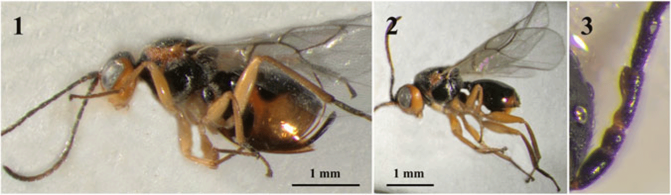
Synergus colombianus (new material from Colombia). 1, Female habitus; 2, male habitus; 3, first male flagellomeres.

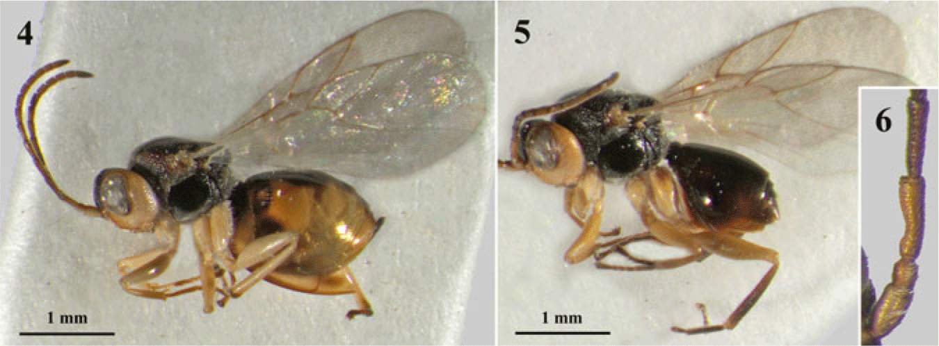
Synergus dawnus. 4, Female habitus; 5, male habitus; 6, first male flagellomeres.

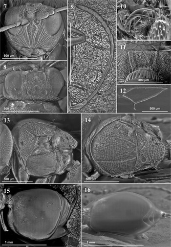
Synergus dawnus. 7, Head in anterior view; 8, head in dorsal view; 9, female antenna; 10, tarsal claw; 11, propodeum; 12, radial cell; 13, mesosoma in lateral view; 14, mesosoma in dorsal view; 15, metasoma in lateral view; 16, metasoma in dorsal view.

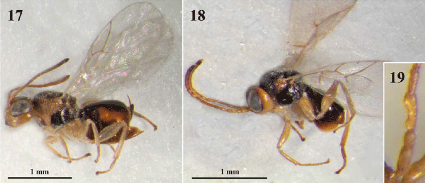
Synergus pedroi (paratypes deposited in UB). 17, Female habitus; 18, male habitus; 19, first male flagellomeres.

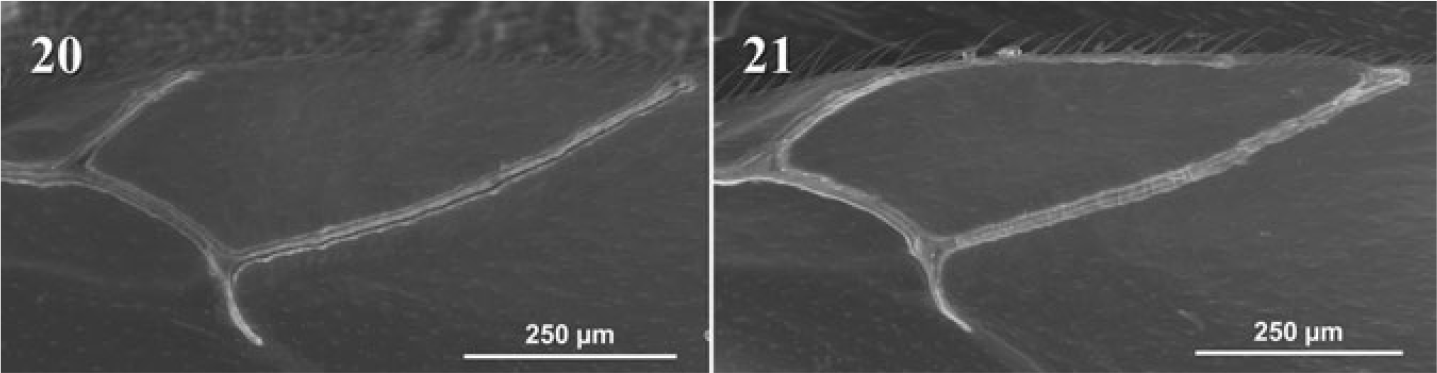
Radial cell variability of Synergus pedroi. 20, Radial cell open; 21, radial cell partially closed.

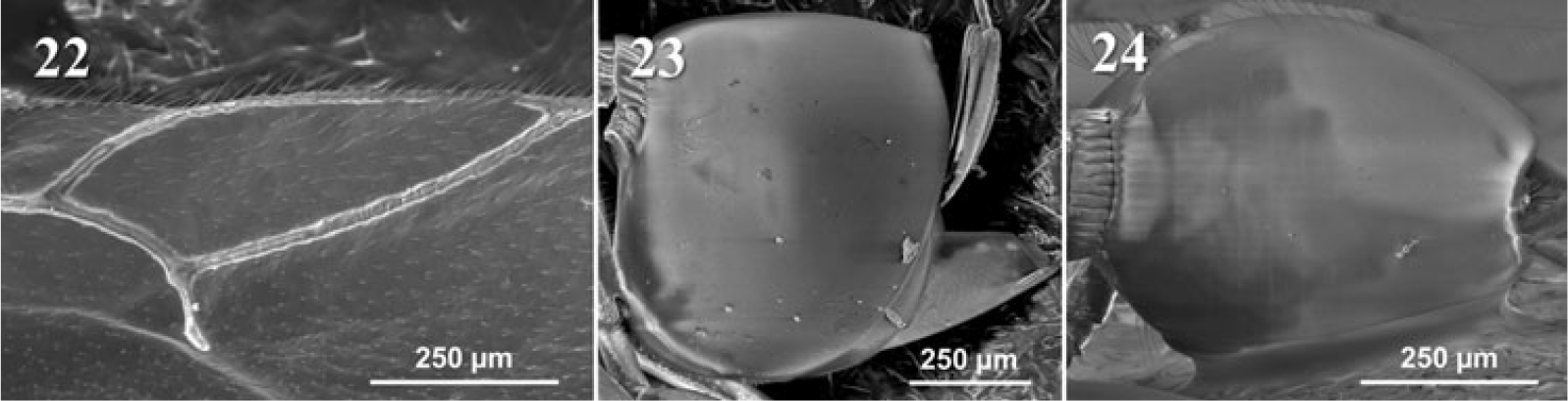
Morphological variations within Synergus pedroi (specimens from La Florida). 22, Radial cell; 23, metasoma lateral view; 24, metasoma dorsal view.

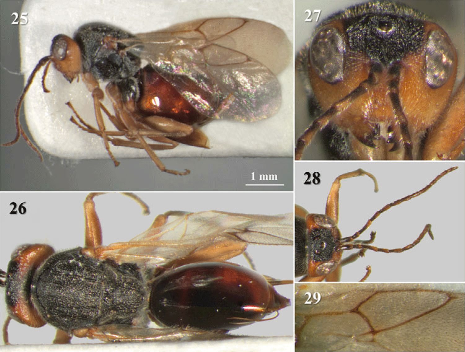
Synergus undescribed species (female). 25, Habitus; 26, dorsal view; 27, head in frontal view; 28, head in dorsal view and antennae; 29, radial cell.

Type and additional material of the species mentioned in this study are deposited in the following institutions and laboratories: IAVH (Instituto de Investigación de Recursos Biológicos Alexander von Humboldt, Villa de Leyva, Boyacá, Colombia; D. Espitia Reina); ICAT (Instituto Colombiano Agropecuario-Tibaitatá, Mosquera, Cundinamarca, Colombia; Pedro A. Rodríguez); MNCN (Museo Nacional de Ciencias Naturales, Madrid, Spain; M. París); and UB (Universitat de Barcelona, Barcelona, Spain; J. Pujade-Villar).
Synergus colombianus Nieves-Aldrey, Reference Nieves-Aldrey2005
Figures 1–3.
Synergus colombianus Nieves-Aldrey, Reference Nieves-Aldrey2005: 501–508. Type material: IAVH (holotype), paratypes in IAVH and MNCN [examined by I.L.-V. and J.P.-V. in Lobato-Vila et al. (Reference Lobato-Vila, Cibrián-Tovar, Barrera-Ruíz and Pujade-Villar2018)].
Material examined. 5♂ and 19♀. New additional material from Colombia deposited in UB with the following collecting data: Via Arauco-La Palma (Arcabuco, Boyacá), 5°44′17″N, 73°24′52″W, 2578 m, ex. Zapatella grahami Pujade-Villar and Melika, 2012, Q. humboldtii, gall collected 8.iv.2019, emerged iv.2019, J. Pujade-Villar: 5♂ and 16♀; same collecting data, but emerged 1–15.v.2019: 3♀.
Diagnosis. Synergus colombianus is characterised by having antennae with 14 flagellomeres in females and 15 flagellomeres in males; flagellomere 1 about as long as flagellomere 2 (Figs. 1, 3); flagellomere 1 of males excavate medially and moderately to strongly expanded apically (Fig. 3); face from round to subtrapezoid-shaped and with genae slightly expanded behind compound eyes; frons weakly wrinkled and punctate; frontal carinae narrow, somewhat branched distally and obscured by wrinkles before reaching lateral ocelli; mesoscutum with dense but weak discontinuous carinae; notauli shallow and narrow but complete, sometimes interrupted by carinae; scutellum from weakly carinate to wrinkled; scutellar foveae subtriangular to oval, shallow, weakly sculptured; circumscutellar carina weak but visible; mesopleurae regularly and densely striate; metasoma deeply dorsodistally incised and posteriorly with a band of minute micropunctures occupying from 1/4 to 1/3 of the metasomal length; radial cell closed, about 2.8 times as long as wide (Figs. 1–2); body mainly black and orange or amber, face and legs yellow except basal half of metacoxae, which is black, and tarsi, which are dark (Figs. 1–2).
Synergus colombianus is morphologically close to Synergus dawnus Lobato-Vila and Pujade-Villar new species (see below), from which can be distinguished by having basal half of metacoxae black (Figs. 1–2) (metacoxae yellow, rarely with a small, dark spot, in S. dawnus, Figs. 4–5); the malar space 0.4 times as long as height of compound eye (0.6 times in S. dawnus, Fig. 7); frons weakly wrinkled (coriaceous, rarely with a few very weak wrinkles, in S. dawnus, Figs. 7–8); areolet small or absent (Figs. 1–2) (visible, however, it is not well delimited by distinct veins, in S. dawnus, Figs. 4–5, 12); flagellomere 1 of males from moderately to strongly expanded apically (Fig. 3) (slightly expanded apically in S. dawnus, Fig. 6), among others (see the identification key).
Natural history.Synergus colombianus was described from specimens that emerged from an unidentified cynipid gall on acorns of Q. humboldtii (Nieves-Aldrey Reference Nieves-Aldrey2005). The additional material presented here was obtained from acorn galls of Zapatella grahami Pujade-Villar and Melika, 2012, which may correspond to the species mentioned by Nieves-Aldrey.
Distribution. Colombia. Boyacá department, above 2500 m (Nieves-Aldrey Reference Nieves-Aldrey2005; and in this work, see the additional material).
Synergus dawnus Lobato-Vila and Pujade-Villar, new species
http://zoobank.org/urn:lsid:zoobank.org:act:0D0526B0-E307-4370-90CC-A04941F9C241.
Type material. Holotype ♀ deposited in IAVH with the following labels: “Guyabal de Siquina, Sitio CAR (Cundinamarca, Colombia), 4°50′36″N, 74°27′43″W” (white label)/“Ex. Zapatella inflata, 2083 m, Q. humboldtii, gall collected 04.i.2013, emerged 20–29.i.2013, P. Rodriguez Col.” (white label)/“Holotype ♀ Synergus dawnus Lobato-Vila and Pujade-Villar, IL-V desig-2019” (red label). Paratypes (14♂ and 12♀) deposited in IAVH (9♂ and 6♀) and UB (5♂ and 5♀): with the same data as the holotype: 5♂ and 5♀ (deposited in UB); “Colombia, Boyacá, Villa de Leyva, La Colorada, 5°38′47″N, 73°30′29″W, 2200m, 23.iv.2014, Z. nievesaldreyi, J. Pujade, M. Torres and J. Cardenas” (white label)/“Instituto Humboldt, Colombia, IAvH-L-146809” (white label, quick response code)/“Paratype ♀ Synergus dawnus Lobato-Vila and Pujade-Villar, IL-V desig-2019” (red label) (1♀; deposited in UB); same collecting data as the previous one, but with the following reference codes: IAvH-E-146798 (1♂); IAvH-E-146799 (1♂); IAvH-E-146800 (1♀); IAvH-E-146804 (1♂); IAvH-E-146808 (1♂); IAvH-E-146811 (1♀); IAvH-E-146813 (1♀); IAvH-E-146816 (1♂); IAvH-E-146819 (1♂); IAvH-E-146820 (1♀); IAvH-E-146822 (1♀); IAvH-E-146826 (1♀) (deposited in IAVH); “Colombia, Boyacá, SFF Iguaque, El Níspero, 5°38′N, 73°31′W, 2730 m, 7–21.xii.2001, Malaise 1, P. Reina M.2581” (white label)/“Instituto Humboldt, Colombia, IAvH-E-147454” (white label, quick response code)/“Paratype ♂ Synergus dawnus Lobato-Vila and Pujade-Villar, IL-V desig-2019” (red label) (1♂; deposited in IAVH); same collecting data as the previous one, but collected 12.xii.2001–19.i.2002, Malaise 2, M.3066, and with the reference code IAvH-E-147459: 1♂ (deposited in IAVH); “Colombia, Boyacá, SFF Iguaque, Qda. Los Mudos, 5°44′N, 73°26′W, 2840 m, 8.vii–24.x.2002, Malaise 2, P. Reina M.116” (white label)/“Instituto Humboldt, Colombia, IAvH-E-147455” (white label, quick response code)/“Paratype ♂ Synergus dawnus Lobato-Vila and Pujade-Villar, IL-V desig-2019” (red label) (1♂; deposited in IAVH).
Diagnosis. This species is characterised by having 14-flagellomere antennae in females and 15 flagellomeres in males; flagellomere 1 about as long as flagellomere 2 (Fig. 9); flagellomere 1 of males weakly excavated medially and slightly expanded apically and basally (Fig. 6); face subtrapezoid-shaped and with genae slightly expanded behind compound eyes (Fig. 7); frons and vertex coriaceous and punctate, rarely with very weak wrinkles; frontal carinae narrow, somewhat branched in the distal half and almost reaching lateral ocelli (Figs. 7–8); mesoscutum with weak discontinuous transversal elements not forming true carinae and with some punctures; notauli complete and narrow, somewhat interrupted by transversal elements; scutellum from weakly carinate to wrinkled; scutellar foveae subtriangular to oval, shallow, weakly sculptured; circumscutellar carina weak but visible (Fig. 14); mesopleurae regularly and densely striate (Fig. 13); in females, metasoma deeply dorsodistally incised and posteriorly with an incomplete band of minute micropunctures occupying at most 1/4 of the metasoma (Figs. 15–16) (about 1/3 in males); radial cell closed, sometimes ambiguously, about 2.8 times as long as wide (Fig. 12); body mainly black and yellow, face and legs light yellow except tarsi, which are darker; metacoxae rarely with a basal small dark spot (Figs. 4–5).
Synergus dawnus is morphologically close to Synergus colombianus (see above), whose main differences have already been commented (see the diagnosis of S. colombianus and the identification key). It is also similar to Synergus pseudofilicornis Lobato-Vila and Pujade-Villar, Reference Lobato-Vila and Pujade-Villar2018 from Mexico (Lobato-Vila et al. Reference Lobato-Vila, Cibrián-Tovar, Barrera-Ruíz and Pujade-Villar2018), but differs from this species by having ocellar-ocular distance about 2.5 times as long as diameter of lateral ocelli (about 1.5 in S. pseudofilicornis); frons and vertex mainly coriaceous, rarely with very weak wrinkles (vertex with some wrinkles in S. pseudofilicornis) and frontal carinae narrow and somewhat branched in the distal half (wide and distally branched, almost reaching lateral ocelli, in S. pseudofilicornis).
Description. Female. Body length 2.4–3.8 mm (n = 13).
Colour (Fig. 4): black and yellow. Face and genae, from light yellow to yellow; frons, vertex and occiput medially black. Antennae yellowish orange or testaceous; tips somewhat darker. Pronotum anterolaterally and anterodorsally black, posterolaterally and posterodorsally usually orange, rarely totally black. Mesoscutum black; scutellum usually black, rarely with orange margins. Mesopleura black. Propodeum black. Metasoma usually with a dorsal black stripe more or less extended laterally and a more or less extended black anterolateral surface; rarely almost completely black. Hypopygium somewhat infuscate. Legs light yellow, metacoxae rarely with a basal small dark spot; all tarsi, and especially metatarsi, infuscate. Wings hyaline, veins yellow.
Head: in anterior view (Fig. 7) subtrapezoid-shaped, about 1.1 times as wide as high, genae slightly expanded behind compound eyes. Face faintly pubescent, lower face with regular fine striae radiating from clypeus and reaching compound eyes and base of toruli; medial carinae sometimes present, but weak. Clypeus sometimes slightly distinct, ventral margin more or less straight, not projected over mandibles. Malar space about 0.6 times as long as height of compound eye. Anterior tentorial pits visible; pleurostomal sulcus absent, epistomal sulcus sometimes slightly impressed. Transfacial line about as long as height of compound eye. Toruli situated mid-height of compound eyes; distance between torulus and compound eye shorter than diameter of toruli; distance between toruli shorter than diameter of toruli. Frons coriaceous and with some small punctures, rarely with very weak wrinkles; frontal carinae narrow, somewhat branched in the distal half and almost reaching lateral ocelli. Head in dorsal view (Fig. 8) is almost 2.0 times as wide as long. Vertex coriaceous, with broad punctures, rarely with weak wrinkles. Post-ocellar distance: ocellar-ocular distance: lateral-ocular distance = 7.0:5.0:3.5 and diameter of lateral ocelli, 2.0. Occiput coriaceous and with some punctures.
Antennae (Fig. 9): with 14 flagellomeres (8.0:4.5:10.0:10.0:9.5:9.0:7.5:7.0:6.0:4.5:4.0:4.0:4.0:8.0); filiform, just slightly broadened apically; pubescence dense and short; placodeal sensilla visible on flagellomeres 3–12. Pedicel about 1.8 times as long as wide; flagellomere 1 about as long as flagellomere 2, flagellomere 2 slightly longer than flagellomere 3. Last flagellomere about 2.7 times as long as wide and 2.0 times as long as flagellomere 11.
Mesosoma: about 1.2 times as long as high in lateral view, including nucha, with short and not dense pubescence (Fig. 13). Ratio of length of pronotum medially/laterally: 0.26. Pronotal plate distinct but incomplete, not reaching the margin of pronotum (Fig. 8). Lateral pronotum from coriaceous to weakly wrinkled; without lateral carina (Fig. 13). Mesoscutum (Fig. 14) almost 1.2 times as wide as long, with weak discontinuous transversal elements not forming true carinae and with some punctures; interspaces from alutaceous to weakly coriaceous; anterior grooves weakly impressed. Notauli complete and narrow, slightly wider and convergent posteriorly, somewhat interrupted by transversal elements. Median groove absent. Parapsidal grooves weakly impressed, almost inconspicuous. Scutellum (Fig. 14) rounded, about as long as wide, from weakly carinate to wrinkled, interspaces from alutaceous to weakly coriaceous; circumscutellar carina weak but visible, obscured by wrinkles; scutellar foveae subtriangular to oval, shallow, weakly sculptured and separated by a narrow carina. Mesopleurae (Fig. 13) regularly and densely striate, interspaces alutaceous; slightly pubescent basally. Metapleural sulcus well defined, reaching about 3/4 parts of mesopleural height. Propodeum (Fig. 11) pubescent, almost smooth; propodeal carinae straight and slightly convergent basally. Nucha sulcate dorsally and laterally.
Legs: tarsal claws with a strong basal tooth (Fig. 10).
Wings: forewings pubescent with short marginal setae; about as long as body length (Fig. 4). Radial cell closed (sometimes ambiguously closed), about 2.8 times as long as wide; areolet visible, but only posterior vein well pigmented (Fig. 12). Rs+M vein inconspicuous. Basal cell with sparsely spaced setae.
Metasoma: about as long as head plus mesosoma, about 1.2 times as long as high in lateral view (Fig. 15). First metasomal segment sulcate dorsally and laterally. Syntergite smooth; anterolateral pubescence composed of a few setae; posteriorly with an incomplete band of minute micropunctures occupying at most 1/4 of the metasomal length (Fig. 15); deeply dorsodistally incised and not pointed (Fig. 16). Following segments and hypopygium minutely and densely punctate (Figs. 15–16). Hypopygial spine slightly longer than wide and with a few lateral setae; without apical setae (Fig. 12).
Male. Similar to female, except for the following morphological traits: body length 1.5–3.1 mm (n = 14). Antennae with 15 flagellomeres (7.0:4.0:12.0:11.0:11.0:10.0:9.0:7.0:6.5:6.0:5.0:5.0:4.5:4.5:7.0); pedicel about 1.3 times as long as wide; flagellomere 1 weakly excavate medially and slightly expanded apically and basally (Fig. 6). Ocellar-ocular distance 1.7 times as long as diameter of lateral ocelli. Malar space 0.5 times as long as height of compound eye. Metasoma posteriorly with a band of micropunctures occupying somewhat more than 1/3 of the metasomal length. Mesosoma and metasoma almost totally black, sometimes just with the posterolateral side of pronotum orange; tibiae sometimes infuscated; antennae wing veins sometimes darker (Fig. 5).
Etymology. Named after Tony Orlando and Dawn, an American pop music group that recorded the song “Tie a Yellow Ribbon Round the Ole Oak Tree” in 1973.
Natural history. Associated with galls of Zapatella inflata Pujade-Villar and Rodríguez, Reference Pujade-Villar and Rodríguez2015 and Zapatella nievesaldreyi Melika and Pujade-Villar, 2012 on Q. humboldtii. A few specimens were caught in Malaise traps.
Distribution. Colombia. Cundinamarca, and Boyacá departments, above 2000 m.
Synergus pedroi Pujade-Villar, Lobato-Vila, and Fernández-Garzón, 2017
Synergus pedroi Pujade-Villar, Lobato-Vila, and Fernández-Garzón in Lobato-Vila et al. (Reference Lobato-Vila, Fernández-Garzón, Reina-Rojas, González and Pujade-Villar2017: 106–113). Type material: IAVH (holotype), paratypes in IAVH, ICAT, and UB (examined by I.L.-V. and J.P.-V. in Lobato-Vila et al. (Reference Lobato-Vila, Fernández-Garzón, Reina-Rojas, González and Pujade-Villar2017)).
Material examined. 11♂ and 6♀. New additional material from Colombia deposited in UB with the following collecting data: Via Arauco-La Palma (Arcabuco, Boyacá), 5°44′17″N, 73°24′52″W, 2578 m, Q. humboldtii, caught in flight, gall collected 8.iv.2019, emerged iv.2019, J. Pujade-Villar: 1♀; Monserrate (Bogotá Distrito Capital, Cundinamarca) (near Universidad Distrital Francisco José de Caldas), 4°36′38.422″N, 74°3′55.548″W, 2650 m, ex. Zapatella petiolata, Q. humboldtii, gall collected 17.iv.2019, emerged iv.2019, J. Pujade-Villar: 5♂ and 2♀; same collecting data as the previous one, but found on catkins of Q. humboldtii: 1♂; same collecting data as the previous one, but caught in flight: 1♂ and 1♀; Colombia, Boyacá, Santuario de Flora y Fauna Iguaque, El Níspero, 5°38′N, 73°31′W, 2730 m, 12.xii.2001–19.i.2002, Malaise, P. Reina, M.3066, Instituto Humboldt, Colombia, IAvH-E-147458: 1♀; same collecting data as the previous one, but collected 19.i–3.ii.2002, M.3067, IAvH-E-147460/IAvH-E-147461/IAvH-E-147462/IAvH-E-147464: 3♂ and 1♀; same collecting data as the previous one, but collected 3–18.ii.2002, M.3068, IAvH-E-147457: 1♂.
Diagnosis.Synergus pedroi is characterised by having antennae with 14 flagellomeres in females and 15 flagellomeres in males; flagellomere 1 about as long as flagellomere 2 in females, about 1.5 in males (Fig. 19); face from round to subtrapezoid-shaped and with genae not expanded behind compound eyes; frons and vertex coriaceous with some punctures; frontal carinae narrow, somewhat branched and not reaching lateral ocelli; mesoscutum with weak discontinuous transversal elements not forming true carinae; notauli complete and shallow, interrupted by carinae; scutellum from weakly carinate to wrinkled; scutellar foveae subtriangular to oval, shallow, weakly sculptured and separated by a narrow carina; circumscutellar carina weak but visible; mesopleurae regularly and densely striate; metasoma dorsodistally incised and with a small posterodorsal patch of micropunctures sometimes somewhat laterally extended, following segments more punctate; radial cell open or partially closed, long, about 3.5 times as long as wide in females, somewhat longer in males; tarsal claws with a strong basal tooth; body mainly black and yellow or reddish yellow (Figs. 17–18).
Synergus pedroi is morphologically similar to Synergus gabrieli Nieves-Aldrey and Medianero, Reference Nieves-Aldrey and Medianero2011 from Panama (Nieves-Aldrey and Medianero Reference Nieves-Aldrey and Medianero2011) and Synergus grahami Lobato-Vila and Pujade-Villar, Reference Lobato-Vila and Pujade-Villar2019 from Mexico (Lobato-Vila et al. Reference Lobato-Vila, Cibrián-Tovar, Barrera-Ruíz, Equihua-Martínez, Estrada-Venegas and Buffington2019), but it clearly differs from this species by having the radial cell open or partially closed (ambiguously closed in these two species). Also, from S. gabrieli by having the metasoma incised (not incised in S. gabrieli), the face from round to subtrapezoid-shaped and the malar space 0.4 times as long as height of compound eye (face trapezoid-shaped and malar space 0.6 in S. gabrieli); and from S. grahami, mainly by having flagellomere 1 and flagellomere 2 subequal in females, and flagellomere 1 about 1.5 times as long as flagellomere 2 in males (flagellomere 1 about 1.5 times as long as flagellomere 2 in both females and males of S. grahami); lower face and metacoxae, yellow (lower face and metacoxae usually with a dark spot in S. grahami); and radial cell about 3.5 times as long as wide (about 3.2 in S. grahami), among others.
Natural history.Synergus pedroi was described from specimens that emerged from galls initiated by different species within Zapatella genus on Q. humboldtii: Z. inflata, Z. nievesaldreyi, and an undescribed species of Zapatella (Lobato-Vila et al. Reference Lobato-Vila, Fernández-Garzón, Reina-Rojas, González and Pujade-Villar2017). The additional material presented here was obtained both from galls of Zapatella petiolata Pujade-Villar and Caicedo, 2017 (which corresponds to the undescribed species mentioned by Lobato-Vila et al. (Reference Lobato-Vila, Fernández-Garzón, Reina-Rojas, González and Pujade-Villar2017)) on Q. humboldtii and Malaise traps. A few specimens were caught on flight or found over catkins of Q. humboldtii.
Distribution. Colombia. Boyacá, Cundinamarca and Santander departments, above 1900 m (Lobato-Vila et al. Reference Lobato-Vila, Fernández-Garzón, Reina-Rojas, González and Pujade-Villar2017; and in this work, see the additional material).
Remarks.Synergus pedroi and Synergus mexicanus Gillette, 1896 are the only two species of Synergus from the New World known to have the radial cell of the forewings open (Lobato-Vila et al. Reference Lobato-Vila, Fernández-Garzón, Reina-Rojas, González and Pujade-Villar2017). However, some of the new specimens of S. pedroi found have the radial cell partially closed, with the radial vein (R1) being very narrow and weakly pigmented in the first 2/3 of its length and disappearing in the last 1/3. This variation has also been observed among specimens composing the type series (Figs. 20–21); so, S. pedroi is characterised by having the radial cell open or partially closed, in addition to what it is said in the original description (Lobato-Vila et al. Reference Lobato-Vila, Fernández-Garzón, Reina-Rojas, González and Pujade-Villar2017).
A few new specimens obtained from galls of Z. petiolata found at La Florida Park (Cundinamarca) (1♂ and 3♀) are considered here as S. pedroi; however, they show some morphological variations that lead us to think they could belong to a different near-related species: the radial cell of the forewings seems to have the last third of R1 vanished (in a way similar to Fig. 21) or very weakly pigmented, thus being very difficult to decide if it is partially or ambiguously closed (Fig. 22); and the metasoma of females appears not incised from the lateral view (Fig. 23) and from slightly to not incised from the dorsal view (Fig. 24), instead of clearly dorsodistally incised. Since we have obtained very few specimens of this morphology, we assign them to S. pedroi until new material is collected.
Synergus undescribed species
Figures 25–29.
Material examined. 1♀ collected in Malaise trap at Acevedo (Huila, Colombia) near Cueva de los Guacharos National Natural Park (1°40′4.44″N, 76°7′31.008″W, 1909 m), 5–10.vii.2016.
Diagnosis. The only female found of this new species is morphologically very similar to Synergus kinseyi Ritchie and Shorthouse, Reference Ritchie and Shorthouse1987 from Guatemala, both being large-sized (females being more than 5 mm long) (Fig. 25) and having antennae with 15 flagellomeres (Figs. 25, 28) (Synergus females usually have antennae with 14 flagellomeres). However, this new species mainly differs from S. kinseyi by having the radial cell a little more than 2.0 times as long as wide (about 3.0 times as long as wide in S. kinseyi) and with the end of the R1 vein curved downwards (Fig. 29) (R1 not curved downwards in S. kinseyi); mesopleura covered with distinguishable striae, thinner and less impressed in the speculum, but visible (speculum finely aciculate to smooth in S. kinseyi); ocellar-ocular distance somewhat more than 3.0 times as long as diameter of lateral ocelli (Fig. 28) (about 2.0 times in S. kinseyi); and median groove short but visible (Fig. 26) (absent in S. kinseyi).
Natural history. Unknown, collected in Malaise trap.
Distribution. Colombia. Huila department, above 1900 m.
Remarks. Currently, the Guatemalan inquiline oak gall wasp fauna includes three of the largest and morphologically most peculiar Synergus known from the New World: S. cultratus Ritchie and Shorthouse, Reference Ritchie and Shorthouse1987, S. mesoamericanus Ritchie and Shorthouse, Reference Ritchie and Shorthouse1987, and S. kinseyi (Ritchie and Shorthouse Reference Ritchie and Shorthouse1987). The type material of these species was examined and commented in Lobato-Vila et al. (Reference Lobato-Vila, Cibrián-Tovar, Barrera-Ruíz, Equihua-Martínez, Estrada-Venegas and Buffington2019). The above diagnosed species is very similar to S. kinseyi, and so extremely different from the other Colombian species within this genus. However, we only captured a female specimen in Malaise trap, so its biology is unknown. Thus, we prefer to wait until new material is found to publish its description.
Key to Synergus from Colombia

Discussion
The Colombian inquiline oak gall wasp fauna is still understudied. To date, only four species belonging to a single genus, Synergus (one of them still undescribed), are known from Colombia. This is in contrast to the higher number of species found in other countries located further north in the New World: more than 60 species within three genera (Synergus, Ceroptres Hartig, 1840 (Hymenoptera: Cynipidae: Cynipinae: Ceroptresini) and Buffingtonella Lobato-Vila and Pujade-Villar, Reference Lobato-Vila and Pujade-Villar2019 (Hymenoptera: Cynipidae: Cynipinae: Ceroptresini)) are known from the United States of America (Burks Reference Burks, Krombein, Hurd and Smith1979; Lobato-Vila and Pujade-Villar Reference Lobato-Vila and Pujade-Villar2019; Lobato-Vila et al. Reference Lobato-Vila, Cibrián-Tovar, Barrera-Ruíz, Equihua-Martínez, Estrada-Venegas and Buffington2019, Reference Lobato-Vila, Cibrián-Tovar, Barrera-Ruíz, Equihua-Martínez, Estrada-Venegas and Tovar-Sánchez2020); 23 species within Synergus and Ceroptres have been described or recorded from Mexico (Pujade-Villar et al. Reference Pujade-Villar, Serrano-Muñoz and Villecas-Guzmán2015, Reference Pujade-Villar, Roca-Cusachs and Lobato-Vila2016; Lobato-Vila and Pujade-Villar Reference Lobato-Vila and Pujade-Villar2017; Reference Lobato-Vila and Pujade-Villar2018, Reference Lobato-Vila and Pujade-Villar2019; Lobato-Vila et al. Reference Lobato-Vila, Cibrián-Tovar, Barrera-Ruíz and Pujade-Villar2018, Reference Lobato-Vila, Cibrián-Tovar, Barrera-Ruíz, Equihua-Martínez, Estrada-Venegas and Buffington2019, Reference Lobato-Vila, Cibrián-Tovar, Barrera-Ruíz, Equihua-Martínez, Estrada-Venegas and Tovar-Sánchez2020); and 10 species of Synergus along with the monotypic Agastoroxenia Nieves-Aldrey and Medianero, 2010 (Hymenoptera: Cynipidae: Cynipinae: Synergini) are known or have been recorded from Panama (Nieves-Aldrey and Medianero Reference Nieves-Aldrey and Medianero2011). In accordance with the above-mentioned, a greater number of inquiline species from Colombia is waiting to be discovered; however, since only one species of oak (Q. humboldtii) occur in this country and just two associated Cynipini genera (Zapatella and Melikaiella) have been found to date, the expected richness of inquilines associated with oak galls is smaller than in areas of the New World north of Colombia, where the presence and diversity of both oaks and Cynipini is significative higher (especially in Mexico).
Currently, only inquilines associated with galls of Zapatella have been recorded from Colombia, but we do not doubt they will be obtained from Melikaiella galls in future collection campaigns as well. Besides, we do not rule out that genera other than Synergus will end up being recorded in this country.
All the Colombian Synergus species known to date show a moderate degree of morphological variability, with S. pedroi probably being the most variable species. Colombia is the southerly limit of Quercus distribution in the New World, so the existence of morphological variability patterns never recorded before among Nearctic species of inquilines are more than probable. However, we do not rule out this variability conceals a greater specific richness, so further morphological studies and molecular analyses should be carried out in order to assess this fact when more fresh material is collected.
Acknowledgements
This study has been carried out thanks to the grant FI-DGR 2019 (Ajuts per a la contractació de personal investigador novell, Agència de Gestió d’Ajuts Universitaris i de Recerca, Generalitat de Catalunya) funded by the European Social Fund. We want to thank Diana Espitia Reina and Jhon Cesar Neita Moreno from Instituto de Investigación de Recursos Biológicos Alexander von Humboldt (IAVH, Villa de Leyva, Boyacá, Colombia) for allowing us to study the type material of S. colombianus during our stay in Villa de Leyva, as well as to examine its collections; and to Miguel Torres (IAVH) for the information provided about the host–inquiline relationships of some of the specimens studied.