Introduction
Work on Tetrahymena showed that telomeres consist of a simple sequence of hexameric repeats of TTGGGG (Blackburn and Gall, Reference BLACKBURN and GALL1978). It was later found that the telomere repeat sequence in human cells is TTAGGG (Moyzis et al., Reference MOYZIS, BUCKINGHAM, CRAM, DANI, DEAVEN, JONES, MEYNE, RATLIFF and WU1988), which is highly conserved, and identical in a vast spectra of life forms, from slime mould to humans (Henderson, Reference HENDERSON1995). The main function of the telomere is to protect chromosomes and maintain genomic integrity by preventing recombination (Blackburn et al., Reference BLACKBURN, CHAN, CHANG, FULTON, KRAUSKOPF, MCEACHERN, PRESCOTT, ROY, SMITH and WANG2000). Meanwhile, Greider and Blackburn (Reference GREIDER and BLACKBURN1985) discovered the ribonucleoprotein enzyme terminal transferase called ‘telomerase’. The ribonucleoprotein complex was found to contain a reverse transcriptase and an RNA template for the synthesis of the repeated telomeric sequence (Shippen-Lentz and Blackburn, Reference SHIPPEN-LENTZ and BLACKBURN1990). Telomerase was found to occur in extracts of immortal human cell lines (Morin, Reference MORIN1989; Counter et al., Reference COUNTER, AVILION, LEFEUVRE, STEWART, GREIDER, HARLEY and BACCHETTI1992) and in approximately 90% of all human tumours studied (Chiu and Harley, Reference CHIU and HARLEY1997). Mean telomere length decreases by 2-3 Kb over the in vitro lifetime of several strains of cultured normal human diploid fibroblasts (Harley et al., Reference HARLEY, FUTCHER and GREIDER1990). However, the telomeres of immortal cells do not shorten with serial passages in vitro. In normal cells, the shortening is progressive and averages 50 bp for each population doubling (Levy et al., Reference LEVY, ALLSOPP, FUTCHER, GREIDER and HARLEY1992), which suggests that telomere length could be a biomarker of somatic cell aging (Allsopp et al., Reference ALLSOPP, VAZIRI, PATTERSON, GOLDSTEIN, YOUNGLAI, FUTCHER, GREIDER and HARLEY1992). These results are not exclusive to in vitro cells but were also shown in vivo in many normal cell types. Thus, telomeres of somatic cells tend to decrease with organism age. Although age is the main determinant of telomere attrition, several factors have been linked to inter-individual variations in telomere shortening dynamics (Delany et al., Reference DELANY, KRUPKIN and MILLER2000; Epel et al., Reference EPEL, BLACKBURN, LIN, DHABHAR, ADLER, MORROW and CAWTHON2004; Kotrschal et al., Reference KOTRSCHAL, ILMONEN and PENN2007). Generally, the factors affecting telomere content can be grouped into two components, hereditary or genetic and epigenetic. At the genetic level, a range of factors such as breed, species, sex, heritability or inheritance and the type of tissue are involved. The epigenetic level includes a wide range of determinants and is therefore sub-categorised into environmental factors and stochastic or undetermined factors. Environmental factors include age, living conditions or habitat and life conditions. Stochastic factors mainly involve stress and stress management mechanisms.
Birds are known for their remarkably slow aging rates and longer life spans compared to similar sized, non-flying mammals, in spite of higher metabolic rates, high body temperatures, and increased blood glucose levels (Monnier, Reference MONNIER1990; Holmes and Austad, Reference HOLMES and AUSTAD1995; Holmes and Ottinger, Reference HOLMES and OTTINGER2003; Munshi-South and Wilkinson, Reference MUNSHI-SOUTH and WILKINSON2010). Telomere shortening is known to occur in chicken somatic tissues in vivo and correlates with advanced developmental age. Chicken telomerase activity is similar to that observed in humans (Taylor and Delany, Reference TAYLOR and DELANY2000; Cho et al., Reference CHO, CHOI and SOHN2005; Jung et al., Reference JUNG, CHO, CHOI, LEE, PARK, JEON and SOHN2006). Thus, chickens represent an emerging model for molecular studies on aging and cellular senescence in addition to being good models for developmental biology, neurobiology, endocrinology and comparative physiology (Swanberg and Delany, Reference SWANBERG and DELANY2005). Telomere biology of the chicken was introduced by Swanberg et al. (Reference SWANBERG, O'HARE, ROBB, ROBINSON, CHANG and DELANY2010). The article reviewed the structure and classification of chicken telomeric arrays and chicken telomerase expression profiles. They described the characterisation of the genes and regulatory sequences for telomerase reverse transcriptase (TERT) and telomerase RNA (TR) in chicken. In this study, we review several factors that affect telomere attrition in chickens, including biological age, breed, gender, type of tissues, stocking density, housing conditions, antioxidant feeding and feed restriction, which are known to affect the telomere length. These factors form the theme of this review and are discussed in great detail.
Genetic determinants affecting telomere length
species and breed
There is considerable variation in telomere content across species, ranging from as low as 20 bp in some ciliates to as high as 2 Mb in chickens (Delany et al., Reference DELANY, KRUPKIN and MILLER2000; Louis and Vershinin, Reference LOUIS and VERSHININ2005). However, there seems to be little correlation between telomere length and the lifespan of a species. Among primates, humans have the shortest telomeres but the longest lifespan (Kakuo et al., Reference KAKUO, ASAOKA and IDE1999), while in various strains of mice, there appears to be no link between lifespan and telomere length (Hemann and Greider, Reference HEMANN and GREIDER2000). In a variety of avian species, however, telomere loss was generally greater in short lived compared with long-lived species (Haussmann et al., Reference HAUSSMANN, WINKLER, O'REILLY, HUNTINGTON, NISBET and VLECK2003). Therefore, a negative link may exist between the lifespan of an avian species and its telomere-shortening rate. Within a species, the breed or race of an individual may also account for much of the variation in different individuals. For example, racial differences in telomere length were reported in humans by Hewakapuge et al. (Reference HEWAKAPUGE, VAN OORSCHOT, LEWANDOWSKI and BAINDUR-HUDSON2008) and Roux et al. (Reference ROUX, RANJIT, JENNY, SHEA, CUSHMAN, FITZPATRICK and SEEMAN2009). Differences were also reported in various breeds of dogs (McKevitt et al., Reference MCKEVITT, NASIR, DEVLIN and ARGYLE2002), swine (Sohn et al., Reference SOHN, JUNG and CHOI2008a) and cattle (Choi et al., Reference CHOI, KIM, CHOE, JEON and SOHN2010). The differences are primarily based on inheritance at the time of conception, determined primarily by the genes inherited from the parents, the race or breed, and the inherent number of telomeric repeats.
In the longitudinal study of chicken breeds, Kim et al. (Reference KIM, SUBRAMANI and SOHN2011) followed 58 Korean Native Chickens (KNC) and 178 Single Comb White Leghorn (WL) chickens from 10 weeks through to 80 weeks under similar living conditions and found significant differences in the mean telomeric DNA amount of lymphocytes between the two breeds (partly including unpublished data). Using quantitative fluorescence in situ hybridisation (Q-FISH) on lymphocytes, the KNC breed showed a significantly higher mean amount of telomeric DNA (1.92±0.58) than the WL breed (1.76±0.57).
sex
Differences in average telomere length between the sexes have been found in several species, with the general trend being shorter telomeres in males compared with females. This might be expected, given that males generally have shorter lifespan than females. Males have shorter telomeres than females in humans (Benetos et al., Reference BENETOS, OKUDA, LAJEMI, KIMURA, THOMAS, SKURNICK, LABAT, BEAN and AVIV2001), rats (Cherif et al., Reference CHERIF, TARRY, OZANNE and HALES2003) and the ant Lasius niger (Jemielity et al., Reference JEMIELITY, KIMURA, PARKER, PARKER, CAO, AVIV and KELLER2007). In humans, there is no difference between the sexes at birth, but telomere length diverges with age (Nawrot et al., Reference NAWROT, STAESSEN, GARDNER and AVIV2004). Thus, it is likely that the different life histories of males and females play a role in their differing telomere lengths (Kotrschal et al., Reference KOTRSCHAL, ILMONEN and PENN2007). Sohn et al. (Reference SOHN, SUBRAMANI, CHO and SON2011b) reported the effect of sex on telomeric DNA quantity of lymphocytes in Korean Native Chickens. Interestingly, no significant sex-specific differences in mean telomeric DNA quantity were found between males (1.90±0.20) and females (1.97±0.21) from 10 to 80 weeks old. The result is quite different in humans, rats and ants; females of those species have longer telomeres than males. They have identified two possible reasons for these results showing no influence of sex on lymphocyte telomeric DNA amount. First, chicken sex chromosomes, Z and W, may play an important role. The ‘W’ is the shortest macrochromosome in the chicken genome. However, the ‘Z’ chromosome is of a size similar to chromosomes 3 or 4 and hence is larger in size than the ‘W’. In chickens, females are heterozygous with ‘ZW’ chromosomes, whereas the males are homozygous with ‘ZZ’ chromosomes. Secondly, the presence of large amounts of interstitial telomere sequences could also contribute to their findings, by negating any differences, even if they existed (Nanda and Schmid, Reference NANDA and SCHMID1994; Sohn and Cho, Reference SOHN and CHO2010). Thus, further investigations should focus on determining the sex differences across a wider range of species to determine if these general trends are consistent across taxa.
tissue types
While every telomere in a cell varies in length, there is a conserved pattern to the length of each individual telomere within a cell (Britt-Compton et al., Reference BRITT-COMPTON, ROWSON, LOCKE, MACKENZIE, KIPLING and BAIRD2006). Within this pattern, there is a considerable amount of individual variation, resulting in a unique ‘telomere profile’ for every individual. Similarly, the rate of telomere loss also varies between cells. Telomere length and the rate of telomere shortening also differ between tissues. Post-mitotic tissues, such as adult brain cells, suffer little or no telomere shortening (Cherif et al., Reference CHERIF, TARRY, OZANNE and HALES2003; Nakamura et al., Reference NAKAMURA, TAKUBO, IZUMIYAMA-SHIMOMURA, SAWABE, ARAI, KISHIMOTO, FUJIWARA, KATO, OSHIMURA, ISHII and ISHIKAWA2007). In chickens, Jung et al. (Reference JUNG, CHO, CHOI, LEE, PARK, JEON and SOHN2006) analysed the amount of telomeric DNA and telomerase activity in cells from the brain, heart, liver, kidney and germinal tissues of Korean Native Chickens in each life stage. They found that the amount of telomeric DNA in chicken cells decreased with age in most tissues. Furthermore, telomere quantity was significantly different among tissues. The telomeres of cells in proliferative tissues such as testis were much longer than liver, brain, heart, blood and kidney cells. Telomerase activity was down-regulated in cells of the brain, heart and liver. Interestingly, gonadal cells showed a constitutive telomerase activity during all stages of life. To conclude, telomere length and telomerase activity in chickens is closely related to cell proliferation and tissue specificity during developmental stages and aging (Venkatesan and Price, Reference VENKATESAN and PRICE1998; Taylor and Delany, Reference TAYLOR and DELANY2000; Delany et al., Reference DELANY, DANIELS, SWANBERG and TAYLOR2003).
inheritance and heritability
In humans, initial telomere length is partly inherited; heritability has been estimated to be as much as 0.44 (Nawrot et al., Reference NAWROT, STAESSEN, GARDNER and AVIV2004; Andrew et al., Reference ANDREW, AVIV, FALCHI, SURDULESCU, GARDNER, LU, KIMURA, KATO and VALDES2006; Njajou et al., Reference NJAJOU, CAWTHON, DAMCOTT, WU, OTT, GARANT, BLACKBURN, MITCHELL, SHULDINER and HSUEH2007; Nordfjall et al., Reference NORDFJALL, ELIASSON, STEGMAYR, MELANDER, NILSSON and ROOS2008). Nawrot et al. (Reference NAWROT, STAESSEN, GARDNER and AVIV2004) found a correlation between human telomere length of fathers and their daughters and between mothers and their offspring of both sexes. They did not find a correlation between fathers and sons, which led them to hypothesise that telomere length is an X chromosome linked trait. However, other studies have found a correlation between telomere lengths of fathers and their offspring, but not between telomere lengths of mothers and their offspring, which suggests that telomere length is paternally inherited (Nordfjall et al., Reference NORDFJALL, LAREFALK, LINDGREN, HOLMBERG and ROOS2005; Njajou et al., Reference NJAJOU, CAWTHON, DAMCOTT, WU, OTT, GARANT, BLACKBURN, MITCHELL, SHULDINER and HSUEH2007). Baird (Reference BAIRD2006) showed that telomere lengths in male germline cells range in size from 8.8 Kb to over 16 Kb, despite expressing telomerase at high levels and so presumably avoiding telomere attrition. This suggests that a large amount of the variation in telomere length in humans could be due to inherent differences in the telomere length of paternal gametes. The degree of inheritance in species other than humans has not been extensively studied. In birds, Horn et al. (Reference HORN, ROBERTSON, WILL, EASON, ELLIOTT and GEMMELL2011) reported that maternal telomere length was correlated with the telomere length of the offspring in the kakapo (Strigops habroptilus), a New Zealand parrot. They found that the telomere length of birds was maternally inherited. This pattern mirrors findings in humans, where telomere length is paternally inherited and there is a stronger correlation between father and daughter than between father and son. In both kakapos and humans, telomere length is inherited from the heterogametic sex, at least in the homogametic offspring. These results suggest that heterogamy plays a role in telomere length inheritance.
Epigenetic determinants affecting telomere length
age
Because telomeres are linked to organismal senescence, one might expect that they shorten with age. While a trend for telomeres to shorten with age in vivo has been found in many species, this trend is not universal. No change in telomere length with age is observed in Drosophila (Walter et al., Reference WALTER, BIESSMANN, BENITEZ, TOROK, MASON and BIESSMANN2007), adult European shags, wandering albatross (Hall et al., Reference HALL, NASIR, DAUNT, GAULT, CROXALL, WANLESS and MONAGHAN2004) and two species of sea urchin (Francis et al., Reference FRANCIS, GREGG, OWEN, EBERT and BODNAR2006). In Leach's storm petrels (Haussmann et al., Reference HAUSSMANN, WINKLER, O'REILLY, HUNTINGTON, NISBET and VLECK2003) and the bristlecone pine (Flanary and Kletetschka, Reference FLANARY and KLETETSCHKA2005), telomere length seems to increase with age. The lack of the expected negative relationship between age and telomere length in these species is partly due to high levels of telomerase expression in adult somatic tissues (Haussmann et al., Reference HAUSSMANN, WINKLER, HUNTINGTON, NISBET and VLECK2007; Haussmann and Mauck, Reference HAUSSMANN and MAUCK2008). A more powerful way of looking at the change in telomere length with age is through longitudinal studies. However, very few of these studies are conducted due to limits in the ranges of tissues that can be studied. For obvious reasons, longitudinal studies on telomeres in the liver or brain are difficult to conduct. Therefore, the trend is to examine telomere dynamics in blood cells. Studies have generally shown a reduction in telomere length with age in humans (Zeichner et al., Reference ZEICHNER, PALUMBO, FENG, XIAO, GEE, SLEASMAN, GOODENOW, BIGGAR and DIMITROV1999), mice (Kotrschal et al., Reference KOTRSCHAL, ILMONEN and PENN2007), domestic cats (Brummendorf et al., Reference BRUMMENDORF, MAK, SABO, BAERLOCHER, DIETZ, ABKOWITZ and LANSDORP2002), European shags (Hall et al., Reference HALL, NASIR, DAUNT, GAULT, CROXALL, WANLESS and MONAGHAN2004), dunlins (Pauliny et al., Reference PAULINY, WAGNER, AUGUSTIN, SZEP and BLOMQVIST2006), cattle (Choi et al., Reference CHOI, KIM, CHOE, JEON and SOHN2010) and chickens (Kim et al., Reference KIM, SUBRAMANI and SOHN2011). Kim et al. (Reference KIM, SUBRAMANI and SOHN2011) investigated the influence of age on chicken telomere quantity using the lymphocytes of 178 Single Comb White Leghorns from 10 weeks through 80 weeks old. They found that the amount of telomeric DNA decreased progressively with age. Thus, the amount of telomeric DNA showed a negative correlation with chronological age. In all species, there is considerable variation in telomere length between individuals of the same age. Longitudinal studies have also shown a significant variation in the rate of telomere loss between individuals followed over the same time period (Zeichner et al., Reference ZEICHNER, PALUMBO, FENG, XIAO, GEE, SLEASMAN, GOODENOW, BIGGAR and DIMITROV1999; Hall et al., Reference HALL, NASIR, DAUNT, GAULT, CROXALL, WANLESS and MONAGHAN2004; Pauliny et al., Reference PAULINY, WAGNER, AUGUSTIN, SZEP and BLOMQVIST2006). Some of this variation, but not all, is expected to be due to the stochastic nature of telomere shortening. This variation between individuals of a similar age negatively affects the R2 values when telomere length is used as an age prediction marker (Tsuji et al., Reference TSUJI, ISHIKO, TAKASAKI and IKEDA2002; Reference TSUJI, ISHIKO and IKEDA2005; Hewakapuge et al., Reference HEWAKAPUGE, VAN OORSCHOT, LEWANDOWSKI and BAINDUR-HUDSON2008; Kim et al., Reference KIM, SUBRAMANI and SOHN2011). More interestingly, telomeres may provide an estimate of the biological age of an individual, rather than being a measure of chronological age (Benetos et al., Reference BENETOS, OKUDA, LAJEMI, KIMURA, THOMAS, SKURNICK, LABAT, BEAN and AVIV2001; Nakagawa et al., Reference NAKAGAWA, GEMMELL and BURKE2004).
There are many theoretical reasons to expect telomere loss to be at its greatest during early growth. Cell turnover will be very high at this point, and oxidative stress levels are also likely to be higher. Both cross sectional and longitudinal studies seem to support this idea. In humans, telomere loss is fastest in infants and slows considerably in adults (Zeichner et al., Reference ZEICHNER, PALUMBO, FENG, XIAO, GEE, SLEASMAN, GOODENOW, BIGGAR and DIMITROV1999). The same relationship has been found in longitudinal studies of zebra finches (Heidinger et al., Reference HEIDINGER, BLOUNT, BONER, GRIFFITHS, METCALFE and MONAGHAN2012) and chickens (Kim et al., Reference KIM, SUBRAMANI and SOHN2011). In zebra finches, Heidinger et al. (Reference HEIDINGER, BLOUNT, BONER, GRIFFITHS, METCALFE and MONAGHAN2012) reported that telomere length in early life predicts lifespan. In dunlins, the average rate of telomere loss in individuals was more than twice as high in young birds than in older birds (Pauliny et al., Reference PAULINY, WAGNER, AUGUSTIN, SZEP and BLOMQVIST2006). Kim et al. (Reference KIM, SUBRAMANI and SOHN2011) analysed the telomere-shortening rates of chickens aged 10 to 20 weeks, 20 to 50 weeks and 50 to 80 weeks. They found that telomere-shortening rates were most rapid during the early stages of life, which is a time of extensive somatic growth and physical maturation. The telomere-shortening rate slowed down in middle age and continued to be at much lower rates with advanced age. Given that most telomere loss occurs early in life, it follows that the rate at which an individual grows will be a key determinant of telomeric loss. Clearly, the conditions experienced by individuals early in life are particularly important for telomere dynamics; however, little work has been conducted to date to test this idea.
stocking density
A major factor in the variation of telomere loss between adult individuals is the amount of stress they experience. In this context, stress refers to physiological stress, which represents a physical response that occurs as a direct effect of a stressor causing an upset in the homeostasis of the body. Generally, commercial chickens are exposed to various husbandry stressors such as high stocking density, unsuitable feeding, high and low rearing temperatures, transportation, etc. (Thaxton et al., Reference THAXTON, DOZIER, BRANTON, MORGAN, MILES, ROUSH, LOTT and VIZZIER-THAXTON2006; Zimmerman et al., Reference ZIMMERMAN, LINDBERG, POPE, GLEN, BOLHUIS and NICOL2006; Delezie et al., Reference DELEZIE, SWENNEN, BUYSE and DECUYPERE2007; Keles et al., Reference KELES, FIDAN, CIGERCI, KUCUKKURT, KARADAS and DUNDAR2010). It is well known that these stressors have adverse effects on the growth and production performance of chickens (De Jong et al., Reference DE JONG, VAN VOORST, EHLHARDT and BLOKHUIS2002; Zulkifli et al., Reference ZULKIFLI, CHE NORMA, ISRAF and OMAR2002; Al-Aqil and Zulkifli, Reference AL-AQIL and ZULKIFLI2009). High stocking density of chickens affects the growth and health of the leg (Shanawany, Reference SHANAWANY1988). In addition, telomere attrition is aggravated by exposure to stress conditions (Von Zglinicki, Reference VON ZGLINICKI2002; Richter and Proctor, Reference RICHTER and PROCTOR2007). In male mice, individuals exposed to crowding stress had a higher rate of telomere loss than controls (Kotrschal et al., Reference KOTRSCHAL, ILMONEN and PENN2007). Beloor et al. (Reference BELOOR, KANG, KIM, SUBRAMANI, JANG, SOHN and MOON2010) investigated the effect of stocking density on telomere length in broiler chickens. They found that the telomeric DNA of chickens housed at high density was significantly reduced compared to chickens housed at low density. Sohn et al. (Reference SOHN, SUBRAMANI, MOON and JANG2012) studied the effects of combined stress of high stocking density and feed restrictions on the amount of telomeric DNA in Single Comb White Leghorn chickens, analysed by quantitative fluorescence in situ hybridisation in the nuclei of lymphocytes. The results showed that the telomere-shortening rate of lymphocytes was significantly higher in the stress group (stocking density of 368 cm2/bird with 75% feeding of the voluntary intake) than the control group (1,350 cm2/bird with feeding ad libitum). These results clearly indicated that stressful conditions such as high stocking density had a negative effect on telomere length. Therefore, it is assumed stress could be associated with telomere attrition.
living or housing conditions
A conventional battery cage for laying hens is the predominant housing system in many countries because this system is the most integrated and intensified housing system for obtaining maximum productivity of laying hens. However, this system has been criticised for undesirable effects on physiology, behaviour, health and even the hen's welfare (Nicol, Reference NICOL1987; Baxter, Reference BAXTER1994; Freire et al., Reference FREIRE, WILKINS, SHORT and NICOL2003; Albentosa and Cooper, Reference ALBENTOSA and COOPER2004). The conventional battery cage was banned in the European Union in 2012 for producing table eggs. Therefore, currently, much concern is focused on the housing system as it relates to the welfare of laying hens (Baxter, Reference BAXTER1994). It is also crucial to determine the relationship between housing systems and physiology of the chicken from the viewpoint of productivity, stress and welfare. Alternative housing systems, including enriched cages, an aviary system, furnished cages and floor pens have been introduced to replace conventional battery cages (Tauson, Reference TAUSON2005). In addition, many studies have been conducted to assess the effects of housing systems on productivity, physiology, stress and the welfare of hens (Taylor and Hurnik, Reference TAYLOR and HURNIK1996; Rodenburg et al., Reference RODENBURG, TUYTTENS, SONCK, DE REU, HERMAN and ZOONS2005; Vits et al., Reference VITS, WEITZENBÜRGER and DISTL2005; Platz et al., Reference PLATZ, HEYN, HERGT, WEIGL and ERHARD2009; Pohle and Cheng, Reference POHLE and CHENG2009; Singh et al., Reference SINGH, CHENG and SILVERSIDES2009; Tactacan et al., Reference TACTACAN, GUENTER, LEWIS, RODRIGUEZ-LECOMPTE and HOUSE2009; Sherwin et al., Reference SHERWIN, RICHARDS and NICOL2010; Lay et al., Reference LAY, FULTON, HESTER, KARCHER, KJAER, MENCH, MULLENS, NEWBERRY, NICOL, O'SULLIVAN and PORTER2011; Tuyttens et al., Reference TUYTTENS, SONCK, STAES, VAN GANSBEKE, VAN DEN BOGAERT and AMPE2011). Some reports suggest that stressors induced by intensive housing systems result in increased susceptibility to mortality and stress responses (Mashaly et al., Reference MASHALY, WEBB, YOUTZ, ROUSH and GRAVES1984; Shini, Reference SHINI2003; Guesdon et al., Reference GUESDON, AHMED, MALLET, FAURE and NYS2006; Singh et al., Reference SINGH, CHENG and SILVERSIDES2009). However, the assessment of individual stress responses according to the housing system is extremely difficult because the birds are exposed to various another environmental conditions (Cook, Reference COOK1991; Puvadolpirod and Thaxton, Reference PUVADOLPIROD and THAXTON2000). To assess the physiological and immunological responses of chickens in stressful conditions, several biological markers such as haematological values, plasma corticosterone levels and the expression of cytokines were introduced (Mashaly et al., Reference MASHALY, WEBB, YOUTZ, ROUSH and GRAVES1984; Hocking et al., Reference HOCKING, MAXWELL and MITCHELL1994; Puvadolpirod and Thaxton, Reference PUVADOLPIROD and THAXTON2000; De Jong et al., Reference DE JONG, VAN VOORST, EHLHARDT and BLOKHUIS2002; Mashaly et al., Reference MASHALY, HENDRICKS, KALAMA, GEHAD, ABBAS and PATTERSON2004; Fassbinder-Orth and Karasov, Reference FASSBINDER-ORTH and KARASOV2006; Kang et al., Reference KANG, KO, MOON, SOHN and JANG2011). Sohn et al. (Reference SOHN, SUBRAMANI, MOON and JANG2012) suggested that telomere length is also a good candidate for an indicator of individual physiological stress. Therefore, Sohn et al. (Reference SOHN, JANG and SON2011a) investigated the effects of housing systems on telomere length as a stress indicator in laying hens. A total of 496 White Leghorn laying hens were randomly allotted into ten conventional battery cages (10 birds/cage and 810 cm2/head) and four floor pens (100 birds/pen and 2,800 cm2/head) with a gender ratio of 9 females to 1 male. The birds were housed in their respective environments for 26 weeks and the blood, kidney and spleen were sampled to analyze telomeric DNA quantity at the age of 40 weeks. They found that birds housed in floor pens, which were almost-natural living conditions, fared better at the telomere level with significantly reduced telomere-shortening rates compared to conventionally raised chickens. This result emphasises that chickens housed in conventional cages have a more stressful life experience than those housed in floor pens.
oxidative stress and antioxidants
It is well established that birds tend to have a longer life spans than mammals of a similar size, despite the fact that birds have higher metabolic rates and levels of energy expenditure, both of which are generally thought to reduce lifespan (Holmes and Austad, Reference HOLMES and AUSTAD1995). Because birds produce fewer molecules of reactive oxygen species (ROS) per molecule of oxygen consumed, they may be less susceptible to ROS (Ogburn et al., Reference OGBURN, AUSTAD, HOLMES, KIKLEVICH, GOLLAHON, RABINOVITCH and MARTIN1998; Holmes et al., Reference HOLMES, FLUCKIGER and AUSTAD2001). Two groups independently found significantly lower production of hydrogen peroxide in pigeons compared with similar sized Norway rats (Ku and Sohal, Reference KU and SOHAL1993; Barja et al., Reference BARJA, CADENAS, ROJAS, PEREZ-CAMPO and LOPEZ-TORRES1994). The increased resistance to ROS in birds could be due to increased levels of antioxidant enzymes. The structure of avian cells may also make them more resistant to oxidative stress than mammalian cells. Mitochondrial membranes in pigeon liver cells have greater resistance to lipid peroxidation than rat liver cells (Pamplona et al., Reference PAMPLONA, PRAT, CADENAS, ROJAS, PEREZ-CAMPO, LOPEZ TORRES and BARJA1996). This relationship between lifespan and vulnerability to oxidative stress may be quite widespread. Given the established link between oxidative stress and telomere loss, this provides further evidence for a link between lifespan and telomere dynamics.
In addition to oxidative stress, the degree of telomere loss in mitotic cells depends on the levels of antioxidant defences (Saretzki and von Zglinicki, Reference SARETZKI and VON ZGLINICKI1999; Von Zglinicki, Reference VON ZGLINICKI2002; Richter and Proctor, Reference RICHTER and PROCTOR2007). Many different antioxidant supplements have been introduced in chickens. Vitamin E has been reported to be an excellent antioxidant that protects cells and tissue from lipoperoxidative damage (Halliwell and Gutteridge, Reference HALLIWELL and GUTTERIDGE1989; Yu, Reference YU1994). The antioxidation properties of vitamin E play a role in the development of the immune response in chickens (Franchini et al., Reference FRANCHINI, CANTI, MANFREDA, BERTUZZI, ASDRUBALI and FRANCIOSI1991; Meydani and Blumberg, Reference MEYDANI and BLUMBERG1993). Vitamin C is well known as a major component of the antioxidant system (Whitehead and Keller, Reference WHITEHEAD and KELLER2003; Abidin and Khatoon, Reference ABIDIN and KHATOON2013). Vitamin C is given as a nutritional intervention to alleviate stress in chickens on the assumption that during stress, vitamin C requirements may exceed the synthesizing ability of the cell or the synthesis may be impaired (Gous and Morris, Reference GOUS and MORRIS2005; Maurice et al., Reference MAURICE, LIGHTSEY, TOLER and CANTY2007). Providing vitamin C may enhance the immune response by modifying corticosteroid synthesis in the adrenal glands (Pardue and Thaxton, Reference PARDUE and THAXTON1984; Mahmoud et al., Reference MAHMOUD, EDENS, EISEN and HAVENSTEIN2004). There is a positive synergistic effect of vitamins E and C on the immune response (Gonzalez-Vega-Aguirre et al., Reference GONZALEZ-VEGA-AGUIRRE, CONTRERAS, KLEIN and BOHMWALD1995; Puthpongsiriporn et al., Reference PUTHPONGSIRIPORN, SCHEIDELER, SELL and BECK2001). Sohn et al. (Reference SOHN, CHO, JANG and MOON2013) investigated the influence of antioxidants on chicken telomeric DNA quantity by dietary supplementation of vitamin C and E in broilers. They randomised 216 of birds into four groups, control, vitamin C supplement (99.8% ascorbic acid, 200 mg/kg feed), vitamin E supplement (50% alpha-tocopherol acetate, 100 mg/kg feed) and combined vitamin C and E supplement, which were fed with diets containing vitamin supplements. They found that the addition of antioxidants to the feed helped alleviate telomeric loss, measured by differences in the mean telomere length between the control and treated groups. Telomere-shortening rate was significantly reduced in the vitamin E supplemented group compared to controls. They also studied the effect of dietary supplementation with the natural plant antioxidants Siberian ginseng and Eucommia on telomere length in broilers and laying hens. In layers, diets containing Siberian ginseng leaf and Eucommia leaf at 0.5% and 1% were given to Hyline Brown commercial laying hens during two periods of age, 20 to 30 wks and 60 to 70 wks (Lee et al., Reference LEE, LEE, KIM, KO, JANG, MOON, CHOI and SOHN2008). The amount of telomeric DNA in the lymphocytes, liver, ovary, heart and lung was analysed. Younger chickens consuming diets containing Siberian ginseng and Eucommia had higher telomeric DNA quantity in lymphocytes than control chickens, whereas no significant differences were detected in all target cells analysed from older birds. In broilers, the same experiment was conducted to investigate the effects of Siberian ginseng leaf and Eucommia leaf in Ross commercial broilers from 4 to 35 days of age (Sohn et al., Reference SOHN, JANG, MOON, KIM, LEE, KO, KANG and KANG2008b). The relative amount of telomeric DNA in the lymphocytes of broilers with dietary antioxidant supplementation was significantly higher than that of control chickens, but no difference were found in the liver, heart and testis. Antioxidants such as Siberian ginseng and Eucommia improved immune activity and telomere length without decreasing the chicken's productive performance. In conclusion, these antioxidants most likely help quench the free radicals generated due to normal physiological processes in the body, which are known to damage the telomeres, thereby accelerating shortening of chromosome ends.
feed restriction
A high density of nutrients and energy in the feed renders broilers more susceptible to various metabolic diseases including ascites, sudden death syndrome and leg abnormalities, resulting in increased mortality and economic loss (Kestin et al., Reference KESTIN, KNOWLES, TINCH and GREGORY1992; Julian, Reference JULIAN1993; Olkowski et al., Reference OLKOWSKI, WOJNAROWICZ, NAIN, LING, ALCORN and LAARVELD2008). Thus, numerous studies have been conducted to find optimum feeding programs without hampering growth, development and productivity. Feed restriction in broilers has been adopted to control body weight, alleviate metabolic diseases and reduce mortality (Zubair and Leeson, Reference ZUBAIR and LEESON1994; Tolkamp et al., Reference TOLKAMP, SANDILANDS and KYRIAZAKIS2005). However, feed restriction sometimes suppresses the systemic immune system (Cook, Reference COOK1991). Jang et al. (Reference JANG, KANG, KO, MOON and SOHN2009) investigated the effect of restricted feed intake on growth performance and immune function in broiler chickens. They reported that 85% feed restriction had a beneficial effect on the expression of some cytokines, including IL-4 and iNOS without changing the growth performance of birds. However, there have been few studies to investigate the effects of feed restriction on telomere length in chickens. Hence, these authors also investigated the effect of restricted feed intake and restricted energy supplied to chickens on telomeric DNA quantity and the telomere-shortening rates (data unpublished). Energy restrictions significantly reduced telomere-shortening rates compared to control chickens. However, chickens subjected to restricted feed intake did not show significantly reduced telomere loss – although such findings have yet to be validated. These results support numerous studies on caloric or food restriction that has been routinely used in optimising reproductive lifespan and general health in poultry (McDaniel, Reference MCDANIEL1983; Sexton et al., Reference SEXTON, RENDEN, MARPLE and KEMPAINEN1989; Miles and Leeson, Reference MILES and LEESON1990; Renema et al., Reference RENEMA, ROBINSON, MELNYCHUK, HARDIN, BAGLEY, EMMERSON and BLACKMAN1994; Walsh and Brake, Reference WALSH and BRAKE1999; Zuidhof et al., Reference ZUIDHOF, ROBINSON, FEDDES, HARDIN, WILSON, MCKAY and NEWCOMBE1995). These results, however, need to be investigated further to understand the underlying mechanisms behind the benefits of calorie restriction. One hypothesis involves the control/quenching of free radicals generated by unknown mechanisms, which would cause less oxidative damage compared to normally fed birds and reduce the rate of growth and telomere loss.
Conclusions
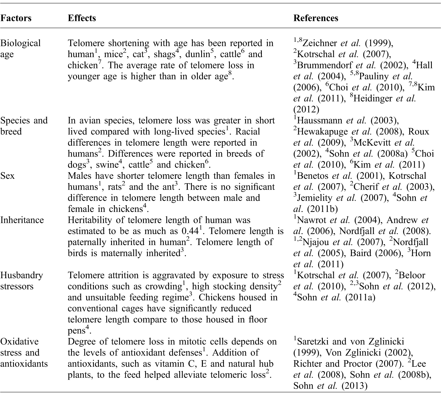
There is increasing evidence that telomere length is linked to senescence. Therefore, telomere length might provide a measure of an individual's aging and also provide an indicator of organismal bioactivity. It is clear that chicken telomere length dynamics are similar to humans and are influenced to a similar extent by various factors that affect telomere length. Thus, this study reaffirms that chickens represent a good model for aging and cellular senescence. Given that there is significant variation in telomere length among individuals, the next obvious question is the effect of this variation. This review article presents a basic picture of the various factors affecting chicken telomere length (Table 1). The most important factor is age. Age directly or indirectly affects telomere length in any individual. Therefore, the telomere-shortening may more accurately represent the influence of a given factor than the mean telomere length. It should also be noted that genetic factors are the only factors affecting telomere quantity until birth. However, after birth, there is a greater interplay between factors acting at both the genetic and the epigenetic level. Therefore, telomere length in general is greatly affected by factors acting at the epigenetic and environmental level, which most likely influences the life span of an individual.
Summary of factors affecting telomere length.

Acknowledgement
This work was supported by a grant from the Next-Generation Biogreen 21 Program (No. PJ007981), Rural Development Administration, Ministry of Agriculture, Food and Rural Affairs, Republic of Korea.