Introduction
Cochlear implant surgery is increasingly being performed through a small incision (less than 3 cm). The benefits of a small incision include reduced hair shaving, smaller scar size and fewer wound complications.Reference Gibson, Harrison and Prowse1–Reference Ray, Gibson and Sanli3 Efforts have been made to maximise surgical exposure during small incision surgery. For instance, the development of instruments such as the Graham retractor has enabled an improved surgical view when drilling the package bed (i.e. the well in the skull for the body of the implant) through a small incision.Reference Obholzer and Graham4
Proponents of this minimal access technique have also described the use of elasticated stay hooks for skin retraction.Reference James and Papsin5, Reference Monksfield and Proops6 These can provide retraction at multiple points in a wound, and retract in a more radial fashion than traditional retractors. In addition, the placement of the hooks can be adjusted to retract deeper tissues as the dissection progresses. They are less bulky than traditional retractors, which helps to maximise exposure, especially when drilling in a confined space. Furthermore, they obviate the need for an assistant and remain static during delicate steps in microsurgery. Finally, the incision can be as small as 2.5 cm in length (less than the width of a cochlear implant); the wound size will have lengthened by the time of implant insertion (associated with the constant traction during the procedure, which causes the wound size to increase).
These elasticated stay hooks can either be clipped to the surgical drapes or to a retractor ring (the latter can be utilised to provide equal retraction in all directions);Reference Monksfield and Proops6 however, both of these methods have disadvantages.
Materials and methods
As an alternative, we describe the use of a laryngeal suspension bar (‘Pete's bar’), in which the bar acts as a fixed structure to which the elasticated stay hooks can be attached.
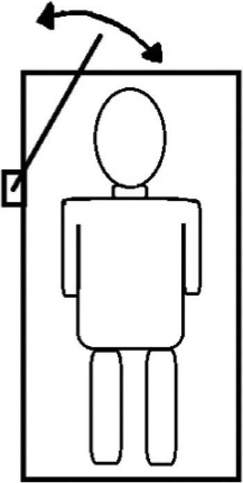
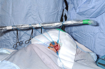
The laryngeal suspension bar is covered with a sterile transparent drape (the material normally used for endoscope light and camera cables). It is attached to the table at the level of the patient's shoulders. The angle and height of the bar is adjusted as required, as indicated in Figure 1. The elasticated stay hooks are then attached to the bar: they are wrapped around the bar twice to prevent slipping and are then secured with clips (Figure 2).
Diagrammatic representation of the laryngeal suspension bar position in relation to the patient's head (arrow shows adjustable angle for optimal retraction).

Intra-operative view showing elasticated stay hooks attached to the laryngeal suspension bar during small incision cochlear implantation.

Discussion
A potential criticism of retractor ring use is that it provides excellent exposure only if the focus of the dissection remains in the centre of the wound throughout the operation. However, the small incision technique relies on the fact that the wound is mobile, and can be retracted anteriorly and posteriorly into position to expose the relevant anatomy, depending on which part of the procedure is being performed.
The authors found that during posterior tympanotomy, cochleostomy and electrode insertion, most of the retraction force was required anteriorly. This force can be achieved by clipping the elasticated stay hooks to the drapes; however, the drapes are prone to move under a degree of tension, resulting in the loss of retraction. In addition, significant soft tissue injury may result from inadvertent application of a clip to underlying facial structures.
Conclusion
When using elasticated stay hooks, we have found the laryngeal suspension bar technique to be safer and more effective for obtaining optimal exposure than a technique whereby the stay hooks are attached directly to the drapes or to a disposable retractor ring. In addition, the bar technique utilises equipment that is already available in most ENT operating theatres, thereby obviating the need to purchase an additional disposable retractor ring (which retails at around £35.00 per unit).
Acknowledgement
The authors wish to acknowledge Peter Sleeman-Barker (senior operating department practitioner (scrub) at the ENT department, John Radcliffe Hospital, Oxford, UK) for his help in developing the laryngeal suspension bar technique.