Introduction
Parasite-induced mortality is a key process in host–parasite relationships, and typically operates in an intensity-dependent manner (Hudson et al., Reference Hudson, Newborn and Dobson1992; Benesh, Reference Benesh2011; Schutgens et al., Reference Schutgens, Cook, Gilbert and Behnke2015), but there are exceptions (Benesh & Valtonen, Reference Benesh and Valtonen2007). As a density-dependent process, parasite numbers in individual hosts at the time of death are important, and have been obtained directly for many vertebrate (e.g. Forrester, Reference Forrester1971; Hayes et al., Reference Hayes, Bailer and Mitrovic1973; Goater & Ward, Reference Goater and Ward1992) and invertebrate (e.g. Wedekind, Reference Wedekind1997; Benesh, Reference Benesh2011; Shostak et al., Reference Shostak, Van Buuren and Cook2015) hosts. An alternative, indirect, approach when dead hosts cannot be examined directly is to study parasite frequency distributions in groups of hosts that survive some mortality event (e.g. Crofton, Reference Crofton1971; Lester, Reference Lester1977; Keymer, Reference Keymer1980). Unfortunately, most macroparasites have aggregated distributions (Crofton, Reference Crofton1971; Shaw & Dobson, Reference Shaw and Dobson1995). These distributions have a long right tail due to a small number of heavily infected hosts, which are also going to be the hosts most susceptible to parasite-induced mortality (Anderson & May, Reference Anderson and May1978; May & Anderson, Reference May and Anderson1978), but which have low expected frequencies for any given intensity (Crofton, Reference Crofton1971). Direct determination of parasite numbers requires fewer assumptions than indirect methods and can better detect subtle changes in the number of hosts in different infection classes as a consequence of parasite-induced mortality.
Invertebrates have many advantages as experimental hosts from both logistical and ethical perspectives (Woolsey et al., Reference Woolsey, Fredensborg, Jensen, Kapel and Meyling2015), particularly for studies where mortality is the endpoint and large sample sizes may be required. Terrestrial arthropods present an interesting situation for studies on host mortality: they host a variety of helminth parasites (Roberts & Janovy, Reference Roberts and Janovy2009) and many are important pests. Moreover, some, such as tenebrionid beetles, are important model systems for host–parasite–environment interactions (Shostak, Reference Shostak2014; Schutgens et al., Reference Schutgens, Cook, Gilbert and Behnke2015; Woolsey et al., Reference Woolsey, Fredensborg, Jensen, Kapel and Meyling2015). However, terrestrial arthropods can desiccate post-mortem, rendering their carcasses difficult to dissect and obtain direct parasite counts (Ungureanu, Reference Ungureanu1972; Keymer, Reference Keymer1980; Schutgens et al., Reference Schutgens, Cook, Gilbert and Behnke2015). Our aim was to develop and evaluate methods for quantifying parasite intensity from otherwise unusable dry carcasses.
A method to rehydrate dry mosquito carcasses and examine them for protozoan and helminth parasites has been described (Ungureanu, Reference Ungureanu1972). It uses a two-stage rehydration process: first, application of a weak detergent or emollient solution (e.g. 1% soap, 0.5% sodium benzoate (NaC7H5O2) or 0.5% potassium hydroxide (KOH)), and then, soaking in saline or tap water. This method does not appear to have been applied to insects, other than Diptera, until recently. A study on parasite-induced mortality (Shostak et al., Reference Shostak, Van Buuren and Cook2015) attempted to rehydrate carcasses of flour beetles Tribolium confusum using 0.5% KOH (2–3 h) followed by water (3 days) prior to dissection for cysticercoids of the rat tapeworm Hymenolepis diminuta. This method was promising from the standpoint of accurate parasite recovery, but was inefficient because dissection was much more time-consuming than when performed on fresh carcasses (Shostak et al., Reference Shostak, Van Buuren and Cook2015).
Our first objective was to determine when the use of a rehydration procedure might even be necessary, by documenting post-mortem drying of carcasses of T. confusum under various conditions. Our second objective was to develop and evaluate various rehydration techniques for accurate parasite recovery. The aim was to identify an optimal technique that would allow for speedy application and ease of dissection, while maintaining the integrity of recovered parasites.
Materials and methods
Maintenance of parasites and hosts
The life cycle of H. diminuta comprises adult parasites that live in the intestine of rats, passing eggs in the faeces that are ingested by tenebrionid beetles. Once ingested, the eggs hatch, releasing oncospheres that grow and develop in the haemocoel. Parasite growth and development in the beetle has been described qualitatively (Voge & Heyneman, Reference Voge and Heyneman1957) and quantitatively (Shostak et al., Reference Shostak, Walsh and Wong2008). There is an initial period of exponential parasite growth and differentiation, with parasites passing rapidly from predominantly stage 2 (unidirectional elongation) at 5 days post-infection (PI), to stage 3 (from first appearance of tripartite body, up to scolex formation) at 6–8 days PI, to stage 4 (withdrawal of scolex, but without capsule development) at 8–9 days PI, followed by a slowing of parasite growth, and final differentiation by 9–10 days PI into stage 5 (mature cysticercoid with a well-developed capsule surrounding a withdrawn scolex), which is the terminal, long-lived stage of development in the beetle.
We maintained H. diminuta by passage between rats (Sprague–Dawley) and flour beetles (T. confusum) using procedures described elsewhere (Shostak, Reference Shostak2009). Mixed-sex beetles (2 months post-eclosion) were fasted for 3–7 days prior to infection. Parasite eggs were collected and purified from freshly collected rat faeces, and pipetted on to multiple oatmeal flakes in a 75-mm diameter glass bowl. Beetle numbers and density, and egg density varied with experimental design, but all exposures were for 24 h, with beetles stored subsequently in a white flour–5% brewer's yeast culture medium (28°C, dark) to permit parasite development.
Experimental design
General procedures
Treatments were assigned at random (Microsoft Excel 2010 random number generator, Microsoft Corporation, Redmond, Washington, USA) either to individual beetles, or to groups of ≤10 beetles in which the groups themselves were formed using a sequential allocation method (Shostak et al., Reference Shostak, Van Buuren and Cook2015). We recognized that no single method of killing beetles could replicate all types of natural beetle mortality. We opted simply for rapid killing by placing beetles for 5 min in a sealed vial containing a cotton swab saturated with chloroform. Although chloroform can bind to tissues by hydrogen bonds, it does not cause irreversible changes to proteins or interfere with rehydration (Puchtler et al., Reference Puchtler, Waldrop, Conner and Terry1968). Beetle carcasses were transferred to 1.5-ml microtubes (Axygen Inc., Union City, California, USA), and either capped immediately or kept open, according to experimental protocol, until used in rehydration tests.
Rehydration treatments were combinations of initially soaking groups of ≤10 beetles for various times in 400 μl distilled water (H2O) containing various concentrations (by weight) of KOH, followed by removal of the KOH and replacement with 400 μl distilled H2O for various times. The treatments were chosen to represent a range of qualitative categories of ‘harshness’ (i.e. increased time and concentration of exposure to KOH) that likely present a trade-off between the speed of the procedure and the condition of material obtained. As ‘mild’ treatments, which we viewed as minor modifications of the protocols in Ungureanu (Reference Ungureanu1972), we tested: H2O for 2 days; 0.5% KOH for 1 h followed by H2O for 2 days; 5% KOH for 1 h followed by H2O for 1 day; 10% KOH for 1 h followed by H2O for 1 day; 0.5% KOH for 1 day. As ‘moderate’ treatments, we tested: 5% KOH for 1 day; 10% KOH for 1 day. As ‘harsh’ treatments, we tested: 5% KOH for 1 day followed by H2O for 5 days; 10% KOH for 1 day followed by H2O for 5 days.
Following rehydration, beetles were transferred to water (for cysticercoid counts only), or saline (9 g sodium chloride (NaCl), 0.2 g potassium chloride (KCl), 0.2 g calcium chloride (CaCl2) per litre) when cysticercoids were also going to be measured, and then dissected. Contents were examined routinely using a stereomicroscope at 25× magnification with transmitted light, and parasites were counted. Parasites were separated from host debris and photographed at 2048 × 1536 pixel resolution using a Coolpix 995 digital camera (Nikon Canada, Inc., Mississauga, Canada) with an Optem eyepiece adapter (Bock Optronics, Etobicoke, Canada). Cysticercoid body lengths were measured from photographs using the method of Shostak et al. (Reference Shostak, Walsh and Wong2006). Qualitative observations on the ease of dissection were recorded.
All chemicals were BDH (VWR International, Radnor, Pennsylvania, USA) unless indicated otherwise. All post-mortem procedures were done under ambient conditions (21–22°C and 19–29% relative humidity), as measured using a Traceable® Jumbo max–min temperature humidity meter (Control Company, Friendswood, Texas, USA).
Post-mortem desiccation of beetle carcasses
The aim of this experiment was to assess how post-mortem storage conditions affect carcass desiccation between host death and examination. Groups of beetles were weighed live (ML204 balance, d = 0.1 mg, Mettler Toledo, Mississauga, Canada), killed, then stored under various test conditions and reweighed at various times 1–49 days post-mortem. We used three storage conditions at ambient temperature to provide a gradient from higher to lower relative humidity: (1) groups of 10 beetles placed immediately post-mortem into 1.5-ml microtubes and capped (approximately 30 volumes of air per beetle), with each tube opened only once after that for reweighing; (2) groups of 50 beetles placed in open 16-ml vials under ambient relative humidity and reweighed daily; (3) groups of 50 beetles placed in open 16-ml vials, but stored within a capped 120-ml vial containing 40 g of the desiccant Drierite (W. A. Hammond Drierite Company, Xenia, Ohio, USA), and reweighed daily. We assumed that changes in beetle mass post-mortem were due to water loss (Rigaux et al., Reference Rigaux, Haubruge and Fields2001).
Post-mortem examination without rehydration
We determined the time post-mortem before a rehydration method would even be needed. Beetles with 2-week-old infections were killed, with three randomly selected for immediate dissection, and three more at each of 1, 3 and 5 days post-mortem after storage in open microtubes under ambient conditions. We removed host debris and photographed all parasites to evaluate general appearance, and to measure cysticercoid body length as a proxy for parasite size. Observed lengths were compared to predicted lengths based on cysticercoids dissected from freshly killed beetles with comparable infection intensity, using raw data available from previous studies (Shostak et al., Reference Shostak, Walsh and Wong2006, Reference Shostak, Walsh and Wong2008).
Rehydration of stage 2–4 parasites
We performed one experiment to evaluate the number and condition of developing parasites following rehydration. Given the rapid development of H. diminuta (Shostak et al., Reference Shostak, Walsh and Wong2008), we predicted that we would need to examine beetles at both days 7 and 8 PI to maximize the chances of obtaining parasites at stages 2–4. Beetles were exposed to parasite eggs, and then assigned at random to groups of ≤10 beetles and stored (28°C, dark) in culture medium. These groups of beetles were assigned at random to one of four treatments: (1) killed on day 7 PI followed by immediate dissection; (2) killed on day 7 PI, sealed in a microtube to dry for 1 week, and then rehydrated and dissected; (3) killed on day 8 PI followed by immediate dissection; (4) killed on day 8 PI, sealed in a microtube to dry for 1 week, and then rehydrated and dissected.
We tested two mild (H2O for 2 days; 5% KOH for 1 h followed by H2O for 1 day) and one moderate (5% KOH for 1 day) rehydration treatments. All three were tested on day 7 PI rehydrated beetles, but the H2O-only treatment produced such poor recovery on day 7 PI that it was not used on day 8 PI. Parasites recovered by dissection of fresh or rehydrated beetles were photographed and categorized by developmental stage (Voge & Heyneman, Reference Voge and Heyneman1957). Confirmation of parasite identity in suspect cases was made by preparing a wet mount and examining at 100× using a compound microscope.
Rehydration of stage 5 parasites
We performed one experiment to evaluate the recovery of parasites following rehydration. Beetles were exposed to parasite eggs and killed on day 14 PI. Control beetles were selected at random for immediate dissection to determine baseline parasite numbers. The remaining beetles were killed, sealed in microtubes containing groups of 10 beetles, and stored until rehydration. Beetles were rehydrated using all treatments, and examination of the replicate groups for each treatment was staggered such that the time between first and last replicate for each treatment spanned 1–4 months post-mortem.
We ran a separate series of experiments to evaluate the condition of parasites that were recovered following rehydration. We tested each rehydration treatment following several different carcass storage conditions: (1) immediately after killing the beetles (0 weeks dried); (2) sealed in microtubes immediately after killing, then rehydrated 1 week later (1 week dried); (3) left in open microtubes under ambient conditions, then rehydrated 2 weeks later (2 weeks dried); (4) sealed in microtubes after natural death from exposure to diatomaceous earth, which kills by desiccating the insect (Korunic, Reference Korunic1998; Shostak et al., Reference Shostak, Van Buuren and Cook2015), then rehydrated after 70 weeks (70 weeks dried). We exposed a different set of beetles for each test. We rehydrated 2–4 beetles per combination of storage condition and rehydration treatment and assessed the condition of the parasites at dissection, where parasites were counted and photographed.
Data analysis
We used non-parametric Kruskal–Wallis or Kolmogorov–Smirnov tests to compare parasite recoveries among rehydration methods, with a Dwass, Steel, Critchlow–Fligner multiple comparison analysis (PROC NPAIR1WAY) in SAS 9.4 (SAS Institute Inc., Cary, North Carolina, USA). The body length of rehydrated cysticercoids relative to the expected length for fresh cysticercoids was tested using analysis of variance, ANOVA (PROC GLM), with a Tukey adjustment for post-hoc testing among treatment groups. Data are presented as mean ± 1 standard deviation (SD) unless indicated otherwise. Results with P < 0.05 were considered statistically significant.
Results
General observations
Even a carcass desiccated to the point of being brittle could be softened sufficiently by the mildest of the rehydration treatments we used so that it could be teased apart in search of cysticercoids, but internal organs formed a network of fibres that obscured and stuck to smaller cysticercoids. We would qualitatively characterize as ‘painstaking’ the effort required to dissect these carcasses to the point where we were satisfied that all parasites present had been detected. By increasing the concentration or duration of KOH exposure, carcasses became more flexible, and internal host structures were digested so that parasites became more visible and floated free; a dissection could be completed in about one-third of the time. Carcasses remained intact even when rehydrated by the harshest treatments; we had no indication that any cysticercoids were lost by a carcass breaking before dissection.
Post-mortem desiccation of beetle carcasses
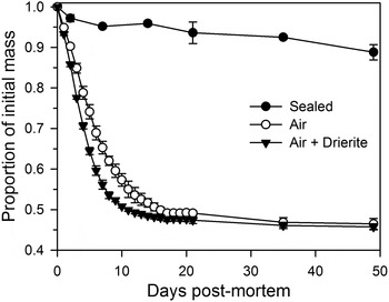
Freshly killed beetles sealed in microtubes lost 0.16 ± 0.024% (SE) of their original body mass day−1 (linear regression, n = 6 groups, R 2 = 0.83, P < 0.001) and after 49 days were still >85% of their fresh mass (fig. 1). Beetles stored while exposed to ambient air lost 5.1 ± 0.12% (SE) day−1 (n = 3 groups, R 2 = 0.99, P < 0.001) for the first 7 days post-mortem, and by day 49 were at 46% of their original mass (fig. 1). Beetles stored while exposed to air conditioned with Drierite lost 6.3 ± 0.36% (SE) day−1 (n = 3 groups, R 2 = 0.98, P < 0.001) for the first 7 days post-mortem, and by day 49 were at 45% of their original mass (fig. 1).
Changes in mass of Tribolium confusum carcasses when sealed immediately post-mortem in microtubes (n = two groups of 10 beetles), exposed to ambient air (n = three groups of 50 beetles) or exposed to air conditioned by Drierite (n = three groups of 50 beetles). Values are mean ± 1 SD.

Post-mortem examination without rehydration
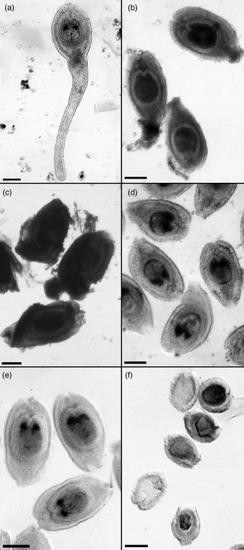
Dissection of beetle carcasses exposed to ambient conditions for 1 day post-mortem was judged qualitatively to be as easy as the dissection of freshly killed beetles. At 3 and 5 days post-mortem, carcass texture was noticeably tougher, though not yet brittle, and separation of cysticercoids from internal organs was difficult. The predicted relationship between cysticercoid body length (y, as log mm) and intensity (x) from freshly dissected beetles was: y = –0.3735–0.0023x (n = 705, R 2 = 0.62). Relative cysticercoid length (r/f, where r = body length of a rehydrated cysticercoid, and f = body length predicted by the formula above for a fresh specimen at the same intensity) varied according to time post-mortem (ANOVA, F 3,435 = 25.2, P < 0.001). Post-hoc testing showed similar relative cysticercoid size when comparing 0–1 day post-mortem (1.024 ± 0.082, n = 282) and when comparing 3–5 days post-mortem (0.962 ± 0.068, n = 157), but relative cysticercoid size at 3–5 days was smaller than at 0–1 day (Tukey test, P < 0.05). A recognizable cercomer was present on 126/149 (85%) cysticercoids from freshly killed beetles (fig. 2a), 43/133 (32%) after 1 day, 19/105 (18%) after 3 days and 1/52 (2%) after 5 days. After 5 days of air drying (fig. 2b), cysticercoids remained recognizable by their size and layered structure, although they were considerably darker and internal details were indistinct.
Stage 5 cysticercoids of Hymenolepis diminuta recovered from Tribolium confusum dissected (a) immediately post-mortem; (b) 5 days post-mortem using no rehydration method; (c) 70 weeks post-mortem after placing in water for 2 days; (d) 70 weeks post-mortem after placing in 5% KOH for 1 day; (e) immediately post-mortem after placing in 5% KOH for 1 day; (f) 2 weeks post-mortem after placing in 5% KOH for 1 day followed by water for 5 days. Bars represent 100 μm.

Rehydration of stage 2–4 parasites
The total number of stage 2–4 parasites recovered from freshly dissected beetles on day 7 PI (n = 100 beetles, 0–45 parasites each) and on day 8 PI (n = 84, 1–52 parasites) was similar (Kolmogorov–Smirnov two-sample test, KS a = 0.579, P = 0.89). Few stage 2 parasites were recovered from any beetles, fresh or rehydrated, and were excluded from further analysis.
Fresh stage 3 and stage 4 parasites were easily recognizable. Rehydrated stage 3 parasites were typically dark brown to opaque and identifiable only by the elongate body outline and presence of a cavity, and they were often broken. Stage 4 parasites were also dark brown to opaque, the cercomer was always broken or missing, and if the scolex was retracted, it was not always visible when using a stereomicroscope and needed confirmation using a compound microscope.
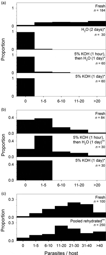
Stage 3 parasites were found on days 7 and 8 PI and those data were pooled to evaluate recovery. All rehydration treatments resulted in significantly lower recovery of stage 3 parasites compared to fresh samples (fig. 3a). Whereas few (6%) freshly dissected hosts were uninfected, almost all (90–100%) rehydrated hosts were categorized as uninfected. Stage 4 parasites were found on day 8 PI only. The number of stage 4 parasites recovered by a mild rehydration treatment (5% KOH for 1 h followed by H2O for 1 day) was indistinguishable from that of fresh samples (fig. 3b). However, a moderate rehydration treatment (5% KOH for 1 day) recovered significantly fewer (≤5) stage 4 parasites (fig. 3b).
Numbers of Hymenolepis diminuta recovered from freshly killed Tribolium confusum compared to recovery from dried carcasses rehydrated by various methods, for (a) stage 3 parasites; (b) stage 4 parasites; (c) stage 5 parasites. Proportions of hosts in various abundance categories for each rehydration treatment were compared to the corresponding control (fresh) distribution (*, P < 0.05; ns, not significant).

Rehydration of stage 5 parasites
Freshly dissected beetles had 0–69 parasites (fig. 3c). Rehydration treatments did not vary in parasite recovery (Kruskal–Wallis test, χ2 8 = 6.94, P = 0.54). Pooled, these rehydrated beetles had 0–87 parasites (fig. 3c), and their distribution was similar to that of fresh beetles (Kolmogorov–Smirnov two-sample test, KS a = 0.879, P = 0.42), with strong overlap of their frequency distributions over the entire range of parasite abundances (fig. 3c).
Treatments differed in ability to rehydrate beetles. Prior to treatment, carcasses floated. Mild rehydration treatments produced a mixture of floating and sunken (some also swollen) carcasses by the end of treatment. Dissection typically took longer than for fresh carcasses, and following dissection the internal organs were present as opaque, sticky debris that obscured cysticercoids and extended the time required to macerate carcasses sufficiently to reveal parasites. Almost all carcasses treated by moderate–harsh methods sank, and had swollen bodies, by the end of treatment. Dissection following rehydration by moderate–harsh methods was much quicker than for fresh carcasses, because most of the internal organs of the beetle were liquefied, leaving cysticercoids clearly visible.
Fresh stage 5 cysticercoids (fig. 2a) were transparent, and had distinct layering and the presence of clusters of granules, as noted by Voge & Heyneman (Reference Voge and Heyneman1957). The cercomer rarely remained attached following any rehydration treatment. Even rehydration using only H2O resulted in just 22/383 (6%) cysticercoids recovered with a cercomer, compared to 85% from fresh dissections. Cysticercoids from beetles rehydrated by mild treatments (fig. 2c) had tissue adhering externally, and were often so dark that the layering and granules were obscured. Moderate rehydration treatments increased overall cysticercoid transparency (fig. 2d–e), but while the granules remained as visible as in fresh cysticercoids, some damage to cysticercoids was evident. The boundaries between layers became slightly less distinct when moderate rehydration was applied to a long-dried host (fig. 2d), and more so when applied to a freshly dead host (fig. 2e). Harsh rehydration treatments resulted in loss of most of the outer layers, leaving only remnants of the tissue immediately surrounding the scolex (fig. 2f).
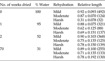
The damage to cysticercoids from moderate–harsh methods was evident from measurements of relative cysticercoid size (table 1). Freshly killed beetles were assumed to be at 100% of their live mass. Beetle water content before rehydration (table 1) after 1 week drying was estimated from the ‘sealed’ group in fig. 1; after 2 weeks, from the ‘air’ group in fig. 1; after 70 weeks, by comparing beetles that died following diatomaceous earth exposure and were stored in sealed microtubes for 70 weeks (0.76 ± 0.19 mg beetle−1, n = 16 determinations) to the mass of freshly killed beetles (2.48 ± 0.03 mg beetle−1, n = 6 determinations). There was a significant interaction between post-mortem drying time and rehydration treatment on relative length (table 1) of rehydrated cysticercoids (ANOVA, F 6,2332 = 139, P < 0.001). No rehydration treatment completely restored cysticercoids to their fresh length (table 1), but mild treatments resulted in cysticercoids that were close; on average, 88–92% of their fresh length. Cysticercoids from freshly killed hosts that also received a harsh rehydration treatment were only 31% of expected fresh length (table 1). Measurements were consistent: coefficients of variation (SD/mean) in relative body length were low (8–20%) for all combinations of time dried and mild–moderate rehydration, and only slightly larger (19–25%) following harsh rehydration (table 1).
Relative lengths of cysticercoids of Hymenolepis diminuta recovered from carcasses of Tribolium confusum dried for various times post-mortem and then rehydrated using mild, moderate or harsh treatments. Percentage water (% Water) was estimated by final carcass mass relative to fresh mass. Relative length, expressed as mean ± 1 SD (n), was calculated as the observed length of each rehydrated cysticercoid relative to the expected length for cysticercoids from a fresh beetle with the same infection intensity.

Discussion
We demonstrated that even beetle carcasses stored under the low relative humidity conditions of our laboratory could be dissected without rehydration up to 1 day post-mortem with no reduction in ease of dissection and no degradation of cysticercoids. Therefore, no rehydration method would be required for dissection within this narrow window of time. Dissection could also be done up to at least 5 days post-mortem (or about 75% fresh host mass) without rehydration, albeit with difficulty and with some degradation of cysticercoids. However, if time of death is unknown, it is important to recognize the influence of pre- and post-mortem storage conditions on carcass water content and rate of drying. For example, Tribolium castaneum can tolerate loss of about two-thirds of its body water content before death, and the rate of post-mortem water loss from a carcass can be 2–4× the pre-mortem rate (Rigaux et al., Reference Rigaux, Haubruge and Fields2001). We found that the rate of initial post-mortem water loss varied more than 30× between beetles sealed in microtubes immediately post-mortem and those stored in the open. For studies in which beetle carcasses are recovered infrequently, or from natural habitats, we conclude that little expectation can be placed on the time of death, and hence water content, of the carcass.
Previous work by Shostak et al. (Reference Shostak, Van Buuren and Cook2015) showed that rehydration of T. confusum and successful recovery of fully developed cysticercoids of H. diminuta was possible. However, given the uncertainty regarding carcass water content noted above, a more important question becomes whether any single rehydration treatment can produce acceptable results over the entire range, from freshly dead to fully desiccated carcasses. With respect to simply obtaining accurate parasite counts, any of the mild–moderate treatments we used were suitable for routine application on carcasses ranging from freshly dead to highly desiccated. Harsh treatments resulted in extensive degradation of cysticercoids, bordering on cysticercoids being unidentifiable and potentially leading to underestimation of parasite numbers. However, with respect to ease of dissection and cysticercoid quality, rehydration treatments exhibited major differences. Progressively harsher conditions (either by length or concentration of KOH exposure) facilitated dissection by softening or digesting host tissues. On the other hand, all rehydration treatments were associated with loss of the cercomer, and progressively harsher treatments appeared to digest cysticercoids from the outside in (fortunately, at least, exhibiting consistency within a treatment). While we had hoped to identify an optimal rehydration treatment for all situations, we could not. However, the opposing outcomes of a progressive increase in ease of dissection, but decrease in parasite condition, when proceeding from milder to more moderate treatments, would permit users of this general method to choose a specific technique that would give a balance between the two outcomes appropriate for the goal of their particular study. If accurate counts alone are required, we recommend a moderate treatment because of increased speed of dissection and visibility of the parasites. If recovery of parasites with minimal damage is also required, we recommend a mild treatment, at the expense of increased time to complete each dissection. We advise against using any harsh treatment.
Rehydration was much less successful for recovering stage 2–4 (pre-infective) H. diminuta, than for fully developed cysticercoids. We speculate that final differentiation of the parasite necessary to deal with the digestive system of its next host also makes it more resistant to damaging effects of the rehydration procedure. Parasite numbers may be underestimated following rehydration if pre-infective forms are present, but given the rapid development of H. diminuta in the intermediate host and lengthy persistence once infective (Voge & Heyneman, Reference Voge and Heyneman1957; Shostak et al., Reference Shostak, Walsh and Wong2008; Shostak, Reference Shostak2014), we would anticipate minimal impact on overall parasite detection.
Our approach to rehydration shows promise as a general method for detecting larval helminths in terrestrial arthropods. In addition to hosting H. diminuta, beetles in the genus Tribolium are also hosts for many other helminths: other cyclophyllidean cestodes such as Hymenolepis nana (Voge & Heyneman, Reference Voge and Heyneman1957), Hymenolepis microstoma and Hymenolepis citelli (Heyneman & Voge, Reference Heyneman and Voge1971), and Raillietina cesticillus (Gordon & Whitfield, Reference Gordon and Whitfield1985); a spirurid nematode Protospirura muricola (Schutgens et al., Reference Schutgens, Cook, Gilbert and Behnke2015); and several species of entomopathogenic rhabditid nematodes (Rumbos & Athanassiou, Reference Rumbos and Athanassiou2012). Other terrestrial arthropod–helminth systems that might benefit from availability of a rehydration method would include formicine ants infected with metacercariae of the plagiorchiform digenean Dicrocoelium dendriticum (Manga-González & González-Lanza, Reference Manga-González and González-Lanza2005). Rehydration may be less successful for parasites that cause host mortality prior to achieving the infective stage, such as the cyclophyllidean cestode Dipylidium caninum in fleas (Marshall, Reference Marshall1967). Finally, the original rehydration protocol (Ungureanu, Reference Ungureanu1972) claimed to detect helminths such as digenean metacercariae and filarial nematodes hosted in more delicate terrestrial arthropods, such as mosquitoes, although the condition of the helminths recovered was not described.
We cannot know how many previous studies have avoided quantifying parasite-induced host mortality because of statements in the literature (e.g. Keymer, Reference Keymer1980; Schutgens et al., Reference Schutgens, Cook, Gilbert and Behnke2015) that helminth counts in dried insect carcasses cannot be determined. The consequences may vary. On the one hand, lack of parasite counts from desiccated hosts may simply lead to increased experimental error, such as when infections of T. confusum with P. muricola could be categorized crudely ante-mortem but not confirmed post-mortem (Schutgens et al., Reference Schutgens, Cook, Gilbert and Behnke2015). On the other hand, lack of direct parasite counts from dead hosts may affect conclusions. Before-and-after frequency distributions from groups of live hosts (Keymer, Reference Keymer1980) suggest a linear relationship between numbers of H. diminuta and survival of T. confusum, whereas actual parasite counts from dead, rehydrated hosts (Shostak et al., Reference Shostak, Van Buuren and Cook2015) indicate that host mortality is intensity-independent when fewer than about 20 parasites are present.
Our study has shown that the rehydration method initially proposed by Ungureanu (Reference Ungureanu1972) for mosquito hosts is robust and should be easily modifiable to deal with other hosts and parasites, and we have identified dissection ease and parasite condition as features that can be balanced by a researcher to meet experimental goals. By permitting accurate determination of parasite number at time of death for individual hosts, new opportunities for studies on parasite-induced mortality of terrestrial arthropod hosts may now be considered or revisited. Experimental designs with much higher resolution of mortality effects are now possible. The need to control parasite establishment precisely in control and treatment groups, which is critical for before-and-after studies, is reduced. Finally, killing and drying of infected hosts intentionally may be considered as a preservation method, and can facilitate large-scale experiments with time-sensitive collection points, where dissection of large numbers of fresh carcasses would be unfeasible logistically.
Financial support
This research received no specific grant from any funding agency, commercial or not-for-profit sectors.
Conflict of interest
None.
Ethical standards
The authors assert that all procedures contributing to this work comply with the ethical standards set by the Canadian Council on Animal Care (Ottawa, Canada) on the care and use of laboratory animals.