Introduction
Prothymosin alpha (PTMA) is a highly acidic (Frangou-Lazaridis et al., Reference Frangou-Lazaridis, Clinton, Goodall and Horecker1988), intrinsically disordered protein (Gast et al., Reference Gast, Damaschun, Eckert, Schulze-Forster, Maurer, Müller-Frohne, Zirwer, Czarnecki and Damaschun1995), which was first extracted from rat thymus and characterized as an immunogenic factor (Haritos et al., Reference Haritos, Goodall and Horecker1984a) but soon detected in a variety of mammalian tissues (Clinton et al., Reference Clinton, Frangou-Lazaridis, Panneerselvam and Horecker1989; Haritos et al., Reference Haritos, Tsolas and Horecker1984b). The presence of a nuclear localization signal and the adoption of a peculiar random coil conformation are amongst the reasons behind its interaction with a number of molecular partners; hence, today PTMA is known to be a very conserved and widely expressed molecule, involved in several and diverse biological processes, such as H1 histone interaction and chromatin remodelling (Karetsou et al., Reference Karetsou, Sandaltzopoulos, Frangou-Lazaridis, Lai, Tsolas, Becker and Papamarcaki1998; Ueda et al., Reference Ueda, Matsunaga and Halder2012), cell death (Enkemann et al., Reference Enkemann, Ward, Trumbore and Berger2000a; Jiang et al., Reference Jiang, Kim, Shu, Zhao, Zhang, Kofron, Donnelly, Burns, Ng, Rosenberg and Wang2003; Malicet et al., Reference Malicet, Giroux, Vasseur, Dagorn, Neira and Iovanna2006; Ueda Reference Ueda2009), transcriptional regulation (Martini et al., Reference Martini, Delage-Mourroux, Kraichely and Katzenellenbogen2000; Karetsou et al., Reference Karetsou, Kretsovali, Murphy, Tsolas and Papamarcaki2002; Martini & Katzenellenbogen, Reference Martini and Katzenellenbogen2003) cancer development (Dominguez et al., Reference Dominguez, Magdalena, Cancio, Roson, Paredes, Loidi, Zalvide, Fraga, Forteza and Regueiro1993; Tsitsiloni et al., Reference Tsitsiloni, Stiakakis, Koutselinis, Gogas, Markopoulos, Yialouris, Bekris, Panoussopoulos, Kiortsis and Voelter1993; Wu et al., Reference Wu, Habib, Mitry, Reitsma, van Deventer and Chamuleau1997; Skopeliti et al., Reference Skopeliti, Voutsas, Klimentzou, Tsiatas, Beck, Bamias, Morak,i, Livaniou, Neagu, Voelter and Tsitsilonis2006; Zhang et al., Reference Zhang, Cui, Lu, Lu, Jiang, Chen, Zhang, Jin, Peng and Tang2014) and, as already alluded to, immunity (Pan et al., Reference Pan, Haritos, Wideman, Komiyama, Chang, Stein, Salvin and Horecker1986; Baxevanis et al., Reference Baxevanis, Thanos, Reclos, Anastasopoulos, Tsokos, Papamatheakis and Papamichail1992; Voutsas et al., Reference Voutsas, Baxevanis, Gritzapis, Missitzis, Stathopoulos, Archodakis, Banis, Voelter and Papamichail2000). Numerous studies have collectively hinted at the functional promiscuity of this small polypeptide and, even at its possible therapeutic applications (Moody et al., Reference Moody, Leyton, Zia, Tuthill, Badamchian and Goldstein2000; Romani et al., Reference Romani, Bistoni, Gaziano, Bozza, Montagnoli, Perruccio, Pitzurra, Bellocchio, Velardi, Rasi, Francesco and Garaci2004; Mosoian et al., Reference Mosoian, Teixeira, Burns, Khitrov, Zhang, Gusella, Klotman and Klotman2007; Mosoian, Reference Mosoian2011) suggesting that there might not be a univocal definition describing its physiological role (Piñeiro et al., Reference Piñeiro, Cordero and Nogueira2000; Hannappel & Huff, Reference Hannappel and Huff2003; Wang and Pan, Reference Wang and Pan2007).
Since 1990 some investigations have enriched this intricate picture by reporting the possible involvement of PTMA in vertebrate reproduction, specifically during spermatogenesis: today, PTMA is known to be expressed by the meiotic and post-meiotic germ cells in all the vertebrate species examined (Ferrara et al., Reference Ferrara, Izzo, Liguori, d’Istria, Aniello and Minucci2009, Reference Ferrara, Izzo, Pariante, Donizetti, d’Istria, Aniello and Minucci2010, Reference Ferrara, Pariante, Di Matteo, Serino, Oko and Minucci2013; Prisco et al., Reference Prisco, Donizetti, Aniello, Locascio, Del Giudice, Agnese, Angelini and Andreuccetti2009); interestingly, in mammals, it is also associated with the acrosomal system of the differentiating spermatids (SPT) throughout spermiogenesis (Ferrara et al., Reference Ferrara, Izzo, Pariante, Donizetti, d’Istria, Aniello and Minucci2010). Finally, the protein localizes inside the inner acrosomal membrane in rat and human spermatozoa (SPZ; Ferrara et al., Reference Ferrara, Pariante, Di Matteo, Serino, Oko and Minucci2013).
To date, no investigation has been led on Ptma during the spermatogenesis of a very common vertebrate model, the teleost Danio rerio, where two paralog genes have been described and characterized during embryogenesis (ptmaa and ptmab; Donizetti et al., Reference Donizetti, Liccardo, Esposito, Del Gaudio, Locascio, Ferrara, Minucci and Aniello2008). The anastomosing tubular organization of the testis and, above all, the fact that the SPZ do not develop an acrosomal system, is very compelling, given the established association between PTMA and the acrosome in mammals. With this in mind, and with the aim of expanding upon the current comparative knowledge, in the present work we assessed Ptma RNA expression and protein localization during adult zebrafish spermatogenesis.
Materials and Methods
Animal care and tissue extraction
Danio rerio (Cypriniformes) were obtained from a local pet shop and kept in activated carbon filtered water at a density of approximately one fish per 2 litres, with a 14 h:10 h light:dark cycle and a temperature of 28.5°C. Animals were sacrificed by immersion in a solution of 0.04% (w/v) MS-222 (Tricaine Methanesulfonate), in accordance with local and international guidelines covering experimental animals. For each animal muscles, intestine, heart, brain, ovary and testes were dissected; all tissues were quickly frozen by immersion in liquid nitrogen and stored at −80°C until RNA or protein extraction; one testis for each animal was fixed in Bouin's fluid for histological analysis. Sprague–Dawley rats (Rattus norvegicus) were housed under definite conditions (12D:12L) and they were fed standard food and provided with water ad libitum. Animals were sacrificed by decapitation under ketamine anesthesia (100 mg/kg i.p.). Additionally, adult rat testes were dissected, frozen and stored at −80°C until protein extraction, and used as a positive control in western blot analyses.
Preparation of total RNA, RT-PCR, cloning, and sequencing
Total RNA from zebrafish tissues was prepared with the procedure described by Sprenger et al. (Reference Sprenger, Konrad, Rischowsky and Gemsa1995) and treated with DNase I recombinant, RNase free (Roche Diagnostics; Monza, Italy) following the manufacturer's instructions. First strand cDNA was synthesized using 3 μg total RNA and 100 U Superscript III RT enzyme as recommended by the manufacturer (Invitrogen, Paisley, UK). As a negative control, the reaction was carried out on the same RNA without using the enzyme (RT−). Three microlitres of the obtained cDNA template were then used for the PCR reaction as described by De Rienzo et al. (Reference De Rienzo, Di Sena, Ferrara, Palmiero, Chieffi Baccari and Minucci2002) and Ferrara et al. (Reference Ferrara, Palmiero, Branno, Pierantoni and Minucci2004). Based on the published sequence of Danio rerio prothymosin alpha a mRNA (ptmaa; GenBank accession no. NM_194376.2) and prothymosin alpha b mRNA (ptmab; GenBank accession no. NM_001098730.1), specific oligonucleotide primers were designed as follows: zfPtmaa for = 5′-CATTTA-GGAAAAATGGCTGACACA-3′ and zfPtmaa rev = 5′-TGAGAACATTTCCAGCAGTGAAGC-3′; zfPtmab for = 5′-TACACAACATTAATTATGGCAGAT-3′ and zfPtmab rev = 5′-GCTCAGCAATATGAAAC-AATCCTT-3′. An appropriate region of Danio rerio Ribosomal Protein Large P0 (rplp0) mRNA (GenBank accession no. NM_031144), was amplified with specific oligonucleotide primers (Rplp0 for = 5′-ATCTCCAGAGGAACCATTGAAA-3′, Rplp0 rev = 5′-AAGCCCATGTCTTCATCAGACT-3′) and used as a control. The amplifications were carried out for 30 cycles, with denaturation at 94°C for 30 s, annealing at 58°C (ptmaa and ptmab) or 62°C (rplp0) for 45 s and extension at 72°C for 45 s, using Taq Polymerase Recombinant kit (Invitrogen; Milan, Italy). The expected RT-PCR product sizes were: 357 bp for ptmaa, 360 bp for ptmab and 497 bp for rplp0. The cDNA fragments amplified with zfPtmaa and zfPtmab primers were purified by QIAGEN gel extraction kit (QIAGEN; Hilden, Germany) and cloned into the pGEM®-T Easy Vector according to the manufacturer's instruction (Invitrogen; Milan, Italy). Sequence determination was performed on both strands with the dideoxynucleotide chain termination method (Sanger & Coulson, Reference Sanger and Coulson1975). The deduced amino acid sequences were compared with entries in the GenBank database.
Tissue quality control and classification of testicular germ cells
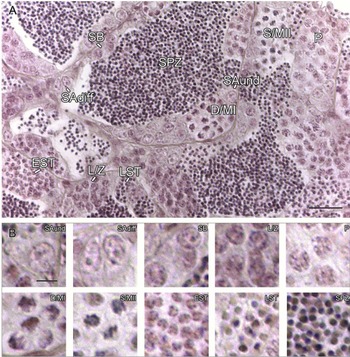
In order to assess the quality of the tissue samples, 5-μm thick testis sections were prepared and a haematoxylin–eosin staining was performed (see Fig. 1). The germ cell types inside the tubules were characterized by their size, number and morphology, following previously reported classifications (Leal et al., Reference Leal, Cardoso, Nóbrega, Batlouni, Bogerd, França and Schulz2009; Rupik et al., Reference Rupik, Huszno and Klag2011; Schulz et al., Reference Schulz, de França, Lareyre, Le Gac, Chiarini-Garcia, Nobrega and Miura2010).
Histology of the adult zebrafish testis. Haematoxylin–eosin staining of a tissue section in which the most representative germ cell types are highlighted (A). (B) Enlargement of the germ cells, which emphasizes their different sizes and morphology (for review see Schulz et al., Reference Schulz, de França, Lareyre, Le Gac, Chiarini-Garcia, Nobrega and Miura2010). SAund and SAdiff: undifferentiated and differentiated A SPG; SB: B SPG. L/Z: leptotene/zygotene primary SPC; P: pachytene primary SPC; D/MI: diplotene/metaphase I primary SPC; S/MII: secondary/metaphase II SPC; EST: early SPT; LST: late SPT; SPZ: spermatozoa. Scale bars represent 20 μm in (A), whereas 5 μm in (B).

In situ hybridization
For the in situ hybridization, randomly chosen 5-μm testis sections (seven sections/animal) were treated following the same conditions as described by Ferrara et al. (Reference Ferrara, Izzo, Liguori, d’Istria, Aniello and Minucci2009), using pGEM®-T Easy containing the ptmaa or ptmab cDNA linearized with either NcoI or SalI (Fermentas; Milan, Italy) to produce a template for antisense or sense probe, using SP6 or T7 RNA polymerases, respectively. The sense (control) and antisense cRNA probes were prepared by in vitro transcription with Dig RNA Labelling kit (Roche Diagnostics, Monza, Italy) as recommended by the manufacturer.
Preparation of total protein extracts and western blot analysis
Testes (from rat or zebrafish) were lysed in a specific buffer [1% NP-40, 0.1% SDS, 100 mM sodium orthovanadate, 0.5% sodium deoxycholate in PBS (phosphate-buffered saline; 13.6 mM NaCl; 2.68 mM KCl; 8.08 mM Na2HPO4; 18.4 mM KH2PO4; 0.9 mM CaCl2; 0.5 mM MgCl2 pH 7.4)] in the presence of protease inhibitors (4 mg/ml of leupeptin, aprotinin, pepstatin A, chymostatin, PMSF, and 5 mg/ml of TPCK). The homogenates were sonicated twice by three strokes (20 Hz for 20 s each); after centrifugation for 30 min at 10,000 g, the supernatants were stored at −80°C. Proteins from testis (100 μg) were separated by 15% SDS-PAGE and transferred to Hybond-P polyvinylidene difluoride (PVDF) membranes (Amersham Pharmacia Biotech; Buckinghamshire, UK) at 280 mA for 2.5 h at 4°C. The filters were treated for 3 h with blocking solution [5% skimmed milk in PBS pH 7.4 containing 0.1% Tween-20 (Sigma-Aldrich Corp., Milan, Italy)] before the addition of the primary antibody (zebrafish Ptmaa polyclonal antibody diluted 1:1000; Ferrara et al., Reference Ferrara, Izzo, Liguori, d’Istria, Aniello and Minucci2009, Reference Ferrara, Izzo, Pariante, Donizetti, d’Istria, Aniello and Minucci2010, Reference Ferrara, Pariante, Di Matteo, Serino, Oko and Minucci2013; Prisco et al., Reference Prisco, Donizetti, Aniello, Locascio, Del Giudice, Agnese, Angelini and Andreuccetti2009) and overnight incubation at 4°C. After washing, the filters were incubated with horseradish peroxidase-conjugated anti-rabbit IgG (DAKO; Glostrup, Denmark) diluted 1:5000 in the blocking solution. The filters were washed again and the immunocomplexes were revealed using the ECL-Western blotting detection system (Amersham Pharmacia Biotech; Buckinghamshire, UK). Results shown are representative of three independent experiments. Antibody reactivity against both Ptmaa and Ptmab was checked and confirmed via bioinformatic analysis: Ptmaa (GenBank accession no. NP_919357.2) and Ptmab (GenBank accession no. NP_001092200.1) sequences were aligned and compared using BLAST Bl2seq tool (http://blast.ncbi.nlm.nih.gov/Blast.cgi) and EMBL-EBI Clustal Omega (http://www.ebi.ac.uk/Tools/msa/clustalo/). The two sequences are highly similar, especially in their N-term regions (aa 1–43), where they show 72% identity. As the whole Ptmaa coding sequence had been injected to raise the immune response in rabbit and to produce the antiserum, both proteins can be recognized. Moreover, the analysis also revealed the same putative molecular weight of about 12 kDa for both proteins.
Immunohistochemistry
For Ptma localization, 5-μm thick testis sections were dewaxed, rehydrated, and processed as described by Ferrara et al. (Reference Ferrara, Izzo, Liguori, d’Istria, Aniello and Minucci2009). In order to check the specificity of the immunoreaction, the controls were treated either by omitting the primary antibody in the incubation reaction or by using the preimmune sera.
Immunofluorescence analysis
Immunofluorescence experiments were carried out following the same protocol as the immunohistochemistry, with some variations. In brief: sections of 5 μm were dewaxed, rehydrated, and then washed in phosphate buffer (0.01 M PBS, pH 7.4). Antigen retrieval was performed by pressure-cooking slides for 5 min in 0.01M citrate buffer (pH 6.0). Non-specific binding sites were blocked with an appropriate normal serum diluted 1:5 in PBS containing 5% (w/v) BSA before the addition of the primary antibody (zebrafish Ptmaa polyclonal antibody; Ferrara et al., Reference Ferrara, Pariante, Di Matteo, Serino, Oko and Minucci2013) diluted 1:50 and overnight incubation at 4°C. After washing in PBS, slides were incubated for 30 min with the appropriate secondary antibody (Alexa Fluor 488 Goat anti-rabbit; Invitrogen; Paisley, UK) diluted 1:500 in the blocking mixture. The slides were mounted with Vectashield + DAPI (Vector Laboratories; Peterborough, UK) for nuclear staining, and then observed under the optical microscope (Leica DM 5000 B+CTR 5000) with UV lamp, and images where viewed and saved with either IM1000 or Leica Application Suite.
Results
ptmaa and ptmab expression in the adult zebrafish tissues
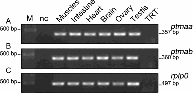
The presence of ptmaa and ptmab mRNAs was assessed by RT-PCR analysis on adult zebrafish tissues (Fig. 2). Amplification products of the expected size (357 bp and 360 bp, respectively) were detected, then cloned and sequenced: the analysis confirmed that both mRNAs are expressed in all of the analysed tissues, consistent with the usually varied pattern of their single-copy ortholog, ptma. As expected, no amplification bands were detected in negative controls (Fig. 2).
Expression of ptmaa and ptmab mRNA in adult zebrafish tissues. Agarose gel electrophoresis of RT-PCR products performed on total extracts from muscles, intestine, heart, brain, ovary, testis. Both ptmaa (A) and ptmab (B) are expressed in all the analysed tissues. The quality of the cDNA samples was checked by amplifying a fragment of the housekeeping zebrafish Ribosomal Protein Large P0 (rplp0) mRNA. M: molecular weight marker (GeneRuler Express DNA Ladder). nc: negative, no-cDNA control. TRT−: testis specific negative control of the RT reaction, performed by omitting the reverse transcriptase. All the other RT− controls were also negative (not shown).

Localization of ptma mRNAs in zebrafish mature testis
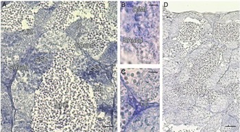
First, in order to check tissue quality and to distinguish the major germ cell types inside the tubules, an haematoxylin-eosin staining was performed on testis sections (Fig. 1). Then, the localization of ptmaa and ptmab transcripts was studied by in situ hybridization analysis (Fig. 3). Both antisense signals were present in the interstitial compartment (Leydig cells; Fig. 3 C) and in the tubules, where they were only detectable in meiotic and post-meiotic cysts. Specifically, no signals were obtained from type undifferentiated A spermatogonia (SPG; Fig. 3 A, B), or from type B SPG (Fig. 3 A), whereas they started to be visible in primary spermatocytes (SPC), such as leptotene/zygotene, pachytene (Fig. 3 A), and diplotene /metaphase I SPC (Fig. 3 B) and SPT (early and late, Fig. 3 A). No staining was detectable in sense-treated sections (Fig. 3 D). Figures refer to ptmaa; the equivalent ptmab data are not shown.
Localization of ptmaa mRNA in the testis of adult zebrafish. Sections were treated with antisense probe (A); high magnification in (B, C); or sense probe as a negative control (D). The blue staining indicates positive cells, which include meiotic SPC [leptotene/zygotene and pachytene primary SPC in (A); diplotene/metaphase I primary SPC in (B)], post-meiotic SPT [early and late in (A)], and Leydig cells (C). Type A SPG (A, B) and type B SPG (A) are both negative. ptmab antisense probes achieved the same result (not shown. SAund: undifferentiated A SPG; SB: B SPG; L/Z: leptotene/zygotene primary SPC; P: pachytene primary SPC; D/MI: diplotene/metaphase I primary SPC; EST: early SPT; LST: late SPT; SPZ: spermatozoa; LE: interstitial Leydig cells. Scale bars represent 10 μm in (A), 5 μm in (B, C), 20 μm in (D).

Ptma expression in adult zebrafish testis
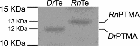
The expression of prothymosin α proteins in the testis was assessed with a polyclonal antibody against zebrafish Ptma (Ferrara et al., Reference Ferrara, Pariante, Di Matteo, Serino, Oko and Minucci2013), which, as verified trough bioinformatic epitope analysis, can recognize both proteins (Ptma from now on). Western blot analysis was performed on total protein extracts from zebrafish testis and rat testis, as a control: bands of the respective expected sizes of 12 kDa for the zebrafish Ptma and 13 kDa for the rat protein were obtained (Fig. 4).
Expression of Ptma in the testis of adult zebrafish. Western blot analysis for Ptma performed on total protein extract from adult zebrafish testis and rat testis as a control. The analysis reveals a single band of the expected 12 kDa size for zebrafish PTMA. DrTe: zebrafish testis; RnTe: rat testis (positive control, Ferrara et al., Reference Ferrara, Izzo, Pariante, Donizetti, d’Istria, Aniello and Minucci2010); DrPtma: zebrafish Ptma band; RnPTMA: rat PTMA band.

Localization of Ptma in zebrafish adult testis
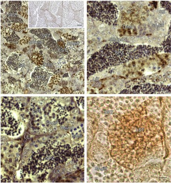
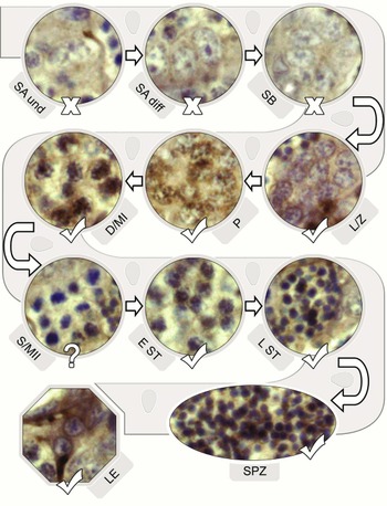
The distribution of Ptma was assessed by immunohistochemistry on sections of adult zebrafish testes, treated with the same polyclonal antibody. The staining was detectable in the cytoplasm of the Leydig cells (Figs 5 A, C and 7). Inside the tubules compartment, no positive signals were detected in any of the gonial stages (Figs 5 A–D and 7); Ptma signal started to be visible in the perinuclear region of leptotene/zygotene primary SPC (Figs 5 B, C and 7); then, it extended its localization to the chromatin region of pachytene and diplotene/metaphase I SPC (Figs 5 A–C and 7). Curiously, the immunopositivity was absent in the short secondary SPC stage (Figs 5 C and 7), then reappeared as nuclear and, possibly, perinuclear in early SPT (Fig. 5 A, C). Finally, it was still moderately visible in counterstained late SPT, then barely discernable in SPZ. Non-counterstained sections (Fig. 5 D) highlighted and confirmed the presence of Ptma in late SPT, but did not help enhance the signal in the small SPZ. Negative controls did not show any staining (Fig. 5 A, inset). The scheme in Fig. 7 recapitulates the progression of Ptma signal during zebrafish spermatogenesis.
Immunolocalization of Ptma in adult zebrafish testis. (A) General appearance of testis sections incubated with anti-zebrafish Ptma antibody and counterstained with haematoxylin. The inset shows a negative control, performed by omitting the primary antibody. (B, C) Higher magnifications. (D) High magnification of non-counterstained slide, which highlights small late SPT staining. All meiotic stages are positive, with the exception of secondary SPC. Post-meiotic germ cells are also positive. Mitotic stages are negative; finally, Leydig cells are positive. SAund and SAdiff: undifferentiated and differentiated A SPG; SB: B SPG. L/Z: leptotene/zygotene primary SPC; P: pachytene primary SPC; D/MI: diplotene/metaphase I primary SPC; S/MII: secondary/metaphase II SPC; EST: early SPT; LST: late SPT; SPZ: spermatozoa; LE: interstitial Leydig cells. Scale bars represent 20 μm in (A–C) and the inset in (A), whereas the scale bar represents 5 μm in (D).

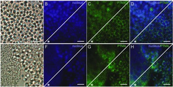
Ptma localization in SPZ by immunofluorescence on zebrafish testis. Two different fields were captured [field 1: (A/D); field 2: (E/H)]. (A, E) Images taken under visible light. (B, F) DAPI-fluorescent nuclear staining (blue). (C, G) PTMA fluorescence (green). (D, H) Merged fluorescence channels (blue/green). Upper triangles of the splitted pictures (B–D, F–H) show unaltered fluorescence; lower triangles, marked by an asterisk (*), were edited by overlaying the fluorescent images with their respective visible fields, in order to enhance the separation between each nucleus and cytoplasm. Ptma fluorescence is present in SPZ, with a possible nucleo-cytoplasmic distribution. SPZ: spermatozoa. Scale bars represent 5 μm.

A schematic illustration of the progression of the germinal line in the tubules, with pictures of selected cysts which describe the pattern of Ptma sub-localization in germ cells during zebrafish spermatogenesis. PTMA-positive cell types are marked with a ‘V,’ the negative types with an ‘X.’ Non-stained secondary SPC are marked by a ‘?.’ SA und and SA diff: undifferentiated and differentiated A SPG; SB: B SPG. L/Z: leptotene/zygotene primary SPC; P: pachytene primary SPC; D/MI: diplotene/metaphase I primary SPC; S/MII: secondary/metaphase II SPC; EST: early SPT; LST: late SPT; SPZ: spermatozoa; LE: interstitial Leydig cells.

Immunofluorescence analysis of Ptma localization in zebrafish spermatozoa
In order to obtain more detailed data on Ptma distribution inside the gametes, immunofluorescence analyses were performed on testis sections, which again confirmed the presence of the protein in the head of luminal SPZ (Fig. 6, upper triangles; refer to the legend for details). Additionally, the lower triangles of each frame in Fig. 6 (marked with an asterisk) were enhanced by merging the fluorescent channels and their respective visible fields, with the purpose of visually increasing the separation between the nucleus and the cytoplasm the cells: these data provide a slight hint about Ptma sub-localization in the head of SPZ, which might be both nuclear and cytoplasmic.
Discussion
PTMA is one of the most acidic proteins ever characterized (isoelectric point: 3.55); it is a very small-sized polypeptide (Haritos et al., Reference Haritos, Goodall and Horecker1984a), which belongs to the group of the intrinsically disordered proteins (Gast et al., Reference Gast, Damaschun, Eckert, Schulze-Forster, Maurer, Müller-Frohne, Zirwer, Czarnecki and Damaschun1995). This factor is reflected by its random coil conformation and its ability to adapt to several molecular partners during physiological interactions (Enkemann et al., Reference Enkemann, Ward, Trumbore and Berger2000a; Papamarcaki & Tsolas, Reference Papamarcaki and Tsolas1994). Such peculiarities, together with the presence of a C-terminal nuclear localization signal, account for its varied nucleo-cytoplasmic distribution (Enkemann et al., Reference Enkemann, Ward and Berger2000b; Martini et al. Reference Martini, Delage-Mourroux, Kraichely and Katzenellenbogen2000; Malicet et al., Reference Malicet, Giroux, Vasseur, Dagorn, Neira and Iovanna2006), and are linked to its participation in several biological processes (Karetsou et al., Reference Karetsou, Sandaltzopoulos, Frangou-Lazaridis, Lai, Tsolas, Becker and Papamarcaki1998, Reference Karetsou, Kretsovali, Murphy, Tsolas and Papamarcaki2002; Voutsas et al., Reference Voutsas, Baxevanis, Gritzapis, Missitzis, Stathopoulos, Archodakis, Banis, Voelter and Papamichail2000; Skopeliti et al., Reference Skopeliti, Voutsas, Klimentzou, Tsiatas, Beck, Bamias, Morak,i, Livaniou, Neagu, Voelter and Tsitsilonis2006; Ueda, Reference Ueda2009), which has been hypothesized to be an indication of molecular mimicry.
The first reports about PTMA involvement in reproduction started with the detection of the transcript and the protein in rat testis (Dosil et al., Reference Dosil, Freire and Gómez-Márquez1990; Rosón et al., Reference Rosón, Gallego, García-Caballero, Heimer, Felix and Domínguez1990), and in 2002 our group characterized an ortholog for the first time in a non-mammalian vertebrate, the amphibian Pelophylax esculentus (Aniello et al., Reference Aniello, Branno, Rienzo, Ferrara, Palmiero and Minucci2002). The comparative studies which have emerged since have highlighted a common expression pattern for PTMA during spermatogenesis, even in species which greatly differ in fertilization and gonad structure: it is now established that in cystic non-mammalian models, such as the cartilaginous fish Torpedo marmorata and in Pelophylax esculentus the protein is associated with both the interstitial Leydig cells and the meiotic and post-meiotic germ cells in the germinal compartment (Ferrara et al., Reference Ferrara, Izzo, Liguori, d’Istria, Aniello and Minucci2009; Prisco et al., Reference Prisco, Donizetti, Aniello, Locascio, Del Giudice, Agnese, Angelini and Andreuccetti2009). This pattern is conserved in the testis of the Mammal Rattus norvegicus, where PTMA is also clearly associated with the acrosome system throughout the stages of SPT differentiation (Ferrara et al., Reference Ferrara, Izzo, Pariante, Donizetti, d’Istria, Aniello and Minucci2010). Finally, recent analyses on rat and human SPZ have shown that PTMA is distributed in the region of the inner acrosomal membrane, suggesting a possible role for the protein in gamete development and function (Ferrara et al., Reference Ferrara, Pariante, Di Matteo, Serino, Oko and Minucci2013).
In the present work we show for the first time Ptma expression in the testis of adult Danio rerio and describe its cellular distribution during spermatogenesis. The male zebrafish, as commonly found in other ‘early’ teleosts, develops anastomosing tubular gonads, in which the germinal compartment branches in loops or tubules that form a connected network (Grier, Reference Grier, Russell and Griswold1993). The tubules are formed by clusters of progressing germ cells enclosed by the cytoplasm of a somatic Sertoli cell, defining the spermatocysts that line the lumina, where the spermatozoa are released (Billard, Reference Billard and Lamming1990; Pudney, Reference Pudney, Payne, Hardy and Russell1996; Parenti & Grier, Reference Parenti and Grier2004; Schulz et al., Reference Schulz, de França, Lareyre, Le Gac, Chiarini-Garcia, Nobrega and Miura2010; Huszno & Klag, Reference Huszno and Klag2012). Peculiarity of these early-teleost SPZ is the lack of an acrosome: the external fertilization is achieved by the mechanical entry of the spermatozoon inside the female egg through a single, funnel-like, micropyle (Hirai, Reference Hirai1988). The study of Ptma in an acrosomeless model is, as such, even more interesting, as the most recent studies in mammals emphasize its association with the acrosomal system.
As already reported, two ptma paralogs are present in zebrafish: ptmaa and ptmab (Donizetti et al., Reference Donizetti, Liccardo, Esposito, Del Gaudio, Locascio, Ferrara, Minucci and Aniello2008). Both transcripts are expressed in all the adult tissues we analysed. This was expected, as they equal the general ubiquity of vertebrate prothymosins. Furthermore, their localization in the testis of Danio rerio is comparable, and it approximately matches the pattern that we and others had previously highlighted in other species (Aniello et al., Reference Aniello, Branno, Rienzo, Ferrara, Palmiero and Minucci2002, Ferrara et al., Reference Ferrara, Izzo, Liguori, d’Istria, Aniello and Minucci2009, Reference Ferrara, Izzo, Pariante, Donizetti, d’Istria, Aniello and Minucci2010; Prisco et al., Reference Prisco, Donizetti, Aniello, Locascio, Del Giudice, Agnese, Angelini and Andreuccetti2009), with a clear predilection for meiotic and post-meiotic germ cells. As for the proteins, given the unavailability of commercial antibodies specific to either Ptmaa or Ptmab, we used an antiserum (Ferrara et al., Reference Ferrara, Pariante, Di Matteo, Serino, Oko and Minucci2013) that, as verified by bioinformatic analysis, can recognize both proteins which share the same weight and are referred to as Ptma during the text. The immunohistochemical data clearly show that what we observed with the mRNAs holds true for the protein profile: in particular, Ptma absence in SPG suggests that it does not participate in the proliferation of staminal/mitotic phases, while its presence in primary SPC and in SPT supports its possible role during meiosis and/or during the subsequent stages of SPT differentiation into mature SPZ. Specifically, soon after its first appearance in the cytoplasm of leptotene/zygotene primary SPC, Ptma extends its localization to the membrane-free chromatin region of dividing cells and, then, it retains its nuclear distribution during SPT differentiation. Its nucleo-cytoplasmic sub-localization suggests that prothymosin α may be involved in chromatin remodeling during spermatogenesis and spermiogenesis. Interestingly, this appears to be roughly equivalent to previous findings in both Torpedo marmorata and Pelophylax esculentus but, instead, it represents quite a notable difference from the cytoplasmic/acrosomal localization in mammals. This discrepancy, as notable as it may appear, might actually reflect a common role for PTMA during fertilization, led by the physiological differences between species. Indeed, as we described in our previous work in mammals, the protein is targeted and then docked to the inner acrosomal membrane during gamete development (Ferrara et al., Reference Ferrara, Pariante, Di Matteo, Serino, Oko and Minucci2013). We hypothesized that the protein may be retained after the acrosome reaction, to be then delivered inside the oocyte during fertilization; there, it might serve as a chromatin remodeler for the pronuclei by interacting with retained H1 histone, as hinted by previous reports that showed PTMA ability to efficiently decondensate human sperm chromatin (Karetsou et al., Reference Karetsou, Martic, Tavoulari, Christoforidis, Wilm, Gruss and Papamarcaki2004). In the acrosomeless zebrafish model, Ptma may bind to the chromatin during germ cells differentiation, thus being already ‘in place’ when the mature spermatozoon is released. This is corroborated by the reported retention of canonical histones, linker H1 histone and variants in zebrafish SPZ (Carrell, Reference Carrell2011; Wu et al., Reference Wu, Zhang and Cairns2011). We suggest that this condition may allow the protein to play the same role as in mammals after the mechanical entry inside the egg.
Although we have been able to detect Ptma signal inside both the nucleus of SPZ and, perhaps, in the cytoplasm, at the moment we have no additional evidence which may confirm the latter. However we can conclude that, in zebrafish as well as in the other species, this small polypeptide is definitely associated with spermatogenesis. Thus, our present study and the matters that are here discussed represent a compelling starting point for a comparative understanding of PTMA functions in vertebrate gamete physiology and reproduction.
Acknowledgements
The authors wish to thank Dr Teresa Chioccarelli, for the critical and helpful discussions and for her support. This work was funded by grants from ‘Ricerca di Ateneo’, Seconda Università di Napoli. The funding institution had no role in design or interpretation of the experiments.