Introduction
Intestinal parasites exert deleterious effects and pose serious health problems in dogs, resulting in retarded growth, generalized ill health, lowered resistance to infectious disease and reduced work efficiency. The major clinical signs of infection are often non-specific, highly confusing and occasionally the hosts suffer from a subclinical asymptomatic course of disease (Bowman, Reference Bowman1999; Taylor et al., Reference Taylor, Coop and Wall2007).
Symptomatology based diagnosis alone is inadequate or impossible. It is further complicated by trickle and multiple infections acquired by the host from the surroundings (Carrol & Grove, Reference Carrol and Grove1986). Sporadic documentation on the impact of intestinal helminth infection on the haemogram of infected dogs has appeared in the literature from India (Mittra & Sasmal, Reference Mittra and Sasmal1984) and from other parts of the world (Saror et al., Reference Saror, Schillhorn Van Veen and Adeyanju1979). In the present study an effort has been made to generate haematological data on canine intestinal helminth infections acquired from the surrounding environment.
Materials and methods
Collection and examination of faecal and blood samples
Faecal samples were collected from 200 dogs (116 males and 84 females) attending the University Veterinary Clinic, Jabalpur for routine examination between December 2008 and November 2009. Dogs aged less than 12 months were classified as young (n = 59) and those above 1 year were taken to be adults (n = 141). Faecal samples were first examined grossly for the presence of gravid segments of tapeworms, followed by their qualitative examination by the centrifugal flotation technique (Sloss et al., Reference Sloss, Kemp and Zajac1994). The eggs were identified by morphology and morphometry (Soulsby, Reference Soulsby1986). In addition, the samples were subjected to quantitative examination by the modified McMaster technique (Sloss et al., Reference Sloss, Kemp and Zajac1994). Based on the presence or absence of parasitic infections, the dogs were divided into parasitized (n = 39) and non-parasitized (n = 161) groups.
For haematological studies, a blood sample (2 ml) was collected from the cephalic vein of each dog into a sterile vial containing ethylenediaminetetraacetic acid (EDTA) as anticoagulant. Haemoglobin (Hb) percentage, packed cell volume (PCV) and total erythrocyte counts (TEC) were estimated by the method described by Feldman et al. (Reference Feldman, Zinkl and Jain2000). Total leucocyte counts (TLC) and differential leucocyte counts in Geimsa-stained smears were monitored by the methods of Natt & Herrick (Reference Natt and Herrick1952) and Ash & Orihel (Reference Ash and Orihel1991), respectively. Erythrocyte indices, i.e. mean corpuscular volume (MCV), mean corpuscular Hb (MCH) and MCH concentration (MCHC), were calculated from haematological values (Feldman et al., Reference Feldman, Zinkl and Jain2000).
Data analysis
Haematological data were statistically analysed using the Wilcoxon–Mann–Whitney test (Snedecor & Cochran, Reference Snedecor and Cochran1994).
Results
Of the 200 dogs coprologically examined, 39 (19.5%) harboured one or more intestinal helminth parasites. Ancylostoma caninum, Dipylidium caninum and Toxascaris spp. were found, respectively, in 33, 7 and 3 samples, with a prevalence rate of 16.5, 3.5 and 1.5%. Of the seven samples positive for D. caninum, six animals discharged mature proglotids and one animal discharged egg capsules in faeces. Polyparasitism was evidenced in four animals (10.25%). Three animals were positive for A. caninum and D. caninum and one animal was positive for A. caninum and Toxascaris infection. The intensity of A. caninum infection was highest, with mean egg counts of 951.43 (standard error 88.66), followed by Toxascaris spp. (283.33, standard error 116.81) and D. caninum (200).
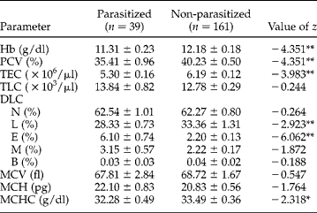
Haematological data generated from parasitized and non-parasitized dog populations are summarized in table 1. The levels of erythrocytic parameters (Hb, PCV and TEC) were significantly lower (P < 0.01) in the parasitized than in the non-parasitized group of animals, whereas group differences for TLC, neutrophils, monocytes and basophils were non-significant. A significant increase (P < 0.01) in eosinophils and decrease in lymphocytes were also observed in parasitized compared to non-parasitized animals.
Mean values of haematological parameters in parasitized and non-parasitized dogs (mean±SE).
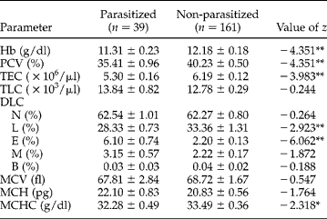
n, Number examined; Hb, haemoglobin; PCV, packed cell volume; TEC, total erythrocyte count; TLC, total leucocyte count; DLC, differential leucocyte count; N, neutrophil; L, lymphocyte; E, eosinophil; M, monocyte; B, basophil; MCV, mean corpuscular volume; MCH, mean corpuscular haemoglobin; and MCHC, mean corpuscular haemoglobin concentration.
Significance: * P < 0.05; ** P < 0.01.
In parasitized dogs, the calculated value for MCV was lower than in non-parasitized animals: however, the difference between the groups was non-significant. The MCHC in parasitized dogs was, however, significantly lower (P < 0.05) than in non-parasitized animals, suggestive of normocytic hypochromic anaemia in the former group of animals.
Discussion
Parasitism is manifested by a wide variety of clinical signs and pathophysiological changes in livestock, including dogs. The combined impact of multiple parasitism makes the situation more complicated and confusing for the clinician on account of non-specific signs. Interestingly, the findings reported in the present paper have revealed a lower prevalence of intestinal helminth infections (19.5%) than in other parts of India (Jani et al., Reference Jani, Jani and Dave1995; Sharma et al., Reference Sharma, Sinha, Sinha, Kumar and Mahtha2006). This could be attributed to a better awareness amongst owners about healthcare and zoonotic significance and easy accessibility to veterinarians. Amongst the parasitized group of animals suffering from multiple helminth infections, the prevalence of cestode (D. caninum) and ascarid infection was negligible. The predominance of hookworm (A. caninum) in the population was in conformity with earlier reports (Yacob et al., Reference Yacob, Ayele, Fikru and Basu2007; Palmer et al., Reference Palmer, Thompson, Traub, Rees and Robertson2008; Little et al., Reference Little, Johnson, Lewis, Jaklitsch, Payton, Blagburn, Bowman, Moroff, Tams, Rich and Aucoin2009). Ancyclostoma caninum reported in this study is a proven zoonotic nematode that can cause creeping eruptions in humans (McCarthy & Moore, Reference McCarthy and Moore2000). The specific use of anti-hookworm drugs in pregnant bitches and prophylactic therapy in pups may further reduce the prevalence rate of the parasite in Jabalpur, India.
Members of the Ancylostomatidae family dwelling the small intestine of the host have been recognized as a major cause of anaemia. Ancyclostoma caninum is a haematophagous nematode, causing traumatic lesions by biting the mucosa: feeding sites continue bleeding for a considerable period of time before the nematode moves to another site and inflicts yet another lesion. These factors could contribute to the significant fall (P < 0.01) in erythrocytic indices in the parasitized group of animals, as documented elsewhere (Mekenna et al., Reference Mekenna, Mepherson and Falconer1975; Mittra & Sasmal, 1984; Naggar, Reference Naggar1994).
A significant increase in eosinophils is a recognized sign of parasitic infection (Saror et al., Reference Saror, Schillhorn Van Veen and Adeyanju1979). Eosinophils are potent effector cells associated with the killing of helminths, as evidenced from their close association with damaged or dead parasites in histological sections and positive correlation with acquired resistance by the parasitized host (Meeusen & Balic, Reference Meeusen and Balic2000; Kirkova et al., Reference Kirkova, Petkov and Goundasheva2005). In massive infections, cell counts may increase 10–30 times. Eosinophils exert their phagocytic function through enzymes in their secretions that injure and kill the parasite in situ (Tizard, Reference Tizard1996).
Profuse infiltration of lymphocytes in the traumatized and oedematous intestinal submucosa at the feeding sites of parasites, as reported elsewhere (Kirkova et al., Reference Kirkova, Petkov and Goundasheva2005), might have contributed to their significant fall in the circulation of the parasitized group of animals. The mobilized lymphocytes in the intestinal submucosa inhibit the activities of deeply penetrating helminths through the immunological response (Tizard, Reference Tizard1996).
In conclusion, intestinal helminths, especially A. caninum, were found to exert deleterious effects on haematological parameters and cause normocytic hypochromic anaemia in a parasitized group of animals. Further studies on the pathophysiology of anaemia in the parasitized dog population seem essential to understand this precisely and to assess the deleterious impact on the health of the host.
Acknowledgements
The authors thank Roshni Tiwari for her kind help in statistical analysis and Dr P.K. Sanyal, Professor and Head, Department of Parasitology, Durg (C. G.) for his review of the manuscript. Thanks are also due to the referees for their invaluable comments.