Infective endocarditis remains a diagnostic and therapeutic challenge associated with a high morbidity and mortality in patients after right ventricular outflow tract valve replacement.Reference Moreillon and Que1 A recent study reported a 5-year cumulative mortality of >50% in healthcare-associated endocarditis,Reference Toyoda, Chikwe, Itagaki, Gelijns, Adams and Egorova2 which is much higher than the mortality associated with many cancers. Clinical observations, which compared the common right ventricular outflow tract prostheses suggest a higher risk of infective endocarditis in patients after implantation of bovine jugular vein conduits than receiving a cryopreserved homografts, occurring at a rate of 3% per patient year.Reference Mery, Guzman-Pruneda and De Leon3,Reference Van Dijck, Budts and Cools4 Furthermore, the use of decellularised pulmonary homografts for pulmonary valve replacement in comparison to “non-decellularised” ones have shown very promising haemodynamical results, with nearly 100% of patients free of endocarditis at 10 -year follow-up and a significantly better freedom from re-operation.Reference Sarikouch, Theodoridis and Hilfiker5,Reference Boethig, Horke and Hazekamp6 The underlying mechanisms leading to a different susceptibility for infective endocarditis in different cardiac graft tissues are not known yet.
Staphylococcus aureus is amongst the most common cause of endocarditis.Reference Moreillon and Que1 The onset of the disease requires bacterial adhesion to the valve endothelium leading to a proinflammatory endothelial cell phenotype, platelet recruitment and activation of the coagulation system.
Aside from the bacterium per se, the pathogenesis of endocarditis involves multiple host factors including tissue damage, inflammation and deposition of platelets and host matrix proteins such as fibrinogen/fibrin, to mention a few. Together these biological responses promote local colonisation by circulating bacteria during boosts of transient bacteraemia. Histological observations raise a concern that fibrin depositions occur on prosthetic valve leaflet tissue even without an ongoing infection.Reference Schneider, Vogt and Boekenkamp7,Reference Pislaru, Pellikka, Schaff and Connolly8 As the native valve endothelium is resistant to bacterial adhesion, endothelial lesions along with the demonstrated fibrin depositions might be a critical precursor of bacteria and platelet attachment. It has been shown that the plasma protein fibrinogen and the platelet receptor αIIbβ3 mediate S. aureus adhesion to bovine jugular vein tissue.Reference Ditkowski, Bezulska-Ditkowska and Jashari9 As the surfaces of the grafts display different binding affinities for proteins, like fibrinogen,Reference Xu, Bauer and Siedlecki10 the decellularisation of the homograft might lead to altered adsorption of plasma proteins. Fabricating a decellularised pulmonary homograft aims to increase its biocompatibility and to cause less activation of the immune system by removing almost all donor DNA during the decellularisation process.Reference Neumann, Cebotari, Tudorache, Haverich and Sarikouch11 In the months following implantation, prosthetic grafts become progressively re-endothelialised by new host cells,Reference Sarikouch, Horke and Tudorache12 of which the course is crucial in terms of the biocompatibility of the valves. The risk of infective endocarditis decreases after re-endothelialisation but does not disappear. The sound endothelium of the valves is naturally resistant to bacterial adhesion,Reference Cahill, Baddour and Habib13 however, it is not clear why re-endothelialised valves are not. Plasma protein-mediated bacterial attachment might be altered on decellularised homograft tissue.
To address these questions, a newly designed low-volume flow chamber has been designed.
In the present study, we investigated in vitro bacterial adhesion to decellularised fresh homograft tissue and compared it to cryopreserved homograft and bovine pericardial patch tissue. Also, a potential contribution of plasma fibrinogen to the interaction between S. aureus and the tissue grafts was addressed.
Materials and methods
In vitro laboratory model to study cardiac tissues
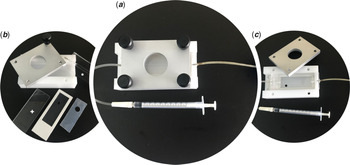
To characterise the propensity of cardiac graft tissues for bacterial attachment, we have developed a new in vitro model of a parallel plate flow chamber (Fig 1). Pieces of the investigated cardiac graft tissues were mounted into this system presented in Fig 1 and submitted to the perfusion of bacterial suspensions at room temperature (details of all used methods are described in the supplementary material).
Schematic presentation of a newly developed low-volume parallel plate flow chamber (in-house design by the Centre for Molecular and Vascular Biology and fabrication by the Department of Cellular and Molecular Medicine, KU Leuven, Belgium). (a) A mounted flow set of dimensions LxWxH: 90 × 50 × 25 mm. (b) Elements of the flow chamber, which are placed in a bottom white frame as presented in (c) a top view. In brief, a thin foil slide with a white frame has a 4 mm circular perforation to allow the exposure of the tissue to the bacterial suspension. The slide has a recess for fitting a rubber gasket with an 8-mm whole, which serves as a holder in which the tissue piece is immobilised during the perfusion. A plastic microscopic slide (+) is used as an additional support for the setup and is placed underneath the tissue holder in the bottom part of the white chamber. The asterisk (*) presents an insert made of a Plexiglas that carries a channel flanked by metal inlet and outlet adaptors connected to the rubber tubes. It is placed on the top of the tissue holder to form a leak-free flow channel right above the central part of the tissue graft.

Graft tissues and bacterial adhesion
Bacterial adhesion and fibrinogen binding were tested for bovine pericardium patch (Supple Peri-Guard®; Synovis Surgical Innovations, St Paul, Minnesota, United States of America), cryopreserved pulmonary homograft (human origin, European Homograft Bank, Brussels, Belgium) and decellularised fresh pulmonary homograft (human origin, Corlife oHG, Hannover, Germany) under shear stress and static conditions, respectively. After overnight incubation with human pooled plasma to provide the fibrinogen binding, graft tissues were mounted into the parallel plate flow chamber and submitted to perfusion.
Bacterial suspensions of the S. aureus strain 8325–4 (suspensions of 107 colony-forming units/mL) were perfused over graft tissues. After serial dilutions, colony-forming units were counted.
Fibrinogen binding to graft tissues
Prepared graft tissues were incubated at 37 °C for 2 hours with fluorescently labelled human fibrinogen. After washing steps, the absolute fluorescence was quantified to express the degree of fibrinogen binding.
Statistical analysis
Non-parametric tests were used to analyse the results as the statistical evaluation revealed that not all data followed a Gaussian distribution. The t-test with the non-parametric Mann–Whitney U test was used to test a statistical significance between groups. Data analyses were performed with GraphPad Prism 8.0 d (GraphPad Software, San Diego, California, United States of America). Values are demonstrated as median and distribution plots using 25–75% percentile boxes with respective min and max binding whiskers. P-values <0.05 were considered significant.
Results
Cryopreserved and decellularised pulmonary homografts display a different propensity for S. aureus attachment in vitro
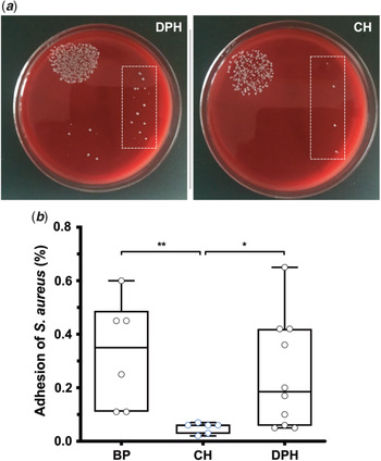
In the present study, we focused on in vitro bacterial adhesion to decellularised pulmonary homograft and compared it to cryopreserved homograft and bovine pericardial patch tissue under shear conditions generated in our laboratory model (Fig 1). We noted that bacterial adhesion to cryopreserved homograft was nearly 6-fold lower than to bovine pericardial patch. Interestingly, decellularised pulmonary homograft displayed a significant increase of bacterial adhesion by about 70% compared to the cryopreserved counterpart (based on median values), which was however characterised by a widespread (Fig 2 b). Values for the decellularised pulmonary homograft and bovine pericardial patch were not significantly different.
Adhesion of Staphylococcus aureus to cardiovascular grafts under shear stress. Perfusion of S. aureus over bovine pericardium patch (BP), cryopreserved pulmonary homograft (CH) and decellularised pulmonary homograft (DPH) (n ≥ 6). (a) Representative Mueller-Hinton blood agar plates with S. aureus colonies, obtained as colony-forming units (CFUs) that bound, respectively, to DPH and CH; dotted squares indicate the same dilution. (b) Degree of bacterial adhesion to BP, CH and DPH. Results are presented as box and whisker dot plots with the upper and lower borders of the box representing the 25 and 75% percentile (upper and lower quartiles). The middle horizontal line represents the median, the upper and lower whiskers the maximum and minimum values of non-outliers. T-test with non-parametric Mann–Whitney U test was applied: *p < 0.05, **p < 0.01.

Figure 2 a shows S. aureus colonies on blood agar plates that bound, respectively, to decellularised pulmonary homograft (left image) and cryopreserved homograft (right image). Dotted squares indicate less colonies obtained for the latter tissue using the same dilution.
Plasma fibrinogen can influence S. aureus attachment to decellularised pulmonary homograft
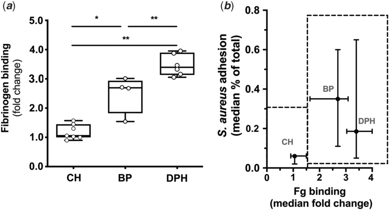
It is known that plasma fibrinogen is an important mediator of bacterial adhesion to endothelial cells and to the subendothelial matrix. We have recently shown that plasma fibrinogen is able to solely support S. aureus adhesion to plasma-coated bovine tissues.Reference Ditkowski, Bezulska-Ditkowska and Jashari9 Therefore, we next aimed to investigate the degree of protein binding to the homograft tissues and evaluate its role in bacterial adhesion. We questioned now if decellularised homografts share similar properties with the cryopreserved counterparts in terms of fibrinogen binding. The fluorescently labelled protein was used in the binding assay, which showed that the bovine pericardial patch had a significantly higher affinity for fibrinogen compared to cryopreserved homograft (p < 0.05) (Fig 3 a). Interestingly, fibrinogen bound also more avidly to decellularised than to cryopreserved homograft or bovine pericardial patch (p < 0.01) (Fig 3 a). Based on this observation, plots combining S. aureus adhesion and fibrinogen binding for the three investigated tissues show that decellularised pulmonary homograft and cryopreserved homograft represent separate groups (Fig 3 b). The first “group” with the cryopreserved tissue presents hardly any affinity for both fibrinogen and S. aureus. The second “group” (decellularised pulmonary homograft and bovine pericardial patch) shows a higher degree of fibrinogen binding and bacterial adhesion. This finding may implicate fibrinogen as a molecule contributing to the bacterial recruitment to the surface of the tissue.
Binding of fibrinogen (Fg) to graft tissues in static conditions. (a) Cryopreserved pulmonary homograft, CH (n = 7), decellularised pulmonary homograft, DPH (n = 6), bovine pericardial patch, BP (n = 4) graft tissues were incubated with fluorescently labelled Fg (30 µg/mL). Values expressing Fg binding are presented as a fold change. Data are demonstrated as box and whisker dot plots with the upper and lower borders of the box representing the 25 and 75% percentile (upper and lower quartiles). The middle horizontal line represents the median and the upper and lower whiskers the maximum and minimum values of non-outliers. t test with the non-parametric Mann–Whitney U test was applied: *p < 0.05, **p < 0.01. (b) Distribution of examined tissues in a high-affinity (BP and DPH) and low-affinity group (CH) is presented by dotted line boxes, based on a degree of their interaction with Fg and S. aureus, respectively, in static and flow conditions. Data are presented as dot plots, representing the median values with whiskers ranging from 25 to 75% percentile for both binding events, respectively.

Discussion
Infective endocarditis remains a severe complication after valve replacement interventions despite advanced treatment options.Reference Cahill, Baddour and Habib13,Reference Que and Moreillon15 The prevalence of this infectious disease is rapidly increasing as a consequence of medico-surgical progresses and population ageing.Reference Cahill, Baddour and Habib13 Although the underlying mechanisms for the different susceptibility of various heart valve tissues for infection remain unclear, some potential factors have been elucidated in vitro.Reference Cahill, Baddour and Habib13,Reference Jalal, Galmiche, Beloin and Boudjemline16 In this study, we addressed the potential role of the plasma protein fibrinogen in mediating S. aureus adhesion to decellularised tissues using a newly constructed flow chamber. Our results reveal that this plasma protein binds well to the decellularised homograft tissue and subsequently contributes to the process of S. aureus adhesion to the graft surface. Interestingly, this propensity of decellularised tissue for fibrinogen binding was significantly higher than that of cryopreserved homograft and bovine pericardial tissues. Fibrinogen plays a crucial role in platelet and S. aureus interaction since as a bridging molecule, it facilitates bacterial recruitment to the tissue surface via the activated platelet receptor α IIb β 3.Reference Ditkowski, Bezulska-Ditkowska and Jashari9,Reference Entenza, Moreillon and Senn17
As a consequence, our data show a relatively higher S. aureus adhesion to decellularised homograft and bovine pericardial tissues than to cryopreserved homograft in vitro. Herewith, we could highlight that the mechanism of bacterial adhesion via fibrinogen plays an important role, also for decellularised biomaterials such as decellularised homografts. In detail, we noted that bacterial adhesion to cryopreserved homograft was nearly 6-fold lower than to bovine pericardium, which supports earlier findings.Reference Ditkowski, Bezulska-Ditkowska and Jashari9
Previous studies showed that differences in bacterial adherence to the bovine jugular vein and cryopreserved homograft tissues cannot be attributed to intrinsic tissue specificities,Reference Veloso, Claes and Van Kerckhoven18,Reference Jalal, Galmiche and Lebeaux19 as the tissue surface itself and the bacterial adhesins (surface molecules) do not seem to have a significant impact on the infection.Reference Veloso, Claes and Van Kerckhoven18 These findings triggered to investigate in detail the role of one of the abundant plasma proteins such as fibrinogen. The exact mechanisms of fibrinogen adsorption to the cardiac tissues are not fully known yet and awaiting further investigation, albeit some important properties like surface chemistry and topography have been elucidated.Reference Xu, Bauer and Siedlecki10,Reference Tsai, Grunkemeier and Horbett20 Decellularisation of biological surfaces can be performed using various protocols,Reference Kasimir, Weigel and Sharma21–Reference Ramm, Goecke and Theodoridis23 leading to different physical and chemical properties of tissue matrices. Thereby, it becomes obvious that all types of surface manipulations of bioprosthetic grafts can influence the biological responses to their surfaces such as the protein adsorption (e.g., fibrinogen), platelet adhesion or blood coagulation.Reference Xu, Bauer and Siedlecki10 Furthermore, commonly used tissue fixatives, like glutaraldehyde to ensure the tissue integrity, may also contribute to altered binding effects. Fibrinogen amongst others acts as a modulating protein for platelet adhesion and clustering by bridging their activated receptor α IIbβ 3.Reference Ditkowski, Bezulska-Ditkowska and Jashari9,Reference Veloso, Ditkowski, Mela, Hoylaerts and Heying24 We developed the new flow chamber, presented in this study, to be able to investigate various tissues using only small amounts of perfusion media with bacteria. By this, the chamber allowed us to study the role of the most abundant plasma protein fibrinogen in bacterial adherence to cardiac graft tissues and will be of use to proceed with investigating the role of platelets in this process occurring on decellularised right ventricular outflow tract tissues. This chamber is superior to the previous design, which required a larger amount of perfusion medium in the circuit and a longer experimental procedure.Reference Ditkowski, Veloso and Bezulska-Ditkowska14
Clinical reports highlight the risk to acquire infective endocarditis after right ventricular outflow tract valve replacement, which seems to be higher after implantation of heterologous bovine jugular vein conduits compared to cryopreserved homograft.Reference Van Dijck, Budts and Cools4,Reference Sharma, Cote, Hosking and Harris25 The recently introduced decellularised pulmonary homografts show a very good haemodynamical performance with a freedom of explanation of >96% which was similar or even higher to retrospective data after cryopreserved homograft and bovine jugular vein conduit implantation.Reference Boethig, Horke and Hazekamp6,Reference Sarikouch, Horke and Tudorache12 Importantly, the decellularised homografts are described with a low risk of associated infective endocarditis.Reference Boethig, Horke and Hazekamp6,Reference da Costa, Etnel and Torres26 Follow-up studies report a low incidence of endocarditis with 96.2% of patients being free from the disease after 10 years, which is similar to the cryopreserved homograft with 97.4%.Reference Boethig, Horke and Hazekamp6
Looking more in detail into the differences between the “cell-free” homograft and cryopreserved homograft, after years of experience, it is postulated that the cryopreserved homograft seems to be more immunogenic than previously thought.Reference d’Udekem27 The main aim of the decellularisation process is to reduce the host immune reaction towards the tissue surface that inherits a great potential to become a more ideal graft. The presence of cellular antigens remaining on bovine jugular vein conduits and cryopreserved homografts may exacerbate inflammatory processes, explaining why potentially the decellularisation could reduce susceptibility for infective endocarditis.
Decellularised homograft tissues retain, however, the capacity for cellular repopulation as recipient endothelial cells are mainly covering the tissue over time.Reference Ramm, Goecke and Theodoridis23 Nevertheless, after implantation, the decellularised homograft tissues contain an exposed extracellular matrix, which might naturally allow for bacterial invasion and thereby facilitating the infiltration of immune-competent cells resulting in the activation of inflammatory cascades. Therefore, this leads to the recommendation of not using the decellularised homografts for valve replacement in case of active endocarditis.Reference Sarikouch, Haverich and Pepper28
In conclusion, our results postulate that fibrinogen plays a role in bacterial recruitment to the surface of decellularised homograft tissue similarly as to the bovine graft tissues in vitro. Future work should essentially involve in vivo models to be able to characterise the role of other players such as platelets in the pathogenesis of infective endocarditis on various cardiac tissues, including the decellularised homografts.
Supplementary Material
To view supplementary material for this article, please visit https://doi.org/10.1017/S1047951120002772
Acknowledgements
We thank Dr. Michael Harder and Dr. Markus Lux, Corlife oHG, Hannover, for providing the decellularised pulmonary homografts and Prof. Dr. Philippe Moreillon and Prof. Dr. J.M. Entenza, Department of Fundamental Microbiology, University of Lausanne, for giving us the bacterial strains.
Financial Support
This work was supported by the Association for European Paediatric and Congenital Cardiology [Junior Research Grant 2018 to BD]; and the Research Fund KU Leuven [OT/14/097 to RH]. RH was sponsored by the Clinical Research Fund of UZ Leuven and the Flemish Research Foundation (FWO).
Conflicts of Interest
None.
Ethical Standards
The research does not involve human and/or animal experimentation.