Origins of coronary arteries from abnormal sites in the aortic root are well known, but there are unresolved problems about their incidence and prevalence, natural history, the mechanisms producing myocardial ischaemia, the need for treatment, and the optimal form of treatment.
There are many different anomalies.Reference Roberts 1 – Reference Poynter, Williams, McIntyre, Brothers and Jacobs 5
Normal variants
A separate origin of the conal branch from the right sinus of Valsalva occurs in 35–50% of people. Separate origins of the left circumflex and left anterior descending coronary arteries from the left sinus of Valsalva occur in about 1% of people.Reference Hoffman 6 These are benign variations that do not cause pathophysiology in the absence of coronary atheroma.
Less frequent but usually benign anomalies
The next most common anomaly of origin is the attachment of the left circumflex coronary artery to the right sinus of Valsalva or the proximal right coronary artery. This anomaly occurs often in patients with tetralogy of Fallot and other congenital heart lesions, but can be isolated. Apart from the risk for coronary atheroma or surgical injury, this anomaly does not usually cause myocardial ischaemia, although sudden death has occurred in a patient with this anomaly.Reference Patterson 7 The anomalous artery arises obliquely from the sinus and may have a slit-like orifice,Reference Ueyama, Ramchandani, Beall and Jones 8 and it is surprising that more complications are not reported.
Rarely, the left anterior descending coronary artery, isolated or as part of a dual left anterior coronary artery, or both it and the left circumflex coronary artery arise from the right sinus of Valsalva.Reference Surucu, Tatly, Deethirmenci and Kurtoethlu 9 , Reference Tuncer, Batyraliev, Yilmaz, Gokce, Eryonucu and Koroglu 10 The aberrant left anterior coronary artery usually passes anterior to the pulmonary outflow tract, but it may pass between the two great arteries or through the ventricular septum. These anomalies, too, are rarely a cause of heart disease in the absence of coronary atherosclerosis. In one patient with effort angina, however, the anomalous left anterior coronary artery left the aorta at an acute angle and then passed between the great arteries; symptoms were relieved by surgery.Reference Surana, Doddamani and Swaminathan 11
Potentially serious anomalies
The major potentially serious anomalies of origin of the coronary arteries are the attachment of the right coronary artery to the left sinus of Valsalva, the attachment of the left main coronary artery to the right sinus of Valsalva, the attachment of one or both major coronary arteries to the posterior – non-coronary or non-facing – sinus of Valsalva, and the single coronary artery that has only one aortic orifice and main stem but then divides to supply branches to the whole heart (see below). A high origin of a main coronary artery above the sinus of Valsalva is often recorded as abnormal, but is uncommon; it is not usually a major threat to health, and will not be discussed further here.
Incidence and prevalence
Ideally, to study the incidence, prevalence, and natural history of these lesions, we would study a large, randomly selected group of subjects of all ages with a diagnostic method that is 100% sensitive and 100% specific. If the anomaly causes many deaths in young patients, then its prevalence will be less in older than in younger subjects. The studies that have been performed using autopsy, echocardiography, cardiac catheterisation, and coronary angiography, and imaged by computerised tomography or magnetic resonance fall short of these ideals.
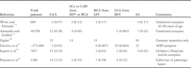
Autopsy studies by experienced pathologists lead to unambiguous diagnoses of these anomalies, but the requirement for random selection of subjects may not be met. In the study by Cheitlin et alReference Cheitlin, De Castro and McAllister 12 , from the Armed Forces Institute of Pathology, there were 19 patients with an anomalous left main coronary artery from the right sinus of Valsalva, but only nine patients in whom the right coronary artery arose from the left sinus of Valsalva. This reversal of the expected ratio (see below) indicates non-random referral of patients. The authors estimated that, over the time of acquisition of these hearts, about 475,000 hearts had been sent to the Armed Forces Institute of Pathology for an incidence of these coronary origin anomalies of 0.01%. Frescura et alReference Frescura, Basso and Thiene 13 studied a small series of autopsies of children with congenital heart disease and found a high incidence of major abnormalities of coronary origins, but this figure is biased by the 13/27 patients who died because of the abnormalities. The studies from a general hospital by Alexander and GriffithReference Alexander and Griffith 14 and from a group of children’s hospitals by Lipsett et alReference Lipsett, Cohle, Berry, Russell and Byard 15 are probably unbiased (Table 1).
Incidence of selected CAA on the basis of autopsies.

AFIP=Armed Forces Institute of Pathology; CAA=coronary artery anomalies; LAD=left anterior descending coronary artery; LCA=left main coronary artery; LCx=left circumflex coronary artery; LSV=left sinus of Valsalva; RCA=right coronary artery; RSV=right sinus of Valsalva; SA=single coronary artery
Percentages in parentheses are related to the total population studied
Congenital coronary arterial fistulae or coronary arteries arising from the pulmonary artery are excluded from this table
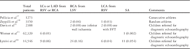
Echocardiography may often miss some of these anomalies. In three series in which children and young athletes were studied,Reference Pelliccia, Spataro and Maron 16 – Reference Davis, Cecchin, Jones and Portman 18 of a total of 6898 subjects, there were three with a left coronary artery arising from the right sinus of Valsalva and four with the right coronary artery arising from the left sinus of Valsalva. These must underestimate the true incidence, because anomalous origin is much more common for the right than the left coronary artery, and the sample sizes are small. A study of 14,546 echocardiograms taken in children seen in a hospital setting for possible heart disease revealed 52 anomalies of coronary aortic origin.Reference Lytrivi, Wong and Ko 19 One large study by Werner et alReference Werner, Wroblewska-Kaluzewska, Pleskot, Tarnowska and Potocka 20 on 62,320 children having diagnostic echocardiography described only six patients with a coronary artery anomaly – an anomalous left circumflex coronary artery. These were routine echocardiography studies, and it is probable that many anomalies were missed (Table 2).
Incidence of selected coronary artery anomalies on the basis of echocardiography in children and young adults.

LAD=left anterior descending coronary artery; LCA=left main coronary artery; LCx=left circumflex coronary artery; LSV=left sinus of Valsalva; RCA=right coronary artery; RSV=right sinus of Valsalva; SA=single coronary artery
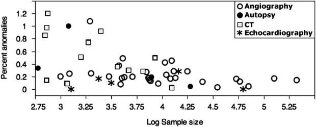
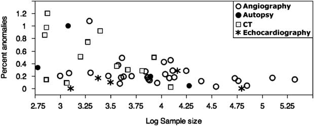
Coronary angiography provides a much larger number of patients but is open to the criticism that most patients with coronary arterial anomalies have angiography because of their symptoms, thus inflating their apparent incidence. This may explain why the incidence is usually higher for angiographic studies than those conducted with echocardiography or autopsy (Fig 1). Computerised tomographic angiography provides more certain diagnosis, but the studies have relatively small sample sizes. A variety of tomographic techniques have been used, such as 64- or 128-slice multidetector computerised tomography, dual-source computerised tomography, or magnetic resonance imaging.
Total of the three major anomalies as a percentage of the total number of patients studied. The horizontal scale is logarithmic (base 10). The percentages are usually lower for data obtained by echocardiography and autopsies than those by angiography; the autopsy exceptionReference Frescura, Basso and Thiene 13 was based on a study on congenital heart disease lesions. As expected, there is more variation in the groups with the smaller sample sizes. Studies conducted by computed tomography tend to have higher values because of small sample size and bias by referral of patients suspected of having anomalies after inconclusive angiography.
When assessing the frequency with which each of these major anomalies occurs, there is bound to be even more variation, but we can get some information by pooling data from several series. In 21 large series, each with over 7000 patients,Reference Tuncer, Batyraliev, Yilmaz, Gokce, Eryonucu and Koroglu 10 , Reference Liberthson, Dinsmore and Bharati 21 – Reference Aydar, Yazici and Birdane 40 104/526,500 patients had an anomalous left coronary artery (0.020%) and 523/526,500 patients had an anomalous right coronary artery (0.099%). In 23 large series with a total of 585,000 patients,Reference Tuncer, Batyraliev, Yilmaz, Gokce, Eryonucu and Koroglu 10 , Reference Liberthson, Dinsmore and Bharati 21 – Reference Neuhaus and Kober 42 148 (0.025%) had a single coronary artery, the proportion being lower than the 11/26,807 (0.0373%) single coronary arteries in autopsied patients,Reference Alexander and Griffith 14 , Reference Lipsett, Cohle, Berry, Russell and Byard 15 and the 33/50,000 (0.066%) found angiographically by Desmet et al.Reference Desmet, Vanhaecke and Vrolix 41 In some angiographic studies, it is likely that single coronary arteries were not recorded. Finally, the most common anomaly – the anomalous left circumflex coronary artery arising from the right sinus of Valsalva or the right coronary artery – occurred in 1074/536,716 (0.200%) of angiograms.Reference Ueyama, Ramchandani, Beall and Jones 8 , Reference Tuncer, Batyraliev, Yilmaz, Gokce, Eryonucu and Koroglu 10 , Reference Liberthson, Dinsmore and Bharati 21 – Reference Aydar, Yazici and Birdane 40
Some of these data are displayed in Supplementary Tables 1 and 2.
The incidence of origin of a main coronary artery from the posterior sinus of Valsalva is extremely rare. Up to 2013, after a MEDLINE search, we found only 30 such patients described. Therefore, they have negligible influence on the total incidence or on the prevalence of these major anomalies of origin.
Omitting the anomalous left circumflex coronary artery, the percentages of major anomalies reported are shown in Figure 1.
On the basis of the studies with larger sample sizes of over 10,000 patients, it appears that the incidence of these major anomalies is under 0.4%, with a possible average of 0.2%. Adding up the percentage incidence of the three major lesions separately gives a total incidence of 0.156%; hence, a range of 0.1–0.2% seems appropriate.
Gross anatomy of anomalies
A recent publication by the Congenital Heart Surgeons Society AAOCA Working groupReference Poynter, Williams, McIntyre, Brothers and Jacobs 5 described four grades on the basis of the relationship of the coronary ostia:
-
1. Two separate non-confluent orifices within the same sinus.
-
2. Two confluent (adjacent) orifices within the same sinus.
-
3. The sinus has a single orifice that bifurcates immediately within the aortic wall into two major coronary arteries.
-
4. The sinus has a single orifice that forms a common trunk that bifurcates into two major coronary arteries outside the aorta.
This description does not include an anomalous coronary artery arising from the non-coronary sinus or the origin of the left circumflex coronary artery from the right sinus of Valsalva.
Grades 1 and 2
A left main coronary artery arising from the right sinus of Valsalva may take one of several pathways after leaving the aorta. In patients in whom the anomaly was unrelated to their illness, about 60% passed anteriorly between the aorta and the right ventricular outflow tract or the main pulmonary artery – inter-arterial course – 18% were retroaortic, 16% passed through the ventricular septum to emerge on the anterior surface of the left ventricle, and 6% ran in front of the main pulmonary artery.Reference Hoffman 6 If the left main coronary artery takes the common inter-arterial course, it often has a long intramural pathway in the aortic wall, its origin may be narrower than normal, it may have a slit-like ostium partly covered by a thin flap, and it may emerge tangentially from the sinus of Valsalva instead of at approximately a right angle to it, or else emerge at right angles but immediately take a sharp bend. Sometimes, the initial portion of the artery is narrowed after leaving the aorta.
If the right coronary artery arises from the left sinus of Valsalva, it usually (>90%) takes an inter-arterial course between the aorta and the main pulmonary artery or the right ventricular outflow tract. Occasionally, it runs retroaortic or anterior to the right ventricular outflow tract. The arterial origin may have an intramural course and arise tangentially from the sinus of Valsalva, and may show localised narrowing as it leaves the sinus of Valsalva or passes between the great arteries.Reference Giroux and Cartier 43 In other patients, the right coronary artery is relatively far from the main pulmonary artery.Reference Angelini 44
A left main coronary artery arising from the posterior sinus of Valsalva frequently has a slit-like orifice, intramural course in the aortic wall, or an acute angulation as it leaves the aortic root. Of the 21 patients with this anomaly, one or other of these abnormal features occurred in 11 patients, eight had no such abnormalities, and in four patients no description was provided. After leaving the aorta, the left main coronary artery passes between the aortic root and the left atrium, and then branches normally. There were six patients with the right coronary artery arising from the posterior sinus of Valsalva, and in none of these were there any other abnormal features.
Grades 3 and 4
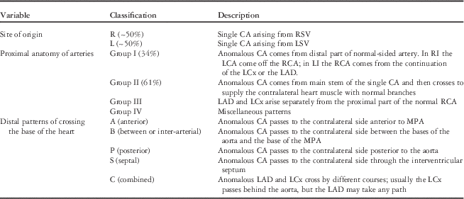
The single coronary artery arises with equal frequency from the right or the left sinus of Valsalva;Reference Hoffman 6 , Reference Ogden and Goodyer 45 rarely, it arises from the posterior – non-coronary – sinus. About 40% have associated congenital heart disease. The main anatomic typesReference Lipton, Barry, Obrez, Silverman and Wexler 46 , Reference Shirani and Roberts 47 are shown in Table 3 in a classification based on three levels: the side of origin (R or L), the proximal anatomy of anomaly (Group I, II, or III), and the distal anatomy where the anomalous artery crosses to the contralateral side of the heart (A,B,P,S, or C). Thus, for example, an anomaly may be classified as RIIB (see Table 3). Occasionally, the right coronary artery arises from the left anterior descending coronary artery; some rarer types are also described.Reference Hoffman 6 , Reference Lipton, Barry, Obrez, Silverman and Wexler 46 , Reference Shirani and Roberts 47 The aberrant branch – left main coronary artery in a single right coronary artery or the right coronary artery in a single left coronary artery – often joins the main stem at the aortic wall (grade 3) and usually runs close to the aortic root so that there is a tangential origin of the aberrant artery. Occasional patients with one of these anomalies of origin have hypoplastic coronary artery branches.
Classification of single coronary artery on the basis of references Reference Lipton, Barry, Obrez, Silverman and Wexler46 and Reference Shirani and Roberts47; table format based on publication by Ay et al.Reference Ay, Aydin, Ay, Inan, Başel and Zeybek 130

CA=coronary artery; LAD=left anterior descending coronary artery; LCA=left main coronary artery; LCx=left circumflex coronary artery; LSV=left sinus of Valsalva; MPA=main pulmonary artery; RCA=right coronary artery; RSV=right sinus of Valsalva; SA=single coronary artery
In the collected literature,Reference Hoffman 6 whether or not symptoms were mentioned, right and left single coronary arteries were about equally represented. About 25% were type I, mostly with a left single coronary artery. About 60% were type II, and of these only 20% had an intra-arterial passage.
Natural history
This is even more difficult to ascertain than the true incidence and prevalence because of the unknown number of asymptomatic subjects. A careful analysis of the issue by Peńalver et alReference Peńalver, Mosca, Weitz and Phoon 48 pointed out that if the prevalence of these anomalies was indeed 0.1–0.2% (1000–2000 per million population), in the United States alone there would be a total of 300,000–600,000 affected people and a yearly addition of 4000–8000 infants born with one of these anomalies. A similar point was made by AngeliniReference Angelini 44 who used somewhat higher figures. There is nowhere near these numbers of clinically significant patients that have been reported – for example, Tables 1–4 record <2500 patients worldwide with these anomalies; hence, their conclusion was that the vast majority of these anomalies were asymptomatic and not a risk to health and life. Not all subjects with these anomalies will have been reported, but with the interest in these anomalies and in coronary artery disease in general there should have been many larger series of patients undergoing surgery than have been reported. The reports of surgical treatment of these anomalies from single centres with the largest numbers of patients were by Mainwaring et alReference Mainwaring, Reddy and Reinhartz 49 who described 50 patients treated over 11 years, with an average of about 5/year; Davies et alReference Davies, Burkhart and Dearani 50 who operated upon 36 patients in 16 years, with an average of 2.25/year; and Brothers et alReference Brothers, McBride and Marino 51 who reported 27 patients operated upon in 5.25 years – with an average of about 5/year. The Congenital Heart Surgeons Society AAOCA Working Group 5 reported 198 patients seen over 11 years among 25 collaborating institutions, with an average of less than one patient per institution per year. On comparing these figures with the number of patients with anomalous left coronary artery arising from the pulmonary artery treated per year, an average of 1.5 patients was reported by Azakie et alReference Azakie, Russell and McCrindle 52 or two patients by Alex-Meskishvili et alReference Alexi-Meskishvili, Nasseri and Nordmeyer 53 for an anomaly with one-tenth to one-twentieth the assumed incidence of anomalous origin of a coronary artery from the aorta.
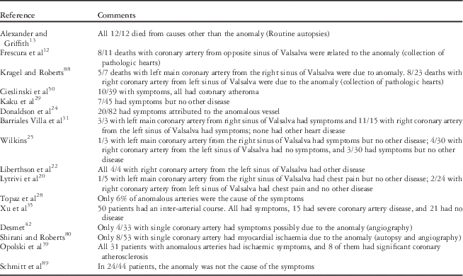
Estimates of proportion of patients with anomalous arteries who had coronary angiography of symptoms or death due to their anomaly and not another cause.

It therefore is of great importance to know whether the incidence of major coronary anomalies of 0.1–0.2% derived from the above studies was inflated by symptomatic patients. Unfortunately, many publications do not distinguish between patients with anomalous arteries who have or do not have other causes for their symptoms. Those who do are summarised in Table 4.
With the exception of the studies by Xu et al,Reference Xu, Zhu, Zhu, Tang and Xu 34 who described that at least 21/50 with an inter-arterial course had no other cause for their symptoms, the study by Opolski et al,Reference Opolski, Pregowski and Kruk 38 who found that about 25% of symptomatic patients with an anomalous left or right coronary artery had unrelated causes for their symptoms, and the autopsy studies on patients with heart disease, less than 25% of the patients with anomalous arteries were studied because of symptoms due to their anomalies, and all the others were found incidentally while studying other diseases. Even if we assume that half of the patients were studied because of symptoms due to their anomaly, that still leaves the prevalence of asymptomatic anomalies at 0.05–0.1%, and suggests that only a minority have symptoms due to their anomaly. This confirms the opinion of Peńalver et al.Reference Peńalver, Mosca, Weitz and Phoon 48 It also confirms the low risk for serious complications of these anomalies observed by Eckart et alReference Eckart, Scoville and Campbell 54 who found only 21 deaths due to a left main coronary artery arising from the right sinus of Valsalva in 6,300,000 army recruits between 1977 and 2001.
If the prevalence of this anomaly is 200/million population (see above), then the population of army recruits surveyed by Eckart et al should have had 1260 subjects with a left main coronary artery arising from the right sinus of Valsalva. If so, the risk of death with this anomaly would be 21/1260=1.67%, and most would be asymptomatic. Even if the prevalence was one-quarter of the figure cited above, the majority of subjects with this anomaly would not have problems from it.
The most lethal form of these anomalies appears to be the left main coronary artery arising from the right sinus of Valsalva, especially if it takes an inter-arterial course. On the basis of the data collected from the literature, 85% of patients with this anomaly who died or had symptoms had an inter-arterial course, whereas only 60% of patients whose clinical course was judged to be unrelated to the anomaly had an inter-arterial course; in these patients, the coronary anomaly was discovered serendipitously.Reference Hoffman 6 This implies that those with an inter-arterial course are more likely to have symptoms. An overlapping series reported by Moustafa et alReference Moustafa, Zehr, Mookadam, Lorenz and Mookadam 55 reached identical conclusions. On the basis of the literature, there are few patients above 30 years of age who died or had symptoms due to a left main coronary artery arising from the right sinus of Valsalva.
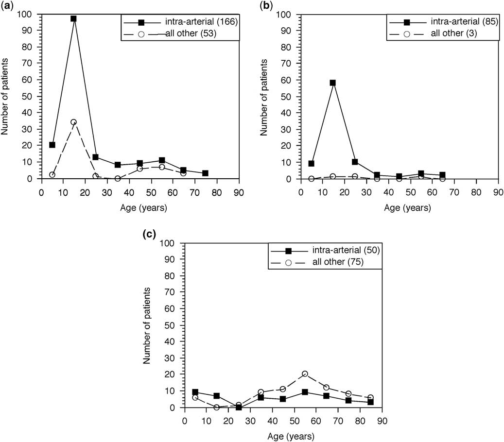
Figure 2 summarises the reports of the age distribution of patients with a left main coronary artery arising from the right sinus of Valsalva. Figure 2a shows those in whom the anomaly caused symptoms, including death (Figure 2a ), and only those who died (Figure 2b ).
Age distribution for left main coronary artery arising from the right sinus of Valsalva. Symptoms due to the anomaly: ( a ) Any symptoms, including death, ( b ) Only deaths, (c) All symptoms, including death, not due to the anomaly, or no symptoms. The numbers in parentheses are the totals for that group. There are far fewer patients here than cited in the tables above because many reports do not list individual ages. In many early studies conducted without autopsy or angiography, it is often difficult to decide if the symptoms were due to coronary artery disease or the arterial anomaly.
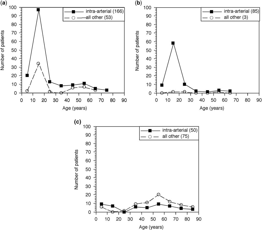
The data shown in Figure 2a and b are not unbiased because they come mainly from reports of one or few patients who were found to have this unusual and interesting anomaly. Nevertheless, there is no reason to think that the age distribution is biased; an older patient with cardiac arrest from this anomaly is probably more likely than a young patient to be the patient of a short report. In addition, cardiac arrest due to a left main coronary artery arising from the right sinus of Valsalva and not passing between the great arteries is unusual enough that it is likely to be reported. Therefore, in these figures the relative numbers per decade and the distribution of the numbers with intra-arterial versus other passage probably reflect the relative differences well. Those in whom the anomaly was found incidental to some other pathology (Fig 2c ) are relatively unbiased; however, when reports of patients whose ages were not mentioned are examined, the proportion with inter-arterial passage is slightly higher.
As shown in Figure 2a and b , most of the deaths occur between 10 and 30 years of age, and almost all deaths are due to inter-arterial passage of the left main coronary artery. The age distribution of those whose symptoms or death were not due to the anomaly (Fig 2c ) is almost flat, and inter-arterial passage does not predominate. The vertical scales are the same in all the figures, and hence reports of symptomatic patients are the majority, probably reflecting selection bias. It is possible that the slight deficit of patients under 30 years of age in Figure 2c is due to their removal because of symptoms. We have no way of knowing whether a large pool of asymptomatic subjects exists, but their rarity suggests that most of these patients come to medical attention when young.
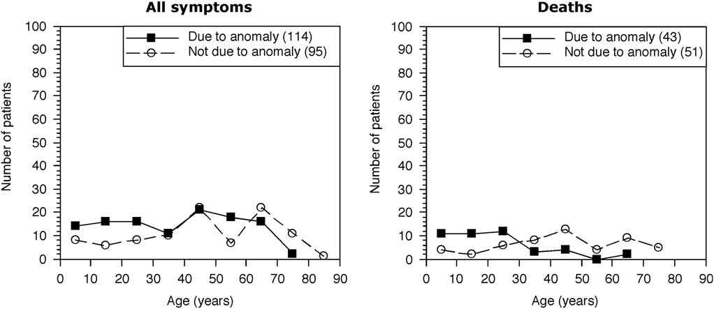
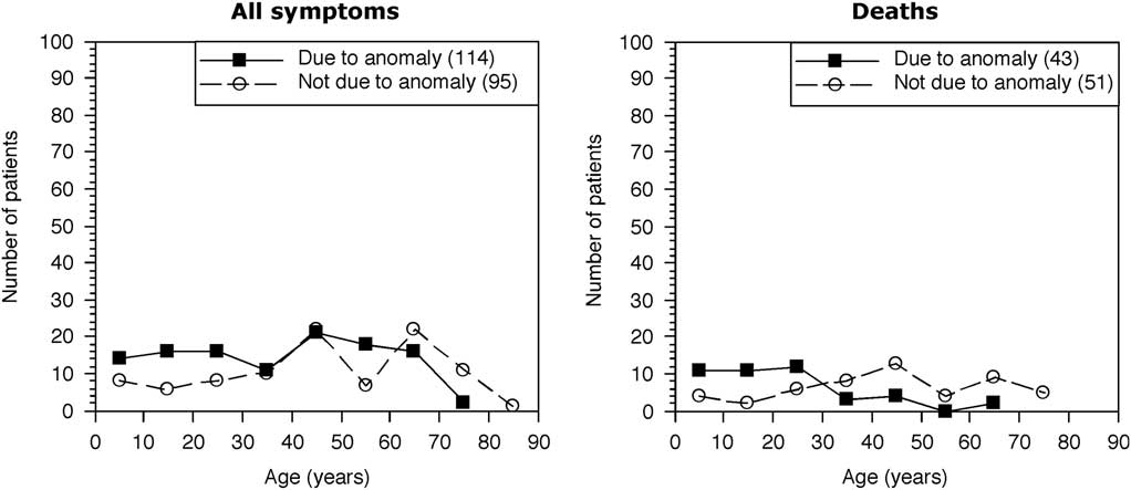
There is less good information about the right coronary artery arising from the left sinus of Valsalva (Fig 3). As described in the literature, by 2013 death or symptoms due to the anomaly were less frequent than those reported in patients with left main coronary artery from the right sinus of Valsalva. There appear to be five to ten times as many patients with an anomalous right than an anomalous left coronary artery, and thus these data strongly suggest that most of the right coronary artery anomalies are benign. Deaths when they occur are predominantly below 30 years of age, but are more spread out than for the anomalous left main coronary artery because of a slightly greater number below 10 years of age. Deaths also occur mainly with respect to strenuous exercise. Those with any symptoms due to the anomaly are spread fairly evenly over all age groups; the higher death rate under 30 years of age may reflect the lesser likelihood of severe exertion over that age.
Age distribution for right coronary artery arising from the left sinus of Valsalva. The numbers in parentheses are the totals for that group. The group listed as symptom not due to the anomaly includes a few with no symptoms.
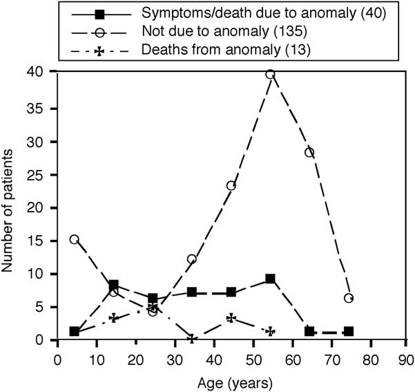
Single coronary arteries appear to be more benign. In three large series, only 17/108 (16%) had ischaemia due to the anomaly. In the data described below, at most 21% had evidence of ischaemia. The age distribution of single coronary arteries that did or did not cause symptoms or death is shown in Figure 4.
Age distribution of patients with a single coronary artery. Numbers in parentheses are totals in each group.
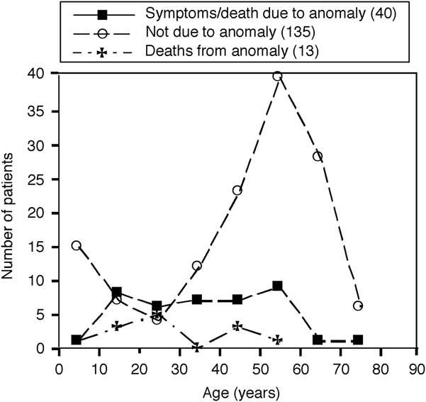
Older patients predominate in those in whom the anomaly did not cause symptoms or death because often the anomaly is discovered during diagnosis and treatment of coronary heart disease. A smaller increase under 10 years of age reflects single coronary arteries to be found coincidentally during diagnosis of congenital heart disease. Those with symptoms or death are spread out evenly across the age groups from 10 to 60 years. Most deaths occurred during or after extreme exertion.
Of those with symptoms or death due to the anomaly, there were 20/131 (15%) with intra-arterial passage and 18/47 (38%) with other anatomic courses. Thus, although the collected literature demonstrates a predominance with intra-arterial passage, the morbidity with intra-arterial passage is significantly lower than for other anatomic variants (χ2, 1.93, p=0.00, odds ratio 0.29 with 95% confidence limits of 0.127–0.661).
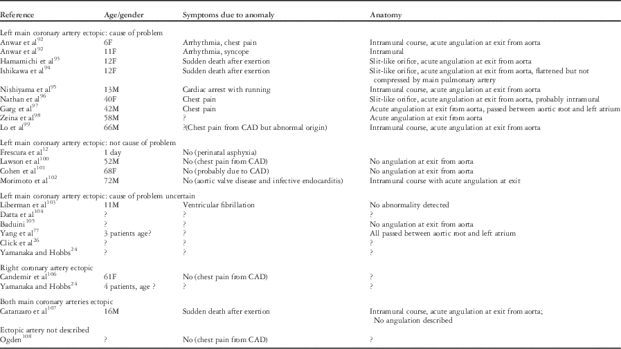
Data for abnormal origin from the posterior sinus of Valsalva are sparse enough to be presented singly in Table 5.
Origin from posterior (non-aortic) sinus of Valsalva.

CAD=coronary artery disease; LMCA=left main coronary artery
Within each group, patients are arranged by ascending age
An ectopic left main coronary artery arising from the posterior sinus, non-facing sinus, of Valsalva is much more often described than an ectopic right coronary artery, but whether this reflects a difference in incidence or is due to selection bias is unknown. All 10 ectopic left main coronary arteries thought to have caused symptoms or death, including one patient with both main coronary arteries ectopic, had one or more abnormal anatomic features – intramural course, acute angulation at exit from aorta, or a slit-like orifice. In one 11-year-old child who developed ventricular fibrillation while running, none of these abnormalities were found. With one exception, a 72-year-old man with infective endocarditis and aortic valve disease, these abnormal anatomic features were not described for those in whom the cause of symptoms was not defined or else due to coronary artery disease or some other disease. Therefore, despite their rarity, these anomalies should be investigated for anatomic or physiologic perfusion abnormalities as for any other form of ectopic origin of a coronary artery.
Pathophysiological mechanisms of symptoms and death
These anomalies cause symptoms and death from myocardial ischaemia. In many older patients, the cause of ischaemia was atherosclerotic narrowing with or without thrombosis in the course of the anomalous artery and differs in no way from coronary atherosclerosis in normally attached arteries.Reference Alexander and Griffith 14 In some studies, there was less atheroma in anomalous than normally attached arteries, whereas in others the anomalous arteries appeared to have accelerated atheroma.Reference Hoffman 6 There was a tendency for more atherosclerosis in the anomalous left circumflex coronary artery arising from the right sinus of Valsalva than that arising normally.Reference Ueyama, Ramchandani, Beall and Jones 8 , Reference Wilkins, Betancourt and Mathur 24 , Reference Click, Holmes, Vlietstra, Kosinski and Kronmal 25 , Reference Leroy, Germain, Bauters, Lablanche and Bertrand 56 , Reference Samarendra, Kumari, Hafeez, Vasavada and Sacchi 57
Even in the absence of coronary atheroma, some patients have classical angina pectoris, although older adults may have atypical chest pain. In a few patients, regional myocardial ischaemia has been shown by scintigraphic or computerised tomography studies. About 20% of autopsies have shown subendocardial scars or even a localised infarct.Reference Basso, Maron, Corrado and Thiene 58 Death may be due to ischaemia-induced ventricular fibrillation, as documented many times.Reference Hoffman 6 Almost all deaths occurred during or just after exercise, and frequently patients had no preceding symptoms.
There are four functional demonstrations of myocardial hypoperfusion and ischaemia. Brandt et alReference Brandt, Martins and Marcus 59 studied a man with angina of effort who had an anomalous right coronary artery arising from the left sinus of Valsalva. During surgery, a graft was placed from the aorta to the right coronary artery after its emergence between the great arteries. Coronary flow reserve as tested by reactive hyperaemia was normal when flow perfused the graft, but was much reduced when only the proximal right coronary artery conducted the flow. In a similar patient, the fractional flow reserve after injecting adenosine into the anomalous right coronary artery was reduced to 0.41.Reference Naruse, Sato and Kakefuda 60 In one patient with single right coronary artery, infusing adenosine reduced the fractional flow reserve in the left anterior descending coronary artery;Reference Coceani, Ciardetti and Pasanisi 61 this patient had apical hypoperfusion abolished after surgery. Reduced fractional flow reserve after infusing adenosine or dobutamine was documented in a 14-year-old girl with the left main coronary artery attached to the right sinus of Valsalva, chest pain, and evidence of apical hypoperfusion on stress testing.Reference Lim, Forsberg, Lee and Kern 62 During surgery, a slit-like orifice was observed and corrected. The reduced myocardial fractional flow reserve indicates that the orifice obstruction was the major cause of the ischaemia.
A more elaborate study was performed by Bartoli et alReference Bartoli, Wead, Giridharan, Prabhu, Koenig and Dowling 63 who took advantage of the incidental finding of a calf with an anomalous left coronary artery from the right sinus of Valsalva with inter-arterial passage. They increased myocardial metabolism and heart rate by infusing dobutamine and arterial blood pressure by infusing phenylephrine. During an increased heart rate, the resistance to flow increased in the left main coronary artery; its flow did not increase as much as the flow in the right coronary artery, and there was increasing ST depression. Elevating arterial blood pressure in normal calves increased the flow in normal left main coronary arteries but decreased it in the anomalous left main coronary artery. Histological changes of acute ischaemia were seen in the territory supplied by the anomalous left main coronary artery.
Schmidt et alReference Schmidt, Klues, vom Dahl, Brenn, Hugel and Hanrath 64 studied two adults with angina pectoris and a single right coronary artery. In one, the left main coronary artery passed anterior to the main pulmonary artery but ran deep in the ventricular muscle. In the other, the left main coronary artery passed between the two great arteries. A Doppler wire demonstrated the localised regions of flow acceleration in each abnormal left main coronary artery remote from its origin, suggesting narrowing and potential restriction to maximal flow. Dauty et alReference Dauty, Piriou, Laprerie, Menu, Dubois and Trochu 65 observed a 22-year-old professional football player with a right coronary artery arising from the left sinus of Valsalva. The artery had a diameter of 3.5 mm at its origin that decreased to 1.6 mm as it passed between the great arteries. There are few reports on the absolute sizes of these abnormal coronary arteries, and no flow studies on them other than those cited above. It is possible that arteries may either become narrower over time or that patients might outgrow the vasodilator capability of these arteries, leading to late onset of symptoms.
Basso et alReference Basso, Maron, Corrado and Thiene 58 postulated that the subendocardial scarring resulted from repetitive episodes of ischaemia, a conclusion that fits the patchy distribution of coronary flow reserve demonstrated in dogs by Coggins et al.Reference Coggins, Flynn and Austin 66 This probably explains the high number of sudden deaths during intensive exercise, but does not completely explain the age incidence of sudden death, which occurs predominantly between 10 and 20 years of age.Reference Hoffman 6 , Reference Moustafa, Zehr, Mookadam, Lorenz and Mookadam 55 It may take several years before there is enough myocardial damage to make the heart vulnerable to ventricular fibrillation. Alternatively, the obstruction to coronary flow gradually increases because of differential growth of the anomalous coronary artery and the aorta so that the obstruction gradually increases in severity, and it is even possible for the flap that sometimes covers the coronary orifice to grow and become more obstructive with time. These considerations have a bearing on diagnosis, because tests for myocardial ischaemia may be normal at one time but abnormal some years later.
Mechanisms of ischaemia
Myocardial ischaemia suggests an obstruction to coronary blood flow that could be either distal, as the anomalous artery passes between the great arteries, peri-aortic due to kinking and angulation of the anomalous artery as it leaves the sinus of Valsalva, or proximal with respect to the abnormal coronary orifice and intramural course. A rarer possibility is diffuse spasm of the artery.
Early observations about the predominance of inter-arterial passage of the anomalous artery among patients who died suggested that during exercise the anomalous artery was compressed between the aorta and the main pulmonary artery or right ventricular outflow tract. One study of an anomalous right coronary artery with multidetector computed tomography reported a 35% decrease in the cross-sectional area of the origin of the right coronary artery in systole in a patient at rest.Reference Ghersin, Litmanovich and Ofer 67 Other investigators have demonstrated narrowing of the inter-arterial portion of the right coronary artery.Reference Giroux and Cartier 43 , Reference Dauty, Piriou, Laprerie, Menu, Dubois and Trochu 65 , Reference Bagur, Gleeton, Bataille, Bilodeau, Rodes-Cabau and Bertrand 68 – Reference Porto, MacDonald, Selvanayagam and Banning 70
During strenuous exercise in normal individuals, systolic and diastolic blood pressures increase in the aorta and pulmonary arteries. Aortic systolic arterial pressures may rise up to about 200 mmHg with severe exercise, and diastolic aortic pressure may increase by 10–20 mmHg.Reference Cumming 71 – Reference Kurl, Laukkanen, Rauramaa, Lakka, Sivenius and Salonen 73 Therefore, with exercise the coronary arteries should be exposed to these higher pressures and should dilate. The mean pulmonary arterial pressures can rise to 30 mmHg,Reference Cumming 71 , Reference Damato, Galante and Smith 74 – Reference Fowler 76 the increase being much less than in the aorta so that the pressure difference between the pulmonary and coronary artery increases during exercise, suggesting less ability of the pulmonary artery to compress the coronary artery. Some deformation of the anomalous arteries in systole might occur because of stretching of the aortic root or tension or pressure changes in the dense connective tissue between the great arteries, but unfortunately there are no reports of the cyclic changes in diameter of these anomalous coronary arteries during exercise. Chaitman et alReference Chaitman, Lesperance, Saltiel and Bourassa 77 described a case of a 49-year-old woman with mitral stenosis and pulmonary hypertension who had no evidence of myocardial ischaemia, despite inter-arterial passage of the left main coronary artery.
If because of some presently unknown anatomic factors the anomalous coronary artery is narrowed in systole, it is still not clear that this is of physiologic importance. As far as we know, almost all the systolic flow in the left coronary artery distends the extramural arteries and little if any perfuses the myocardium. If it does, it is confined to the superficial myocardium because the subendocardium is perfused only in diastole.Reference Flynn, Coggins and Goto 78 Furthermore, those anomalous arteries that run in the upper part of the septum and are always exposed to high pressures during systole have a very low incidence of symptoms and death. Add to this list the fact that some patients develop myocardial ischaemia and even die suddenly with anomalous arteries that do not pass between the great arteries, so that compression between the great arteries is an unlikely mechanism for the observed myocardial ischaemia in most patients.
An alternative mechanism is a proximal obstruction associated with the slit-like coronary orifice, sometimes an overlying tissue flap, or narrowing of the artery as it runs in the aortic wall. In one study,Reference Kaushal, Backer and Popescu 79 patients were more likely to have symptoms if the intramural pathway was long (10 mm) than when it was short (5 mm). They did not, however, measure the luminal diameters.
It is possible that during exercise the increased systolic pressure in the aortic wall narrows the intramural artery further, or at least prevents the artery from dilating, thereby impeding the increased coronary blood flow that is required by the increased myocardial work.
The slit-like ostium, tissue flap, intramural course, and kinking of the artery as it leaves the sinus of Valsalva are found with the anomalous right coronary artery arising from the left sinus of Valsalva, with the left main coronary artery, which runs from the right sinus of Valsalva between the great arteries, or with the left main coronary artery arising from the posterior sinus of Valsalva.
Lipton et alReference Lipton, Barry, Obrez, Silverman and Wexler 46 observed that in four patients with a single coronary artery the cross-sectional area of the single main coronary artery was about 46% of the combined cross-sectional area of two normally attached coronary arteries. It is possible that in some patients the diameter might be even smaller and restrictive at times when high coronary flows are needed.
Treatment
Medical treatment
Conservative treatment with beta-adrenergic blockers, nitrates, angiotensin-converting enzyme blockers, or calcium blockers, as well as limiting exercise, has been effective for some older patients with a right coronary artery arising from the left sinus of Valsalva. Kaku et alReference Kaku, Shimizu and Yoshio 28 described 44 patients with a mean age about 56 years, most of whom had symptoms including syncope and about half had scintigraphic evidence of local myocardial ischaemia. During follow-up for 2 months–14.5 years (mean 5.66 years), there were no deaths due to the anomaly. Ouali et alReference Ouali, Neffeti, Sendid, Elghoul, Remedi and Boughzela 36 treated 20 adults (mean age 53 years) for a mean of 34 months without complications, but there were only three patients with a right coronary artery from the left sinus of Valsalva. Berdoff et alReference Berdoff, Haimowitz and Kupersmith 80 described seven patients with chest pain, all but one becoming asymptomatic on follow-up; the treatment was not described. No patient in these series had an anomalous left main coronary artery arising from the right sinus of Valsalva. Opolski et alReference Opolski, Pregowski and Kruk 38 treated 21/24 symptomatic patients – 20 with an anomalous right coronary artery, two with an anomalous left main coronary artery, and one each with anomalous left circumflex coronary artery or left anterior descending coronary artery – conservatively for 5–44 months with no deaths due to the anomaly but with substantial residual symptoms including chest pain and syncope. A 45-year-old and a 61-year-old patient with a right coronary artery arising from left sinus of Valsalva became asymptomatic after treatment with beta-adrenergic blockers,Reference Barbou, Schiano and Lahutte 81 , Reference Sato, Matsumoto and Komatsu 82 one such patient aged 68 years became asymptomatic with no anti-anginal treatment,Reference Bekedam, Vliegen, Doornbos, Jukema, de Roos and van der Wall 83 and a 51-year-old man with a left main coronary artery arising from the right sinus of Valsalva and taking a long intra-septal course became asymptomatic after slowing of his heart rate with Ivabradine (Procorolan®, Servier Laboratories, Neuilly-sur-Seine, France).Reference Mogensen, Grande, Kober and Kofoed 84
Medical treatment is inappropriate for children or young adults who are usually managed by exercise restriction or surgical correction of the anomaly.Reference Brothers, Gaynor, Paridon, Lorber and Jacobs 85
Surgical treatment
Coronary artery bypass
Some of the earliest operations bypassed the potential obstructions by using a saphenous vein or internal thoracic artery graft,Reference Bett, OʼBrien and Murray 86 – Reference Reddy, Takahashi, Beckles and Filsoufi 89 but competition between the graft and the native flow path may lead to graft failure.Reference Davies, Burkhart and Dearani 50 , Reference Fedoruk, Kern, Peeler and Kron 90 – Reference Botman, Schonberger and Koolen 92
Surgery on origin of anomalous artery
The type of surgery depends on the specific anatomy:
-
a. No intramural course; separate orifices in the sinus of Valsalva.
-
b. Slit-like orifice, intramural course, kinking, or narrowing at exit from aorta; these features are typical of an anomalous coronary artery arising from the wrong sinus of Valsalva and taking an inter-arterial course.
-
c. Both right and left coronary arteries share a common orifice.
-
d. Single orifice and single main coronary artery that then divides outside the aorta.
Direct implantation
Direct implantation may be performed for group a, especially if the orifices are well separated.Reference Mustafa, Gula, Radley-Smith, Durrer and Yacoub 93 – Reference Desgue, Cuttone, Babatasi, Labombarda and Massetti 97 It is usually unsuitable for the other anatomic types because of insufficient tissue to create a button around the orifice or the need to avoid kinking. However, it is not clear that this anatomy causes proximal obstruction.
Reconstruction of the orifice
The operation most often performed for group b is to remove any proximal obstruction by unroofing the intramural portion of the artery. In the large series reported by Poynter et al,Reference Poynter, Williams, McIntyre, Brothers and Jacobs 5 this operation was performed in 87% of the patients. Unroofing eliminates any flap and the slit-like orifice, widens the narrowed intramural artery and prevents its compression during systole, and eliminates kinking. The procedure is uncomplicated if the intramural artery is distal to the commissure. If it is not, then the commissure must be detached and later resuspended, and aortic regurgitation may result.Reference Mainwaring, Reddy and Reinhartz 49 , Reference Kayalar, Burkhart, Dearani, Cetta and Schaff 98 , Reference Mumtaz, Lorber, Arruda, Pettersson and Mavroudis 99 To avoid this complication, some surgeons create a neo-ostium between the distal portion of the intramural artery and the commissure and then obliterate the portion of the artery between the commissure and the original ostium.Reference van Son and Mohr 100
The operative mortality is virtually zero, but occasional short-term complications occur, as after any cardiotomy (see Table 3 by Peńalver et alReference Peńalver, Mosca, Weitz and Phoon 48 ). Of more concern are long-term potential complications such as stenosis of the neo-orifice, loss of endothelial function, residual myocardial ischaemia,Reference Brothers, McBride and Seliem 101 and premature atherosclerosis in these arteries. Follow-up has not been very long, with the two longest series extending to 7 and 9 years.Reference Mainwaring, Reddy and Reinhartz 49 , Reference Wittlieb-Weber, Paridon, Gaynor, Spray, Weber and Brothers 102 In the Mayo Clinic experience,Reference Davies, Burkhart and Dearani 50 the average follow-up was 1.1 years; one patient was doing well 14 years after surgery, and only 1/36 patients had symptoms. However, Wittlieb-Weber et alReference Wittlieb-Weber, Paridon, Gaynor, Spray, Weber and Brothers 102 followed 24 patients after surgery, with no late deaths. They noted, that 54% reported the same symptoms as pre-operatively, but with no signs of ischaemia, and that four patients developed mild aortic insufficiency. Long-term results remain to be evaluated.
Chest pain in children is less likely to be ischaemic in origin and more likely to be of non-specific chest wall origin than it is in adults. It is therefore possible that the anomalous coronary artery in children is often detected by echocardiography performed for non-specific chest pain that will not be relieved by surgery of the anomalous artery.
Similar relief of the proximal obstruction may be obtained by stenting the orifice. In the 1990s, interventional cardiologists learnt how to perform balloon angioplasty to insert a stent for atherosclerotic lesions of anomalous coronary arteries, and in 2000 Doorey et alReference Doorey, Pasquale, Lally, Mintz, Marshall and Ramos 103 reported placing stents in the proximal portions of the coronary artery in four patients with a left main coronary artery arising from the right sinus of Valsalva and in nine patients with a right coronary artery arising from the left sinus of Valsalva. One stent implanted early in the series developed compression of its midsegment. In a short follow-up, all patients did well with normal perfusion scans. Since then, at least six other patients have had successful implantation of a stent into the anomalous right coronary artery.Reference Bagur, Gleeton, Bataille, Bilodeau, Rodes-Cabau and Bertrand 68 , Reference Porto, MacDonald, Selvanayagam and Banning 70 , Reference Hariharan, Kacere and Angelini 104 , Reference Jennings, van Gaal and Banning 105 Little follow-up information is available, and the long-term outlook for stents is uncertain; for this reason, stents are not currently indicated for children.
Surgically reconstructing the orifice of the anomalous coronary artery abolishes symptoms; hence, it strongly favours the concept that the obstruction to flow is proximal and not between the great arteries. The success of proximal stents does not have the same implications, because the stents are 10–20 mm long and hold open not only the region of the orifice, but also the inter-arterial portion of the anomalous artery.
Single ostium or single coronary artery
Sometimes the coronary artery with an anomalous origin shares a single orifice with the normal artery or common trunk and does not have an intramural course in the aortic wall (type c),Reference Desgue, Cuttone, Babatasi, Labombarda and Massetti 97 or else there is a single coronary artery that bifurcates outside the aorta (type d). Removing and reimplanting the ectopic artery into its appropriate sinus of Valsalva may be difficult because there may not be enough tissue to provide a large button for implantation.Reference Kayalar, Burkhart, Dearani, Cetta and Schaff 98 , Reference Jaggers and Lodge 106 The options are then to translocate the pulmonary artery (see below) or to perform a distal coronary artery bypass graft. In one patient,Reference Shah and Redmond 107 the orifice of the single artery was narrowed, and enlarging this relieved the symptoms.
The role of distal obstruction was reconsidered after pulmonary artery translocation was introduced. Rodefeld et alReference Rodefeld, Culbertson, Rosenfeld, Hanley and Thompson 108 operated on an 11-year-old boy with right and left coronary arteries arising from a single ostium in the left sinus of Valsalva, the right coronary artery then passing between the great arteries. Reimplantation was not possible because there would not have been a large enough button of tissue after removing the right coronary artery from the common orifice. The main pulmonary artery was transected just below the pulmonary arterial bifurcation and then reanastomosed to the left pulmonary artery, thus opening up the space between the aorta and main pulmonary artery beyond their attachments to the heart; the right pulmonary artery was patched to prevent stenosis. A variant of this technique is to transect the aortic root as well, widen the proximal part of the anomalous artery beyond the intramural segment with a patch, and then proceed with the pulmonary artery translocation.Reference Alphonso, Anagnostopoulos and Nolke 109 , Reference Karl, Provenzano and Nunn 110 With this technique, both proximal and perhaps distal obstructions will be relieved, although it is unclear whether the benefit is because of more than creating the neo-orifice.
Is the anomaly causing myocardial ischaemia?
These patients may come to medical attention for a number of symptoms: resuscitation after cardiac arrest, syncope or ventricular arrhythmias during or just after strenuous exercise, or angina of effort. Chest pain may be atypical but requires investigation if it is recurrent with exertion and no localised chest abnormalities are found. From the literature, atypical chest pain seems to be more common in older adults. Congestive heart failure is rare.
At other times, coronary anomalies may be found incidentally while the patient is being investigated by imaging techniques for other cardiac problems.
Symptomatic patients
In addition to a careful history and physical examination, the patients need an electrocardiogram to look for ST changes suggestive of myocardial ischaemia and for arrhythmogenic abnormalities such as the long or short QT interval, Brugada syndrome, or abnormalities consistent with arrhythmogenic right ventricular dysplasia.Reference Fontaine, Fontaliran and Hebert 111 – Reference Romero, Mejia-Lopez, Manrique and Lucariello 113
Imaging begins with an echocardiography–Doppler examination that has three objectives:
-
1. Exclude other cardiopulmonary pathology such as hypertrophic cardiomyopathy, aortic stenosis, or severe pulmonary hypertension.
-
2. Look for regional wall motion abnormalities.
-
3. Examine the origins of the coronary arteries. If these are ectopic the possibility of restrictive flow needs to be determined (see below). If they do not appear to be ectopic but the history is compatible with an ectopic coronary artery, further imaging with magnetic resonance imaging or fast computerised tomography is mandatory because of the known lack of sensitivity of echocardiography. Special care is needed for imaging intramural passage of the anomalous artery because most standard computerised tomography or magnetic resonance imaging sequences do not show intramural vessels well.Reference Angelini and Flamm 114 It may even be necessary to examine the origin of the artery by intravascular ultrasound, although relatively few cardiologists have expertise in this form of investigation.
The origins of the coronary arteries may be restrictive because of an ostial flap, slit-like orifice, narrowed and long intramural course in the aortic wall, narrowing or kinking as the artery leaves the aorta, or the tangential arterial exit that leaves the aorta at an acute angle. Such restrictions are associated with relative hypoperfusion, especially when myocardial flow needs to be increased markedly with exercise. Any site of localised flow acceleration in the coronary artery suggests a restriction that may cause hypoperfusion at maximal flow.
In the absence of visible restriction to flow, regional hypoperfusion should be assessed. This can be performed by single-photon emission computed tomography with Thallium201 or TechnetiumReference Mumtaz, Lorber, Arruda, Pettersson and Mavroudis 99m or by positron emission tomography that has advantages over single-photon emission computed tomography.Reference Bateman, Heller and McGhie 115 The patient is examined during exercise and then while resting to look for redistribution. Alternatively, coronary flow reserve in the abnormal artery may be measured by echocardiography–Doppler examination at rest and then after exercise or after increasing myocardial blood flow by infusing dobutamine or dipyridamole. It is important to compare normal and ectopic arteries to show their difference. Farhad and MurphyReference Farhad and Murthy 116 provided a detailed discussion of methods, mechanisms, and dosages.
Dobutamine increases myocardial contractility and heart rate, consequently increasing myocardial work and oxygen consumption. It is used as a substitute for exercise and has been shown to be free of serious complications when used in adults with myocardial ischaemia from coronary atheroma. It may, however, cause a coronary steal and regional myocardial ischaemia. Even without a steal, failure of flow to increase appropriately may lead to regional wall motion abnormalities. The agent is given by infusion at doses of 10–50 μg kg−1min−1.
Vasodilatation without exercise can be achieved by infusing adenosine at a dose of 140 μg kg−1min−1 for 4 min. Adenosine acts on all four purinergic receptors in the arterial wall to cause vasodilatation without an increase in myocardial work or oxygen consumption. Minor side effects are common, and the agent should be avoided in patients with reactive airway disease, sick sinus syndrome, or first- or second-degree heart block. Adenosine is rapidly metabolised. As an alternative, dipyridamole is infused at a rate of 0.14 mg kg−1 min−1 for 4 min. This agent inhibits the reuptake of adenosine into cells and also inhibits its breakdown by adenosine deaminase.Reference Chen, Lo, Chao and Su 117 In patients, it may cause a coronary steal and chest pain, and mild side effects are common, but this effect can be reversed immediately by 75 mg of intravenous aminophylline in adults. Regadenoson (Lexiscan®, AstellasPharma, Tokyo, Japan) has a much greater affinity for A2A receptors – the main receptors for coronary vasodilatation – than for the other adenosine receptors, and hence has fewer side effects. It is given as an intravenous bolus – 0.4 mg in 5 ml saline over 10 s – followed by a 5-ml saline flush.Reference Chen, Lo, Chao and Su 117
It is beneficial to use a combination of these tests, because an important large study of patients showed that any single test may not indicate ischaemia when the others do.Reference Osaki, McCrindle, Van Arsdell and Dipchand 118 Furthermore, the stress electrocardiogram may show ischaemia at one time but not at another.Reference Brothers, Carter, McBride, Spray and Paridon 119
If the ectopic artery is anatomically restrictive or shows a reduced coronary flow reserve, then treatment of the anomaly is required when the obstruction is severe enough (see below). If there is neither anatomic nor functional restriction of flow, then the patient may be observed with or without medical treatment such as nitrites, beta-adrenergic blockers, or calcium blockers.
Patients with incidental finding of an ectopic coronary artery
If an ectopic coronary is found incidentally, much the same course of investigation should be pursued. These patients will probably not have symptoms to suggest that the anomaly is causing myocardial ischaemia. Just as for symptomatic patients, the anatomy of the origin of the ectopic coronary artery must be defined, and if it is normal then the flow capacity of that ectopic artery must be evaluated. Either anatomic or functional flow restriction requires correction of the anomaly, but in their absence only observation is required.
Who should be treated?
Finding an anatomic site of flow restriction or reduced coronary flow reserve is by itself insufficient evidence to proceed to surgery, just as it is not absolute indication for treatment of coronary atheroma. It is the degree of obstruction and hypoperfusion that matters. If the severity of obstruction is not obvious to the naked eye, many cardiologists have determined myocardial fractional flow reserve or coronary flow reserve to assess the severity. The myocardial fractional flow reserve method involves measuring pressures with a pressure-sensitive wire in the proximal coronary or aorta (Pa), beyond the stenosis (Pc), and right atrial pressure as an estimate of coronary venous pressure (Pv). The measurements are made during maximal coronary vasodilatation.Reference De Bruyne, Paulus and Pijls 120 , Reference Pijls, van Son, Kirkeeide, De Bruyne and Gould 121
 $${\rm Myocardial}\,{\rm fractional}\,{\rm flow}\,{\rm reserve}={{Pc{\minus}Pv} \over {Pa{\minus}Pv}} \approx {{Pc} \over {Pa}}$$
This method assumes normal microvascular function and linear relationships that are often absent.Reference Spaan, Piek, Hoffman and Siebes 122 Nevertheless, a myocardial fractional flow reserve ratio of less than 0.75 is a good predictor of the need for stenting a narrowed coronary artery.Reference De Bruyne, Pijls and Kalesan 123 , Reference Tonino, De Bruyne and Pijls 124 This method has been applied successfully in children with Kawasaki disease,Reference Ogawa, Ohkubo and Fukazawa 125 and the cut-off values for myocardial fractional flow reserve in children were also 0.75. These authors also found that a coronary flow reserve of less than 2 had the same significance. Even if the coronary flow reserve can be measured non-invasively, the results are not independent of basal coronary flow. The rationale, values, and limitations of all these methods are described clearly by Canty.Reference Canty 126 Recent studies by Sen et alReference Sen, Asrress and Nijjer 127 , Reference Sen, Escaned and Malik 128 have suggested that a ratio similar to the myocardial fractional flow reserve can be measured without the need for vasodilatation. It is even possible to measure stenosis resistance with a wire containing both pressure and flow sensors,Reference Meuwissen, Chamuleau and Siebes 129 but good standards are not yet available.
With this background, any evidence of anatomic obstruction requires evaluation of its severity. Naturally, if there is associated evidence of regional ischaemia, then hypoperfusion has been confirmed.
Specific anomalies
If there is a left main coronary artery arising from the right sinus of Valsalva and passing between the great arteries in a young athlete, many physicians recommend surgical correction, especially if there is a slit-like orifice, intramural passage, or narrowing or kinking of the artery as it leaves the aorta. These features predict a high risk for sudden death during exercise, and warrant treatment.
If there is a left main coronary artery arising from the right sinus of Valsalva and not passing inter-arterially, then most physicians would look for evidence of significant anatomic obstruction to the coronary flow or evidence of hyoperfusion or myocardial ischaemia before recommending operation, because the risk of symptoms from these other courses is not high and because the long-term complications of the surgery are unknown. The same issue arises with a single coronary artery, although as pointed out above the flow restriction might be because of the size of the single coronary orifice. Should the patient be older, have vague symptoms, and not be involved in strenuous activity, medical treatment has been recommended. As mentioned above, no late deaths have occurred in a limited series of such patients.
If the anomaly is a right coronary artery arising from the left sinus of Valsalva, the decision is more difficult. If the data on the high incidence of asymptomatic anomalies are confirmed, then few of the anomalous right coronary arteries threaten health. Emphasis then must be placed on the anatomy of the orifice, with any signs of significant restriction, or on the functional tests of myocardial ischaemia. If these are present, then surgical correction is recommended. Once again, the more severe the symptoms and evidence of ischaemia, the more likely is it that surgery will be needed.
An ectopic left main coronary artery arising from the posterior sinus of Valsalva may often cause serious problems, and should be investigated as a potential cause of myocardial ischaemia. The risk is much less for a right coronary artery arising from the posterior sinus of Valsalva. Nevertheless, any anomaly of coronary arterial origin associated with symptoms related to myocardial ischaemia needs to be carefully assessed by expert imaging and if necessary by studies to detect hypoperfusion.
Physicians usually seek evidence of myocardial ischaemia by scintigraphic tests with and without exercise; hence, consideration should be given to the alternative of evaluating coronary flow reserve with transthoracic, trans-oesophageal, or intravascular Doppler techniques. The association of symptoms with a reduced flow reserve in the anomalous artery – coronary flow reserve <2 or myocardial fractional flow reserve <0.75 – might be taken to indicate the need for surgery.
Failure to find evidence of coronary flow restriction or myocardial ischaemia with any of the major anomalies suggests that surgery is not required at the time, but does not preclude the need for surgery in the future. One such patient with mild symptoms was followed with annual stress tests from the age of 8 years until she was 14 years when a positive stress test was first observed; surgery corrected the problem.Reference Lim, Forsberg, Lee and Kern 62 Anatomic abnormalities such as ostial flaps and narrowing or kinking of the artery might progress. After all, a patient who drops dead while exercising strenuously at the age of 18 years probably had no trouble performing the same strenuous exercise at the age of 17 years. Therefore, if one of these anomalies is identified in an asymptomatic patient, there should be follow-up with imaging and testing for regional myocardial ischaemia. This will result in many patients having unnecessary tests, but until we have better methods of detecting those at risk this will be the only way to reduce the death rate from these anomalies.
Conclusion
We can now detect in some patients an anomalous coronary artery that is causing ischaemia and is a threat to life; however, in many other patients the cause of the symptoms remains uncertain and our treatment is at best an educated guess. There is a need for extensive further study of these anomalies.
Acknowledgements
The author is grateful for helpful discussions with Dr David Teitel, Dr Paolo Angelini, and Dr Jane Newburger.
Financial support
This research received no specific grant from any funding agency, commercial or not-for-profit sectors.
Conflict of interest
None