Introduction
Cystic echinococcosis is a zoonotic infection caused by the larval, or hydatid cyst, stage in the life cycle of Echinococcus granulosus. The definitive hosts are canids, whereas wild and domestic ruminants or humans act as intermediate hosts (Soulsby, Reference Soulsby1982; Moro & Schantz, Reference Moro and Schantz2009). Hydatid cysts can occur in all organs and tissues of the body, especially the liver and lungs (Soulsby, Reference Soulsby1982; Moro & Schantz, Reference Moro and Schantz2009). Host reaction to the presence of cysts is variable, with severe reactions causing the death of the parasite by degeneration. The severity of this reaction appears to be related the size and weight of the larval form known as the metacestode, which in turn results in the destruction of tissues within infected organs (Turgay & Üstün, Reference Turgay, Üstün, Altıntaş, Tınar and Çoker2004). There also seems to be an association between hydatid cyst infections and bacteria at some localization sites (Aslam et al., Reference Aslam, Bhaila, Nadeem and Fadoo2005; Ziino et al., Reference Ziino, Giuffrida, Bilei and Panebianco2009) and some metacestode antigens can enter the circulation to induce an immune response (Turgay & Üstün, Reference Turgay, Üstün, Altıntaş, Tınar and Çoker2004).
After non-specific inflammatory reactions, the acute-phase response (APR) occurs in the body and this response includes changes in the concentrations of plasma proteins known as acute-phase proteins (APPs). While albumin and transferrin concentrations decrease in plasma, C-reactive protein, serum amyloid A (SAA) and haptoglobin concentrations increase (Kushner & Mackiewicz, Reference Kushner, Mackiewicz, Mackiewicz, Kushner and Baumann1993; Eckersall, Reference Eckersall1995). SAA is discharged from the liver (Gruys et al., Reference Gruys, Obwolo and Toussaint1994), bones (Landman et al., Reference Landman, Gruys and Bwars1994; Ceron et al., Reference Ceron, Eckersall and Subiela2005; Sevimli et al., Reference Sevimli, Misirlioğlu, Polat, Yalçın, Akkoç and Uğuz2005) and abomasum (Dilda et al., Reference Dilda, Pisani, Rahman, Modina, Tessaro, Sartorelli, Ceciliani and Lecchi2012), while haptoglobin is discharged from lungs, the spleen and adipose tissue (Ebersole & Cappelli, Reference Ebersole and Cappelli2000), and from the forestomach and abomasum (Dilda et al., Reference Dilda, Pisani, Rahman, Modina, Tessaro, Sartorelli, Ceciliani and Lecchi2012). APPs are important parameters in terms of herd health and the diagnosis of diseases (Petersen et al., Reference Petersen, Nielsen and Heegard2004). Also, haptoglobin and SAA are important APPs for cattle, and their serum concentrations increase in hosts with toxic, parasitic (Eckersall, Reference Eckersall1995; Ganheim et al., Reference Ganheim, Hoglund and Waller2004), bacterial and viral (Eckersall, Reference Eckersall1995; Petersen et al., Reference Petersen, Nielsen and Heegard2004) diseases. APP synthesis is induced by the release of pro-inflammatory cytokines such as interleukin (IL)-1, IL-6 and tumour necrosis factor-α from macrophages and monocytes in the inflammation site (Beutler & Cerami, Reference Beutler and Cerami1986; Heinrich et al., Reference Heinrich, Castell and Andus1990; Burton et al., Reference Burton, Honor, Mackenzie, Eckersall, Markham and Horney1994; Sevimli et al., Reference Sevimli, Misirlioğlu, Yağcı, Bülbül, Yilmaztepe and Altunbaş2008). In addition, APPs occur in association with oxidative stress and abomasal tissue damage (Maden et al., Reference Maden, Ozturk, Bulbul, Avci and Yazar2012).
Cytokines discharged from helper T cells (Th1 and Th2) have an important role for the formation of the host immune response in hydatid cyst infection. IL-2, interferon-γ and lymphotoxins are discharged from Th1 cells and IL-4, IL-5, IL-6 and IL-10 are discharged from Th2 cells (Finkelman et al., Reference Finkelman, Pearce, Urban and Sher1991; Allen & Maizels, Reference Allen and Maizels1996; Mosmann & Sad, Reference Mosmann and Sad1996). Levels of serum IL-4, IL-5 and IL-6 are increased in humans infected with hydatid cysts (Riganò et al., Reference Riganò, Profumo, Teggi and Siracusano1996, Reference Riganò, Profumo and Siracusano1997). Cytokines discharged from Th2 cells are increased in mice subjected to experimental secondary hydatid cyst infection (Al-Qaoud & Abdel-Hafez, Reference Al-Qaoud and Abdel-Hafez2008; Haniloo et al., Reference Haniloo, Ghasemi, Shikhi and Ghavami2008), as found in humans.
C-reactive protein from APPs has been investigated in humans infected with hydatid cysts (Refik et al., Reference Refik, Mehmet and Durmaz2005; Arıdoğan et al., Reference Arıdoğan, Kaya, Çetin, Taş and Demirci2009) and a positive relationship was determined between IL-6 and C-reactive protein (Refik et al., Reference Refik, Mehmet and Durmaz2005). IL-6, nitric oxide and C-reactive protein may be useful markers in monitoring hydatid cyst management and evaluating surgical stress (Refik et al., Reference Refik, Mehmet and Durmaz2005). To date, there has been no research published related to SAA and haptoglobin in humans and animals.
In this study, the internal organs of hydatid cyst-infected cattle were examined parasitologically, microbiologically and histopathologically. The study aimed to determine the effect of hydatid cysts and microbial agents on the APR in cattle.
Materials and methods
Collection and examination of samples
A total of 35 slaughtered cattle, comprising 11 males and 24 females, of varying ages were used in the study. Clinical findings such as pain, redness, slackness, diarrhoea and fever were not seen in these cattle. Blood samples were taken into vacutainers without heparin during slaughter, the internal organs of the cattle were examined post-mortem for possible infections and faecal samples were collected. For histopathological and microbiological assessments, liver and lung tissues of 27 cattle diagnosed with hydatid cysts during organ examination were evaluated as the study group, while the liver and lung tissues of eight cattle that were not diagnosed with any lesions were evaluated as the control group. Samples of organs and faeces collected in aseptic conditions were placed into sterile plastic bags and numbered, and were brought to the laboratory in a cool box on ice.
Parasitological and histochemical procedures
Cyst numbers were determined and the diameter, at the widest site, of each cyst was measured in the laboratory. The number of cysts was recorded according to organ distribution. The outer wall of each cyst was sterilized with pure alcohol. In the case of non-degenerate cysts, the cyst fluid was collected into a sterile syringe, and, in the other cases, sterile instruments were used. The cyst wall was incised with a sterile scalpel blade, and the content was transferred into a sterile Petri dish. The germinal layer was put in glycerin between two layers of microscope glass. Then drops of cyst fluid, content of the cyst in Petri dishes and germinal layer were examined microscopically for the presence of a protoscolex. The presence of a protoscolex, either attached to the germinal epithelium in the form of a brood capsule or in the cyst fluid, was considered indicative of fertility. Cysts that contained no protoscolex, as well as being heavily supurative or calcified, were considered infertile (Paredes et al., Reference Paredes, Godoy, Rodriguez, Garcia, Cabezón, Cabrera, Jiménez, Hellman, Sáenz, Ferreira and Galanti2011). This application was achieved for all hydatid cysts determined in livers and lungs. After the parasitological procedure, tissue samples were taken from the remaining lesion area for histopathological and microbiological assessments. For this purpose, a cyst with a diameter smaller than 1 cm and a cyst with a diameter larger than 1 cm were chosen in organs that had more than two cysts. Following the selection of tissue samples for microbiological analysis, samples were transferred to 10% buffered formalin solution for histopathological examination.
All faecal samples were examined macroscopically and microscopically. Benedict's sedimentation was used for trematode infections, saturated water flotation for cestodes and nematodes, and the Baermann–Wetzel method for lungworms. In faeces found positive for gastrointestinal nematodes, egg numbers/g faeces were calculated using the McMaster method to detect the severity of infection. Faecal cultures were performed to diagnose these nematodes in terms of genus (Urquhart et al., Reference Urquhart, Armour, Duncan, Dunn and Jennings1987; Hansen & Perry, Reference Hansen and Perry1990).
For histopathological assessments, tissue samples were processed by routine methods, and 5 μm sections were taken. Tissues were stained with haematoxylin and eosin. Immunohistochemistry was performed by the streptavidin–biotin–peroxidase method in order to identify IL-6 (sc 1265, Santa Cruz, Biotechnology Inc., Dallas, Texas, USA) and secondary antibody (sc 2053, Santa Cruz). 3,3'-Diaminobenzidine (DAB) was used as the chromogen in all tissue sections. In the present study, due to the lack of cow-specific reagents, polyclonal primary antibodies of mammalian origin were used (Dionissopoulos et al., Reference Dionissopoulos, Steele, AlZahal, Plaizier and McBride2012). The evaluation criteria used in histopathological examination were applied in terms of positive or negative staining for IL-6.
Microbiological and immunological procedures
A total of 31 liver and 18 lung samples of 27 cattle infected with hydatid cysts were evaluated as the study group. Liver and lung samples without any macroscopic lesions were assessed as the control group. For bacterial isolation, inoculation from the organs was made to general, selective and differential media by sterilized swabs. For this, Columbia blood agar including 7% sheep blood, modified charcoal–cefoperazone–deoxycholate (mCCDA)–Preston agar, Sabouraud dextrose agar and MacConkey agar were used. Petri dishes were incubated in aerobic, anaerobic and microaerobic conditions. After the incubation, growing colonies were examined macroscopically, for colony morphology, haemolysis and pigment production, and microscopically, for Gram stains, morphology and motility examination. The identification of bacteria was made at the generic or species level using standard biochemical tests (Holt et al., Reference Holt, Krieg, Sneath, Staley and Williams2000, Quinn et al., Reference Quinn, Markey, Carter, Donnelly and Leonard2002).
SAA, haptoglobin and IL-6 levels in the serum samples were analysed with the enzyme-linked immunosorbent assay (ELISA) method. In this study, SAA (CSB-E08592b; Cusabio, Wuhan, China), haptoglobin (CSB-E08585b, Cusabio) and IL-6 (CSB-E12899b, Cusabio) were provided commercially.
Data analysis
Data were analysed by using the statistical program SPSS version 16.0 (SPSS Inc., Chicago, Illinois, USA). All values are presented as mean ± standard error (SE). Differences in the concentrations of IL-6, SAA and haptoglobin in control and hydatid cyst groups were analysed using a t-test. One-way analysis of variance (ANOVA) followed by a Tukey test was used to evaluate the differences in the concentrations of IL-6, SAA and haptoglobin in infected cattle to the location of cyst in the liver, lung and control groups. The Kruskal–Wallis test was applied to analyse the differences in the concentrations of IL-6, SAA and haptoglobin in cattle infected with hydatid cyst (Hc), bacteria (Ba), Trichostrongylus sp. (Tr) and Trichostrongylus sp. + bacteria (TrBa), compared with the control group. Data were considered to be significantly different at P< 0.05.
Results
Hydatid cysts were found in the liver of 12 and the lungs of 5 cattle, with both organs being infected in 10 cattle. The majority of cysts were found in the liver and these were unilocular in 18 of 22 (81.8%) livers examined, whereas 4 of 22 (18.2%) were multivesicular. Up to 11 of 15 (73.3%) lungs were unilocular, with 4 of 11 (26.7%) harbouring multivesicular cysts. All hydatid cysts were considered to be sterile since protoscoleces were neither observed in cyst fluids nor in the cyst contents and germinal layers. A mix of parasitic infections, including gastrointestinal nematodes such as Trichostrongylus sp., occurred in the faecal samples of six cattle but these were at a low level of between 50 and 100 eggs/g.
A total of 49 organ samples, being 31 liver and 18 lungs, with hydatid cysts were evaluated. Microbial growth was observed in 24 organ samples. This growth was found in 14 (45.1%) liver samples and in 10 (55.5%) lung samples. From the samples, micro-organisms belonging to eight different genera were isolated: Corynebacterium, Streptococcus, Mannheimia, Staphylococcus, Campylobacter, Escherichia, Micrococcus and Candida. The isolated species from liver samples were Staphylococcus aureus, Escherichia coli, Corynebacterium spp., Campylobacter spp. and Candida spp. From the lungs, the most commonly isolated species were Candida spp., followed by Streptococcus spp., Mannheimia haemolytica, Corynebacterium spp., Micrococcus spp. and S. aureus. It was observed that the diversity of isolated bacteria increased when the number of cysts increased, except for caseified cysts in the sampled cattle. Bacterial growth was not found in the control group.
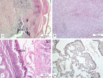
No histopathological changes were observed in the control lung and liver tissues (fig. 1D and 1B, respectively). Granulomatous inflammation was observed in the lungs and liver of the infected group. In the interstitium of the lung, areas surrounded with a fibrous capsule were seen. These areas contained a cuticular, or lamellated, layer and a germinal layer of the cyst with central necrosis and calcification. The outer layer of the fibrous capsule, or intersititium, was also necrotic and contained dense calcification. Mononuclear cells, together with fewer numbers of neutrophils and eosinophils, were seen over and surrounding the capsule (i.e. the cuticular and germinal layers). Several foreign-body giant cells were also observed in this area. The epithelium of the bronchi and bronchioli was desquamated and mononuclear cells were seen in the lumen of these structures and surrounding them. Alveoli were emphysematic (fig. 1C).
Necrosis and inflammatory cell infiltrations (arrowed) in (A) the liver and (C) lung of cattle infected with hydatid cysts, for comparison with (B) control liver and (D) lung.

Similar cystic structures were also seen within the liver parenchyma. In addition, mononuclear cells and some neutrophil infiltrations were observed around the vessels and bile ducts. Peri-acinary, mid-zonal and peri-portal fibrosis and caseified necrosis were also observed (fig. 1A).
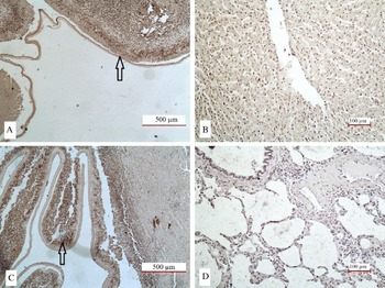
In the lung, strong immunoreactivity for IL-6 was observed in the capsule of the cyst, in the inflammatory cells beneath the capsule, giant cells and the epithelium of bronchi and bronchioli (fig. 2C). In the liver, strong immunoreactivity for IL-6 was observed in the capsule of the cyst, in the inflammatory cells beneath the capsule, giant cells and the bile ducts (fig. 2A). Control slides of lung and liver tissues revealed no staining (fig. 2D and 2B).
Strong immunoreactivity (arrowed) for interleukin-6 (IL-6) in (A) the liver and (C) the lung of cattle infected with hydatid cysts, for comparison with (B) control liver and (D) control lung.

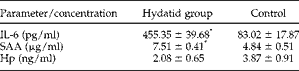
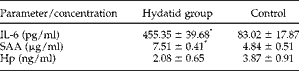
SAA concentrations in serum were 4.84 ± 0.51 and 7.51 ± 0.41 μg/ml in the control and hydatid cyst groups, respectively (P< 0.001). IL-6 concentrations in serum were 83.02 ± 17.87 and 455.35 ± 39.68 pg/ml in the control and hydatid cyst groups, respectively (P< 0.001). Haptoglobin concentrations in serum were 3.87 ± 0.91 and 2.08 ± 0.65 ng/ml in the control and hydatid cyst groups, respectively (P>0.05) (table 1).
Concentrations (mean ± SE) of interleukin-6 (IL-6), serum amyloid A (SAA) and haptoglobin (Hp) in cattle infected with hydatid cysts compared with controls; *significant differences (P<0.001).
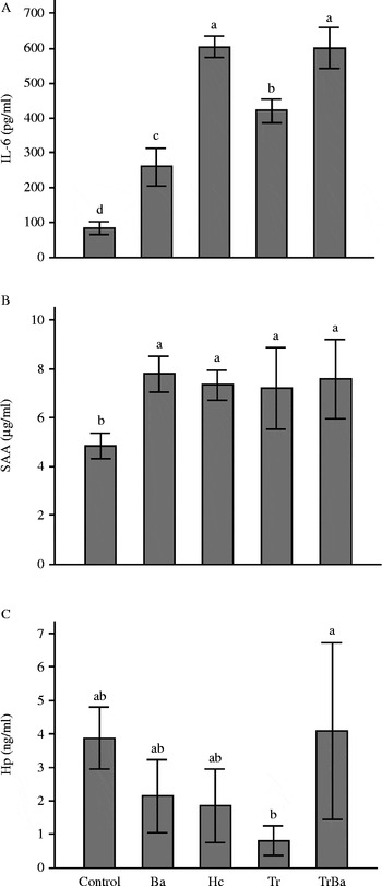
In cattle infected with Trichostrongylus sp., haptoglobin levels decreased whereas those of SAA (P< 0.05) and IL-6 (P< 0.001) increased (fig. 3). In cattle infected with hydatid cysts in the liver only, lung only, and liver and lung, serum IL-6 (P< 0.001) and SAA (P< 0.05) concentrations increased, while haptoglobin levels decreased compared with the control group (table 2). Serum SAA and haptoglobin levels of cattle where only microbial growth had been determined, showed more increase compared with cattle infected with hydatid cysts only. However, this increase was not statistically significant (fig. 3). IL-6 levels were highest in the cattle where hydatid cysts had been detected in the serum, followed by cattle infected with bacteria and Trichostrongylus sp. (P< 0.001) (fig. 3). IL-6 and SAA levels of cattle in which hydatid cysts had been found in the lungs were higher compared with those of cattle whose livers were infected with hydatid cysts. This result was not found to be statistically significant (table 2).
Using enzyme-linked immunosorbent assay (ELISA), the concentrations of (A) serum interleukin-6 (IL-6), (B) serum amyloid A (SAA) and (C) serum haptoglobin (Hp) in cattle infected with hydatid cyst (Hc), bacteria (Ba), Trichostrongylus sp. (Tr) and Trichostrongylus sp. + bacteria (TrBa), compared with controls. a,b,c,d, mean values with dissimilar supercript letters are significantly different (P< 0.001 in (A) and P< 0.05 in (B) and (C)).

Concentrations (mean ± SE) of interleukin-6 (IL-6), serum amyloid A (SAA) and haptoglobin (Hp) in infected cattle relative to the location of cysts in the liver and lungs.

a,b,c Mean values with dissimilar supercript letters are significantly different (P< 0.05).
Discussion
Hydatid cysts chronically infect intermediate hosts, leading to granulomatous inflammation (Thompson, Reference Thompson, Thompson and Lymbery1995). It has been suggested that clinical and radiological findings are not unique and parasitological examinations do not reveal the parasite adequately (Hira et al., Reference Hira, Bahr, Shweiki and Behbehani1990) and hence specific blood parameters are important in the diagnosis of hydatid cyst infection. The APR is the reaction shown by the body towards trauma, neoplasia (Gruys et al., Reference Gruys, Obwolo and Toussaint1994), bacterial, viral (Ganheim et al., Reference Ganheim, Hulten, Carlsson, Kindahl, Niskanen and Waller2003) and parasitic infections (Ganheim et al., Reference Ganheim, Hoglund and Waller2004), and stress (Alsemgeest et al., Reference Alsemgeest, Kalsbeek, Wensing, van Der Kolk, van Ederen, Kim, Westers and Gruys1994, Reference Alsemgeest, Lambooy, Wierenga, Dieleman, Meerkerk, van Ederen and Niewold1995). In this study, 27 cattle infected with hydatid cysts at post-mortem examination were selected. SAA, haptoglobin and IL-6 values, which are specific in infectious and non-infectious conditions, were examined in the cattle serum, and microbiological examination of cystic liver and lung samples was made. In the present study, blood was not examined to determine bacterial and viral infections, as our facilities were insufficient for these examinations. However, it has been suggested that SAA and haptoglobin are more useful in distinguishing acute and chronic inflammations than haematological tests, for example, neutrophil counts and leucocyte counts (Solter et al., Reference Solter, Hoffmann, Hungerford, Siegel, St. Denis and Dorner1991; Skinner & Roberts, Reference Skinner and Roberts1994; Horadagoda et al., Reference Horadagoda, Knox, Gibbs, Reid, Horadagoda, Edwards and Eckersall1999). Therefore in the present study cattle were chosen according to anamnesis, clinical findings and post-mortem examination. However, future studies should assess bacterial and viral infections in the blood in order to eliminate systemic infection.
Haptoglobin and SAA are important APPs in cattle, being present at the minimum level or not being detected in healthy animals (Ganheim et al., Reference Ganheim, Hulten, Carlsson, Kindahl, Niskanen and Waller2003). Also, haptoglobin and SAA are increased in acute infections compared with chronic disease (Horadagoda et al., Reference Horadagoda, Knox, Gibbs, Reid, Horadagoda, Edwards and Eckersall1999). They reach peak levels in the first week after infection and decline to normal levels in the second week (Eckersall et al., Reference Eckersall, Lawson, Bence, Waterston, Lang, Donachie and Fontaine2007). SAA, unlike other APPs, can also be stimulated in more sensitive conditions such as stress, as well as disease states (Alsemgeest et al., Reference Alsemgeest, Lambooy, Wierenga, Dieleman, Meerkerk, van Ederen and Niewold1995). SAA increases moderately in the inflammatory condition when compared with haptoglobin (Horadagoda et al., Reference Horadagoda, Knox, Gibbs, Reid, Horadagoda, Edwards and Eckersall1999). Similarly, in this study, SAA was a more effective indicator of hydatid cyst infection compared with haptoglobin. This result might have originated from the sustained hydatid cyst infection.
Haptoglobin is a protein commonly studied in cattle, binding to free haemoglobin in the circulation and also having an indirect antibacterial effect (Salonen et al., Reference Salonen, Hirvonen, Pyorala, Sankari and Sandholm1996). Haptoglobin increases in diseases caused by bacteria (Salonen et al., Reference Salonen, Hirvonen, Pyorala, Sankari and Sandholm1996), viruses (Hofner et al., Reference Hofner, Fosbery, Eckersall and Donaldson1994) and parasites (Jungerson et al., Reference Jungerson, Jensen, Riber, Heegard, Petersen, Poulsen, Billehansen and Lind1999; Glass et al., Reference Glass, Craigmie, Springbett, Preston, Kirvar, Wilkie, Eckersall, Hall and Brown2003; Matijatko et al., Reference Matijatko, Mrljak, Kıs, Kucer, Forsek, Zivicnjak, Romic, Simec and Ceron2007; Ulutaş et al., Reference Ulutaş, Voyvoda, Ulutaş and Aypak2008) and decreases in haemolysis (Weidmeyer & Solter, Reference Weidmeyer and Solter1996). It can increase 100-fold after stimulation (Eckersall, Reference Eckersall2000). In the present study it was found that as the number of cysts increases in the liver, the serum level of haptoglobin also increases. The increase in SAA seems in accordance with previous studies (Alsemgeest et al., Reference Alsemgeest, Lambooy, Wierenga, Dieleman, Meerkerk, van Ederen and Niewold1995) except in the case of inflammatory conditions such as stress and transport.
In goats infected with Fasciola spp., Trichuris spp. and Trichostrongylidae sp. and displaying clinical symptoms, serum SAA, haptoglobin and C-reactive protein levels increase (Ulutaş et al., Reference Ulutaş, Voyvoda, Ulutaş and Aypak2008). In our study, haptoglobin level decreased while serum SAA and IL-6 levels increased in cattle infected with Trichostrongylus sp. This decrease may be related to the low level of Trichostrongylus sp. infection of 50–100 eggs/g, the small number of cattle or the absence of cattle with clinical symptoms. IL-6 and SAA levels were found to increase in cattle with cysts in the lungs compared with cattle with cysts in the liver, although this was not statistically significant. Thus SAA, although appearing to be an indicator of inflammatory responses, does not reflect the severity and distribution of inflammatory damage.
Ali & Dawood (Reference Ali and Dawood2012) isolated S. aureus and Corynebacterium pyogenes from liver, and Streptococcus spp. and C. pyogenes from lungs in a study in which they examined secondary bacterial invasions in the liver and lungs of cattle with hydatidosis. In the present study microbial agents belonging to Corynebacterium, Streptococcus, Mannheimia, Staphylococcus, Campylobacter, Escherichia, Micrococcus and Candida species were identified, with more bacterial diversity occurring in the lungs compared with the liver, suggesting that the lungs are vulnerable to bacterial infection (Quinn et al., Reference Quinn, Markey, Carter, Donnelly and Leonard2002). Moreover, when the number of cysts increased, an increase occurred in bacterial diversity except for caseified cysts. Serum haptoglobin and SAA are thought to be important biomarkers for diagnosis in experimental bacterial and natural diseases in ruminants (Nielsen et al., Reference Nielsen, Jacobsen, Andersen, Niewold and Heegaard2004; Ceciliani et al., Reference Ceciliani, Ceron, Eckersall and Sauerwein2012). Generally, levels of haptoglobin and SAA increased in the presence of bacterial infection in studies investigating the use of APPs for diagnosis (Gronlund et al., Reference Gronlund, Hulten, Eckersall, Hogarth and Waller2003; Petersen et al., Reference Petersen, Nielsen and Heegard2004; Eckersall et al., Reference Eckersall, Lawson, Bence, Waterston, Lang, Donachie and Fontaine2007). In our study, while the SAA level increased relatively in the serum of cattle in which bacteria were isolated from the liver and lungs, haptoglobin was decreased. Haemolysin, which is an effective factor in the pathogenity of some isolated micro-organisms, could cause the decrease in haptoglobin level in animals (Weidmeyer & Solter, Reference Weidmeyer and Solter1996).
The APR is stimulated by cytokines, e.g. IL-1, IL-6 and tumour necrosis factor-α, released from monocytes and macrophages at the infection site (Dinarello, Reference Dinarello1984; Beutler & Cerami, Reference Beutler and Cerami1986). This response is induced by cytokines released from Th1, i.e. IL-2, interferon-γ and lymphotoxins, and Th2, i.e. IL-4, IL-5, IL-6 and IL-10, in the Echinococcus infection as well as in other helminth infections (Finkelman et al., Reference Finkelman, Pearce, Urban and Sher1991; Allen & Maizels, Reference Allen and Maizels1996; Mosmann & Sad, Reference Mosmann and Sad1996). The levels of serum IL-5 and IL-6 increased in a study carried out on human patients infected with hydatid cysts (Riganò et al., Reference Riganò, Profumo, Teggi and Siracusano1996). Cytokines released from Th2 cells increased in mice in which hydatid cyst infection was induced experimentally (Haniloo et al., Reference Haniloo, Ghasemi, Shikhi and Ghavami2008). It has been emphasized that cytokines released from Th2 cells were very clear in the early phase of the infection (Baz et al., Reference Baz, Richieri, Puglia, Nieto and Dematteis1999). Similarly, cytokines released from Th2 cells increased in rats that were infected experimentally with Fasciola hepatica (Tliba et al., Reference Tliba, Sibille, Boulard and Chauvin2002).
The IL-6 level increases in many diseases of cattle, such as paratuberculosis (Abendaño et al., Reference Abendaño, Juste and Alonso-Hearn2013), infertility with bacterial infections (Price & Sheldon, Reference Price and Sheldon2013), metabolic stress in the peri-parturient period (Trevisi et al., Reference Trevisi, Amadori, Cogrossi, Razzuoli and Bertoni2012) and subclinical endometritis (Ghasemi et al., Reference Ghasemi, Gonzalez-Cano, Griebel and Palmer2012). Similarly, the highest level of IL-6 was found in the serum of cattle detected with microbiological growth (P< 0.001) in our study.
Until now, no studies had been performed in which both tissue and serum levels of IL-6 were examined in animals infected with hydatid cysts. In the present study, in which we assessed the IL-6 level in the tissue and serum, a significant increase was observed in the IL-6 level of infected cattle when compared with the control group (P< 0.001). SAA increased with the increase of IL-6, although the level of haptoglobin decreased. In an experiment carried out in mice (Riley et al., Reference Riley, Dixon, Jenkins and Ross1986), macrophage, eosinophil and neutrophil infiltrations were observed around the injection site 3 days after intraperitoneal protoscolex injection. In previous studies (Chiou et al., Reference Chiou, Wang, Chang, Liu, Jeng, Cheng, Jou and Pang2001; Sakamoto & Cabrera, Reference Sakamoto and Cabrera2003; Bacciarini et al., Reference Bacciarini, Gottstein, Pagan, Rehmann and Grone2004), neutrophil, eosinophil, foreign-body giant cell and mononuclear cell infiltration and necrosis were observed in the adventitial layer around the lamellar layer during the histopathological examination of hydatid cysts. In the present study, similar sights were observed, both in liver and lungs.
Research combining parasitological, microbiological and histopathological assessment with serum SAA, haptoglobin and IL-6 determination in relation to hydatid cyst infection has not been performed previously. In the present study, an extensive investigation was carried out on the serum and tissue samples of cattle infected with hydatid cysts, in order to develop a diagnostic aid in the clinical environment. While the concentration of SAA increased, haptoglobin decreased, but the level of serum IL-6 increased. No statistical relationship was observed between the SAA, haptoglobin or IL-6 values of cattle with positive or negative bacterial growth results. However, the levels of IL-6 were relatively higher in the serum of cattle with bacterial growth. Therefore, increases in this parameter should not be evaluated at only one point. IL-6 and SAA may be collectively considered as valuable and helpful biomarkers in hydatid cyst infections.
Acknowledgements
We thank Dr Aziz Bülbül and Dr I. Taci Cangül for their scientific help.
Financial support
This work was supported by the Commission of Scientific Research Projects of Afyon Kocatepe University, project number 10.VF.03.
Conflict of interest
None.