Introduction
A series of distinct Ca2+ transients is localized to the early cleavage furrows during the embryonic cell divisions of a variety of fish species from the order Cypriniformes, including: medaka (Oryzias latipes; Fluck et al., Reference Fluck, Miller and Jaffe1991; Webb et al., Reference Webb, Fluck and Miller2011); zebrafish (Danio rerio; Chang & Meng, Reference Chang and Meng1995; Webb et al., Reference Webb, Lee, Karplus and Miller1997); rosy barb (Puntius conchonius; Webb & Miller, Reference Webb, Miller, Krebs and Michalak2007); and mummichog (Fundulus heteroclitus; Webb & Miller, Reference Webb, Miller, Krebs and Michalak2007). In these large meroblastically cleaving embryos, cell division can be divided, from both a morphological and a Ca2+ signalling perspective, into a number of post-mitotic sequential steps; cleavage furrow positioning, furrow propagation, furrow deepening, and furrow apposition, such that each step is accompanied by its own distinctive and required Ca2+ transient (Webb et al., Reference Webb, Lee, Karplus and Miller1997; Lee et al., Reference Lee, Webb and Miller2003, Reference Lee, Webb and Miller2006). Taking zebrafish as an example, the furrow positioning Ca2+ transient is the briefest of the four signals lasting ~60 s (at 28°C; Lee et al., Reference Lee, Webb and Miller2006), before it extends and develops into the furrow propagation signal. The latter takes the form of two slow (~0.5 μm/s), shallow, linear Ca2+ waves, which over a period of ~120 s extend from the lateral extremities of the furrowing signal arc out to the edges of the blastodisc (Webb et al., Reference Webb, Lee, Karplus and Miller1997; Lee et al., Reference Lee, Webb and Miller2006). The furrow deepening Ca2+ signal follows the furrow propagation signal and this is the longest lasting signal of all the cytokinetic Ca2+ signals. Similar to the propagation signal, the deepening Ca2+ signal takes the form of a pair of slow Ca2+ waves that travel (at ~0.5 μm/s) from the apex of the blastodisc (i.e., the site of the furrow positioning signal) out towards its edges along the same tracks as the propagation signal, but this time it also accompanies the ingressing furrow as it cuts down through the blastodisc (at ~0.1 μm/s) to meet the rising yolk cell that constitutes the blastodisc floor (Webb et al., Reference Webb, Lee, Karplus and Miller1997; Lee et al., Reference Lee, Webb and Miller2003; Webb et al., Reference Webb, Fluck and Miller2011). Due to the dimensions of the blastodisc and the velocity of the slow Ca2+ waves, it takes ~500 s from the appearance of the furrow deepening signal at the blastodisc apex until the leading edges of the furrow deepening signal reach the margin of the blastodisc and the top of the yolk cell (Webb et al., Reference Webb, Lee, Karplus and Miller1997). The first furrow deepening process effectively divides the blastodisc in two, forming a pair of daughter cells each with its own nucleus. Furrow deepening is then followed by furrow apposition (or zipping), which is essential for maintaining the embryo as a compact entity during the embryonic cell division cycles during early development (Fluck et al., Reference Fluck, Miller and Jaffe1991; Jesuthasan, Reference Jesuthasan1998). The Ca2+ signal that accompanies apposition lasts for ~200 s (Webb et al., Reference Webb, Lee, Karplus and Miller1997). In terms of the magnitude of the intracellular Ca2+ rise during the first set of cytokinetic transients, an approximate five-fold increase above the resting level (i.e., from ~100 nM to ~500 nM) has been reported (Webb et al., Reference Webb, Lee, Karplus and Miller1997). Thus, for deepening and apposition to be completed it takes ~700 s, (i.e., ~12 min) of sustained elevated [Ca2+]i in the localized regions of the blastoderm involved in the cytokinetic process. When this is added to the ~60-s and ~120-s durations of the positioning and propagation signals, respectively, it totals ~15 min to complete the cytokinetic portion of the first cell division cycle (at ~28.5°C).
The goal of this report was to further investigate which molecular components of a cell's Ca2+ signalling toolkit (Berridge et al., Reference Berridge, Lipp and Bootman2000) might be involved in generating and maintaining a sustained intracellular Ca2+ signal for a period of ~12 min during furrow deepening and apposition. There is good evidence to suggest that the endoplasmic reticulum (ER) acts as the primary Ca2+ store for generating all of the cytoplasmic Ca2+ transients in zebrafish embryos via release mainly through IP3Rs (Chang & Meng, Reference Chang and Meng1995; Webb et al., Reference Webb, Lee, Karplus and Miller1997; Créton et al., Reference Créton, Speksnijder and Jaffe1998; Lee et al., Reference Lee, Webb and Miller2003, Reference Lee, Webb and Miller2006). We hypothesized that such a sustained and dynamic Ca2+ release for ~12 min was likely to deplete the ER Ca2+ store before furrow deepening and apposition was completed, unless it was somehow replenished. Thus, Ca2+ toolkit elements participating in store-operated Ca2+ entry (SOCE) to refill the ER were obvious candidates for investigation.
SOCE is a process whereby a decrease in the ER Ca2+ content (sensed by stromal interaction molecule 1; STIM1), activates Ca2+ entry into the cytoplasm across the plasma membrane (PM) via Orai1, a protein that functions as a pore-forming subunit of the SOCE complex, and/or via a member of the canonical transient receptor potential (TRPC) channel family, another type of Ca2+ channel located in the PM (Parekh & Putney, Reference Parekh and Putney2005; Cheng et al., Reference Cheng, Ong, Liu and Ambudkar2010). It has been proposed for a number of cogent reasons that SOCE shuts down during cell division (Smyth & Putney, Reference Smyth and Putney2012). However, data supporting this suggestion have been mainly derived from small, flat tissue culture cells, such as HeLa cells and proliferating rat mast cells (Preston et al., Reference Preston, Sha'afi and Berlin1991; Tani et al., Reference Tani, Monteilh-Zoller, Fleig and Penner2007), where the size and geometry, as well as the post-cytokinesis separation of cells, might not require the sustained Ca2+ signals required during furrow deepening and apposition during cytokinesis of large embryonic cells.
We have previously reported that during furrow deepening and apposition in zebrafish, TRPC1 – reported to be a component of SOCE (Cheng et al., Reference Cheng, Ong, Liu and Ambudkar2010; Li et al., Reference Li, Chen, Zhou, Xu and Yu2012) – is co-localized with the region of elevated [Ca2+]i in the cleavage furrow (Chan et al., Reference Chan, Chen, Hung, Miller, Shipley and Webb2015). TRPC1 has previously been reported to be inhibited by SKF 96365 and by low concentrations of 2-APB (Bomben & Sontheimer, Reference Bomben and Sontheimer2008). We showed that treatment of zebrafish embryos with these two TRPC1 antagonists had no major effect on furrow positioning or propagation (or the Ca2+ signals associated with them), but they did inhibit the furrow deepening process (as well as the associated Ca2+ signal), resulting in the regression of the cleavage furrow (Chan et al., Reference Chan, Chen, Hung, Miller, Shipley and Webb2015).
In this new report, we show that, in addition to TRPC1, two other key components associated with SOCE, i.e., STIM1 and Orai1, are also localized to the deepening furrow during embryonic cleavage in zebrafish. These new data support our proposition that although SOCE might not be required during cell division in small, flat tissue culture calls, it does appear to play a crucial role in helping to sustain the long-lasting localized elevated [Ca2+]i required during the early cleavages of large fish embryos.
Materials and methods
Zebrafish maintenance and embryo collection
Wild-type zebrafish (Danio rerio), AB strain, were maintained at 28.5°C on a 14 h light/10 h dark cycle to stimulate spawning (Westerfield, Reference Westerfield1994) and their fertilized eggs were collected as described elsewhere (Webb et al., Reference Webb, Lee, Karplus and Miller1997). Embryos were dechorionated via treatment with protease (Sigma-Aldrich Co., LLC, St. Louis, MO, USA) at 1 mg/ml in Danieau's solution (17.4 mM NaCl, 0.21 mM KCl, 0.18 mM Ca(NO3)2, 0.12 mM MgSO4.7H2O, 1.5 mM HEPES, pH 7.2), as described previously (Webb & Miller, Reference Webb and Miller2013).
Immunolabelling embryos with antibodies to STIM1 or Orai1
Dechorionated embryos were maintained on a heated ThermoPlate (Tokai Hit Co., Fujinomiya, Shizuoka-ken, Japan), to keep the rate of development consistent for all experiments. Batches of embryos at 35 min post-fertilization (mpf), 40 mpf, 45 mpf, 50 mpf and 55 mpf were fixed at 4°C overnight using 4% paraformaldehyde and 4% sucrose in phosphate-buffered saline (PBS; pH 7.3; Westerfield, Reference Westerfield1994). Embryos were washed with PBS and then with PBS containing 0.1–0.2% Tween-20 (PBST), after which they were transferred to PBST containing 1% dimethyl sulfoxide (PBSTD) and incubated for 1 h in the dark at room temperature. Embryos were then incubated in blocking buffer (PBSTD containing 10% goat or rabbit serum and 1% BSA) for 2 h at room temperature, after which they were incubated at 4°C overnight with one of the following primary antibodies: rabbit anti-STIM1 (N-terminal; Sigma-Aldrich Co., LLC), goat anti-STIM1 (internal region; LifeSpan BioSciences, Inc., Seattle, WA, USA), or rabbit anti-Orai1 (Proteintech, Chicago, IL, USA), all at a dilution of 1:50. The embryos were then washed with wash buffer (a 1:10 dilution of blocking buffer) before they were incubated with either Alexa Fluor 488-conjugated goat anti-rabbit IgG antibody or Alexa Fluor 488-conjugated rabbit anti-goat IgG antibody (Life Technologies, Thermo Fisher Scientific Inc., Waltham, MA, USA), depending on the host species of the primary antibody. Both secondary antibodies were used at a dilution of 1:200 in blocking buffer and embryos were incubated for 3 h at room temperature. The embryos were then washed with wash buffer and incubated overnight at 4°C with Alexa Fluor 568-tagged phalloidin (Life Technologies, Thermo Fisher Scientific Inc.) at 1:50 in blocking buffer. They were then washed extensively with PBST and finally with PBS prior to confocal microscopy.
Dual-immunolabelling embryos with antibodies to STIM1 and Orai1
Embryos were fixed and washed as described previously. They were then incubated in a donkey serum-based blocking buffer (PBSTD containing 10% donkey serum and 1% BSA) at room temperature for 2 h, after which they were incubated overnight at 4°C with the goat anti-STIM1 antibody (LifeSpan BioSciences, Inc.) at a dilution of 1:50 in donkey blocking buffer. Embryos were then washed with donkey blocking buffer diluted 1:10 in PBSTD and incubated at 4°C overnight with Alexa Fluor 488-conjugated donkey anti-goat IgG antibody (Abcam, Cambridge, MA, USA; at a dilution of 1:200 in donkey blocking buffer). Embryos were then washed thoroughly with PBSTD, after which they were blocked again, this time with goat serum-based blocking buffer (PBSTD containing 10% goat serum and 1% BSA) for 2 h at room temperature. After blocking, embryos were incubated at 4°C overnight with the anti-Orai1 antibody, described above. They were then washed with goat blocking buffer diluted 1:10 with PBSTD, and incubated with an Alexa Fluor 546-conjugated goat anti-rabbit IgG (Thermo Fisher Scientific Inc.; at a dilution of 1:200 in goat blocking buffer). The embryos were then washed extensively with PBST and finally with PBS prior to visualization via confocal microscopy.
Confocal microscopy
Confocal images were acquired using a Nikon C1 laser scanning confocal system mounted on a Nikon Eclipse 90i microscope using Nikon Fluor 20X/0.5 NA and 40X/0.8 NA water immersion lenses. The green fluorescence was captured using 488 nm excitation and 515/530 nm emission, whereas the red fluorescence was captured using 543 nm excitation and a 570 nm long pass emission filter. Images were acquired with embryos in an animal pole orientation, and figures were prepared using Corel Graphics Suite (Corel Corp., Ottawa, ON, Canada).
Results and Discussion
Localization of STIM1 and Orai1 in zebrafish embryos undergoing cytokinesis during the first two cell division cycles
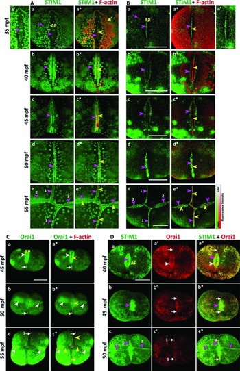
Figure 1 shows representative animal pole (AP) views of dividing zebrafish embryos showing the localization of STIM1 and Orai1 at the first and second cleavage furrows during the first two cell division cycles. Figure 1 A, B shows a representative series of images (collected every 5 min from 35 mpf to 55 mpf) where a number of confocal sections are projected to generate each single composite image. Embryos were immunolabelled with antibodies that recognize Fig. 1 A the N-terminal or Fig. 1 B an internal peptide sequence of the STIM1 protein. The images show the localization of STIM1 alone (Fig. 1 Aa–Ae, Ba–Be), and both STIM1 and F-actin (Fig. 1 Aa*–Ae*, Ba*–Be*), the latter being used to show the localization of STIM1 in relation to known F-actin-rich structures in the blastodisc, i.e., the contractile band (see yellow arrowheads in Fig. 1) and the pericleavage F-actin enrichments (PAEs; Chang & Meng, Reference Chang and Meng1995; Urven et al., Reference Urven, Yabe and Pelegri2006; Li et al., Reference Li, Webb, Chan and Miller2008; Webb et al., Reference Webb, Goulet, Chan, Yuen and Miller2013; see yellow arrow in Fig. 1 Aa*). For the embryos labelled with the antibody recognizing the N-terminal region of STIM1, the images clearly show that there is relatively little STIM1 labelling in the furrow region at ~35 mpf (see Fig. 1 Aa, Aa′), i.e., during furrow positioning and propagation, but this becomes a lot more prominent during furrow deepening at ~40–45 mpf (see pink arrowheads in Fig. 1 Ab, Ab*, Ac, Ac*) and furrow apposition at ~50 mpf (see pink arrowheads in Fig. 1 Ad, Ad*). At the end of the first cell division and the start of the second (Fig. 1 Ae, 1Ae*), STIM1 labelling can be observed in both the apposed first cleavage furrow (see pink arrowheads labelled ‘1’) and in the forming second furrows (see pink arrowheads labelled ‘2’). Indeed, the level of STIM1 labelling was more obvious during the earliest stages of the formation of the second cleavage furrow than it was at approximately the same stage of the first furrow (compare Fig. 1 Ae – pink arrowheads labelled ‘2’ – with Fig. 1 Aa pink arrowhead). In addition, with this antibody, STIM1-labelled patches (or puncta) occurred in the non-ingressing regions of the blastodisc on either side of the cleavage furrow (see pink arrows in Fig. 1 Aa–Ae, Aa*–Ae*). Similar (but smaller) STIM1 ‘puncta’ have also been described in various cells in culture, including in activated T cells (Barr et al., Reference Barr, Bernot, Srikanth, Gwak, Balagopalan, Regan, Helman, Sommers, Oh-hora, Rao and Samelson2008); in PANC1, RAMA37 and HeLa cells on depletion of ATP (Chvanov et al., Reference Chvanov, Walsh, Haynes, Voronina, Lur, Gerasimenko, Barraclough, Rudland, Peterson, Burgoyne and Tepikin2008); and in HEK293 cells following depletion of the ER stores via treatment with thapsigargin (Prakriya & Lewis, Reference Prakriya and Lewis2015). In addition, it has been reported that these puncta correspond to the accumulation of STIM1 in distinct regions of junctional ER, which is located several nanometers from the plasma membrane (Wu et al., Reference Wu, Buchanan, Luik and Lewis2006).
Localization of STIM1 and Orai1 in embryos undergoing cytokinesis of the first and second cell division cycles. (A, B) Animal pole views of dividing zebrafish embryos to show the localization of STIM1 on the sides of the cleavage furrow during cleavage. Representative confocal stacks of single optical sections that were projected as single images [from n = 8 embryos from experiments conducted on four separate occasions (A) and from n = 4 embryos from experiments conducted on four separate occasions (B)], to show the localization of (Aa′, Aa–Ae, Ba′, Ba–Be) STIM1, and (Aa*–Ae*, Ba*–Be*) both STIM1 and F-actin, from an animal pole view. Embryos were immunolabelled with antibodies that recognize (A) the N-terminal region or (B) an internal peptide sequence of the STIM1 protein. (Aa′, Ba′) Higher magnification views of the furrows shown in (Aa, Ba), respectively, which were enhanced (by adjusting the intensity), to emphasize the localization of STIM1 in the furrow. (C) Representative confocal stacks (from n = 4 embryos from experiments conducted on three separate occasions), to show the localization of (Ca–Cc) Orai1, and (Ca*–Cc*) both Orai1 and F-actin, in the deepening and apposing cleavage furrow of the first cell division cycle, and the propagating furrow of the second cell division cycle. (D) Representative confocal stacks (from n = 4 embryos from experiments conducted on four separate occasions), to show the localization of (Da–Dc) STIM1, (Da′–Dc′) Orai1, and (Da*–Dc*) both STIM1 and Orai1, from an animal pole view. The pink arrowheads indicate STIM1 localization in the propagating, deepening and apposing cleavage furrow whereas the pink arrows indicate punctate STIM1 labelling in the other regions of the blastodisc. The yellow arrowheads and arrows indicate F-actin labelling in the contractile band and the pericleavage F-actin enrichments located on either side of the furrow, respectively. The white arrowheads indicate Orai1 localization in the propagating and deepening cleavage furrows whereas the white arrows indicate concentrated Orai1 labelling at either end of the contractile band in the furrow. The numbers, ‘1’ and ‘2’ refer to the apposing and positioning furrows of the first and second cell division cycles, respectively. AP, animal pole. Scale bars all represent 200 µm except in panels Aa′ and Ba′, where they are 100 µm.

As this is the first time that STIM1 has been labelled in early zebrafish embryos, we used a second antibody to STIM1 (see Fig. 1 B) to confirm the pattern of localization of the first antibody used. Whereas the first antibody recognized the N-terminal region of STIM1, this second antibody recognized an internal peptide sequence of the protein. In addition, whereas the N-terminal antibody was raised in rabbit, the internal peptide antibody was raised in goat; thus different secondary antibodies were used to bind the primary antibodies. Our results indicated that the two antibodies did not show exactly the same results; for example, the antibody recognizing the internal peptide sequence gave a more punctate pattern of localization of STIM1 than did the one that recognized the N-terminal region. This might be due to differences in the primary antibodies, or the fact that different secondary antibodies were used. However, the overall pattern of localization for both of these antibodies was very similar, such that during the first cell division, STIM1 labelling was less obvious in the furrow during propagation (Fig. 1 Ba, Ba′, Ba*) and more pronounced during deepening and apposition (Fig. 1 Bb–Bd, Bb*–Bd*). The use of two primary antibodies thus provides convincing evidence that STIM1 is localized to the deepening cleavage furrow in the large cells of zebrafish embryos.
Figure 1 C illustrates a representative (n = 4) series of single confocal sections that were projected as a composite stack to show the localization of Orai1 (Fig. 1 Ca–Cc), and both Orai1 and F-actin (Fig. 1 Ca*–Cc*), in the deepening and apposing cleavage furrow of the first cell division cycle, as well as in the propagating furrows of the second cell division cycle. Figure 1 D shows a representative (n = 4) series of single confocal sections that were projected as single images to show the localization of STIM1 alone (Fig. 1 Da–Dc), Orai1 alone (Fig. 1 Da′–Dc′), and both STIM1 and Orai1 (Fig. 1 Da*–Dc*) in the deepening and apposing cleavage furrow of the first cell division cycle, as well as in the propagating furrows of the second cell division cycle.
Images illustrating STIM1 and Orai1 when merged show that during furrow deepening at ~40 mpf (see Fig. 1 Da*; i.e., when SOCE is most likely to be required), there is a high degree of overlap. At 50 mpf, however, when deepening and apposition are complete, the extensive overlap is no longer seen (see Fig. 1 Dc*) as the presence of Orai1 labelling diminishes. This suggests a close association of these SOCE components from both a spatial and temporal perspective.
In both Fig. 1 C, D, the white arrowheads indicate Orai1 localization in the propagating and deepening cleavage furrows, whereas the white arrows indicate concentrated Orai1 labelling in distinct regions at the leading ends of the contractile band within the cleavage furrow. We have previously reported that the zone of elevated [Ca2+] lingers at the leading (i.e., lateral) ends of the cleavage furrows when they reach the edge of the blastodisc (Webb et al., Reference Webb, Lee, Karplus and Miller1997). Furthermore, a similar pattern of localization of calmodulin (CaM) and the phosphorylated form of myosin light chain 2 (MLC2) has also been reported at the leading ends of the contractile band (see Figs 1 E, F and 2 in Webb et al., Reference Webb, Goulet, Chan, Yuen and Miller2013). In the case of both of these proteins, we previously suggested that due to the biphasic nature of the assembly of the contractile apparatus in the embryonic cells of zebrafish, it is the lateral ends of the contractile band that generate the most force and therefore might require more precise regulation (Webb et al., Reference Webb, Goulet, Chan, Yuen and Miller2013). Thus, our visualization of Orai1 at the lateral ends of the cleavage furrow brings three elements of the Ca2+ signalling toolbox (i.e., Orai1, CaM and MLC2), together in the right place and at the right time to promote this penultimate stage (i.e., the separated daughter cells still have to appose), in embryonic cell division.
When our new STIM1 and Orai1 data are combined with our previously published TRPC1 data (Chan et al., Reference Chan, Chen, Hung, Miller, Shipley and Webb2015) and cytokinetic Ca2+ signalling patterns (see fig. 2 of Webb et al., Reference Webb, Lee, Karplus and Miller1997 and Fig 2 B), they clearly indicate that three of the key elements that have been reported to be involved in SOCE – i.e., STIM1, Orai1 and TRPC1 (Cheng et al., Reference Cheng, Ong, Liu and Ambudkar2010) – are highly localized to the region of sustained elevated [Ca2+]i that is required for cleavage furrow deepening and apposition. SOCE-mediated sustained elevations of [Ca2+]i have been reported to help regulate other cellular functions, such as for the completion of T-cell activation (Iezzi et al., Reference Iezzi, Karjalainen and Lanzaveccia1998; Lewis Reference Lewis2001), cell-fate choice decisions in B cells (Scharenberg et al., Reference Scharenberg, Humphries and Rawlings2007), and insulin secretion from β-cells (Henquin et al., Reference Henquin, Mourad and Nenquin2012). Thus, our new data support our previous suggestion that SOCE might also contribute to the Ca2+-mediated regulation of furrow deepening and apposition in the large embryos of Cypriniformes fish species.
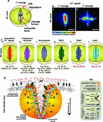
Summary of the proposed role of store-operated Ca2+ entry (together with Ca2+ release from intracellular stores) during cleavage furrow deepening in zebrafish embryos. (A) Schematic to show the morphology of an embryo (in animal pole (AP) orientation) during cleavage furrow deepening. (B) Pseudocolor images to show representative aequorin-generated Ca2+ signals during (Ba) furrow deepening of the first cell division cycle, and (Bb) during furrow apposition of the first cell division and furrow positioning/propagation of the second cell division in embryos when viewed in an animal pole (AP) orientation. The numbers, ‘1’ and ‘2’ refer to the first and second cell division cycles, respectively. Scale bars, 200 µm. (C) Schematic AP views of an embryo to show: (Ca) the Ca2+ signals; and the localization of (Cb) the endoplasmic reticulum; (Cc) microtubules; (Cd) TRPC1, (Ce) STIM1, and (Cf) Orai1, during furrow deepening of the first cell division. (D) Schematic side (facial) view of an embryo during furrow deepening to show how SOCE might contribute (along with Ca2+ release from the endoplasmic reticulum via IP3Rs) to help maintain the high levels of Ca2+ required for furrow deepening (modified from fig. 7 of Chan et al. Reference Chan, Chen, Hung, Miller, Shipley and Webb2015).

Figure 2 A is a schematic to show the morphology of an embryo during deepening of the first cell division, and Fig. 2 B shows representative examples of the localized aequorin-derived cytokinetic Ca2+ signals generated during furrow deepening of both the first (panel 2Ba) and second (panel 2Bb) cell division cycles. A schematic showing the furrow deepening Ca2+ signal of the first cell division is shown in Fig. 2 Ca. This is accompanied by a series of schematics showing the localization of the ER (Lee et al., Reference Lee, Webb and Miller2003, Reference Lee, Ho, Wong, Webb and Miller2004), microtubules (Lee et al., Reference Lee, Ho, Wong, Webb and Miller2004; Li et al., Reference Li, Lee, Webb and Miller2006), and TRPC1 (Chan et al., Reference Chan, Chen, Hung, Miller, Shipley and Webb2015), as well as STIM1 and Orai1 in the deepening cleavage furrow (Fig. 2 Cb–Cf). ER reorganization was previously demonstrated (via immunolabelling with an anti-calnexin antibody) to just precede the appearance of furrow positioning/propagation, and then to accompany furrow deepening and apposition of the first and second cleavage furrows (see fig. 9B of Lee et al., Reference Lee, Webb and Miller2003, and fig. 7A of Lee et al., Reference Lee, Ho, Wong, Webb and Miller2004). The linear reorganization of the ER both before and during the first and second cell divisions nicely match the localization of both STIM1 (see Fig. 1 A, B, D) and Orai1 (see Fig. 1 C, D), with the former being localized to the ER membrane (Liou et al., Reference Liou, Kim, Heo, Jones, Myers, Ferrell and Myer2005) and the latter being localized to the plasma membrane (Gwack et al., Reference Gwack, Srikanth, Feske, Cruz-Guilloty, Oh-Hora, Neems, Hogan and Rao2007). We have previously reported that TRPC1 is localized in the cleavage furrow during deepening of the first cell division cycle (see fig. 6D of Chan et al., Reference Chan, Chen, Hung, Miller, Shipley and Webb2015). Furthermore, treatment of zebrafish embryos with SKF 96365 or 2-APB (two general SOCE antagonists) had no effect on mitosis, or furrow positioning and propagation, but did result in an inhibition of furrow deepening (Chan et al., Reference Chan, Chen, Hung, Miller, Shipley and Webb2015). Thus, these data together with our new findings support our proposition for a functional role for SOCE in sustaining elevated [Ca2+]i during furrow deepening and apposition in the early cell division cycles in zebrafish embryos.
Our data in zebrafish would appear at first to contradict a number of previous reports, which suggest that SOCE is inactivated during the division phase of the cell cycle (Arredouani et al., Reference Arredouani, Yu, Sun and Machaca2010; Smyth and Putney, Reference Smyth and Putney2012). However, this conclusion was derived from data gathered mainly from small, flat tissue culture cells such as HeLa cells (Preston et al., Reference Preston, Sha'afi and Berlin1991; Smyth et al., Reference Smyth, Petranka, Boyles, DeHaven, Fukushima, Johnson, Williams and Putney2009); the cultured rat mast cell line, RBL-2H3-M1 (Tani et al., Reference Tani, Monteilh-Zoller, Fleig and Penner2007); COS-7 cells (Russa et al., Reference Russa, Ishikita, Masu, Akutsu, Saino and Satoh2008); HEK239 cells (Boustany et al., Reference Boustany, Katsogiannou, Delcourt, Dewailly, Prevarskaya, Borowiec and Capiod2010); and the bovine brain endothelial cell line, t-BBEC117 (Kito et al., Reference Kito, Yamamura, Suzuki, Yamamura, Ohya, Asai and Imaizumi2015). In these small cells, the size, volume, and geometry, as well as post-cytokinetic cell separation, suggest that sustained intracellular Ca2+ signals might not be necessary. Indeed, sustained cytosolic Ca2+ elevations (localized or otherwise) have never been visualized during cytokinesis in these cell types. In addition, the requirement for sustained Ca2+ via SOCE does not appear to be necessary to generate the short-duration Ca2+ transients required for furrow positioning and propagation during embryonic cell division in zebrafish (Chan et al., Reference Chan, Chen, Hung, Miller, Shipley and Webb2015). However, such SOCE-generated Ca2+ signals are required for furrow deepening and apposition in these large-volume embryonic cells. The early steps in cell division (i.e., furrow positioning and propagation) in large embryonic cells might therefore more closely resemble cell division in tissue culture cells with respect to the amount of Ca2+ required to generate the necessary signal, i.e., there is sufficient Ca2+ in the ER store to generate the signal without stimulating SOCE (Chan et al., Reference Chan, Chen, Hung, Miller, Shipley and Webb2015). Furthermore, the relatively low level of STIM1 labelling in the cleavage furrow at 35 mpf, i.e., during furrow propagation (see Fig. 1 Aa, 1Aa′, 1Ba, 1Ba’) also suggests that SOCE is not required for the first two steps of Ca2+ signal generation during zebrafish cytokinesis.
It has also been reported that SOCE does not occur during meiosis in large Xenopus laevis oocytes (Machaca & Haun, Reference Machaca and Huan2000; Courjaret & Machaca, Reference Courjaret and Machaca2012). In this case, however, it was suggested that the absence of SOCE might help to prevent an influx of Ca2+, which might prematurely trigger egg activation prior to fertilization. This, therefore, might be a special case, which is unrelated to cytokinesis, where prolonged Ca2+ transients have been reported to occur. In addition to a number of Cypriniformes fish species, such transients have also been visualized in holoblastically-cleaving Xenopus (Webb & Miller, Reference Webb, Miller, Krebs and Michalak2007).
We have previously reported the organization of a furrowing deepening microtubule array (FDMA), located perpendicular to the plane of the deepening cleavage furrow in zebrafish (Li et al., Reference Li, Lee, Webb and Miller2006). The positive ends of the FDMA microtubules are oriented toward the deepening cleavage furrow and act as tracks to transport VAMP2-decorated vesicles, via the action of a Ca2+-sensing kinesin-like protein kif23 (a plus-end directed motor protein), to the deepening and expanding furrow membrane, where they fuse as part of the essential membrane remodeling process required to complete cell division in these large egg cells (Li et al., Reference Li, Webb, Chan and Miller2008). The exact contents of the VAMP2-decorated vesicles have yet to be identified, but it has been suggested that they might contain new cell membrane components and extracellular matrix proteins, such as cadherins that contribute to daughter cell apposition (van Roy & Berx, Reference van Roy and Berx2008; Halbleib & Nelson, Reference Halbleib and Nelson2016). The dynamic appearance of both Orai1 and TRPC1 in the deepening cleavage furrow leads us to suggest that these essential SOCE proteins might be localized via distinct microtubule-based mechanisms. In the case of Orai1, we suggest that a possible mechanism might be via vesicular transport along the FDMA, followed by vesicular fusion with the ingressing furrow wall. Indeed, it has been previously reported from HEK293 cells that TRP channel proteins undergo rapid vesicular translocation to, followed by insertion into the plasma membrane (Bezzerides et al., Reference Bezzerides, Ramsey, Kotecha, Greka and Clapham2004).
During activation of SOCE, the dynamic localization of STIM1, whether within the ER (or a sub-compartment of the ER), or between the ER and the plasma membrane, as well as its relationship with microtubules, is still somewhat controversial. Some reports suggest that microtubules do play a facilitative role in the SOCE signalling pathway by optimizing the localization of STIM1 (Smyth et al., Reference Smyth, DeHaven, Bird and Putney2007), whereas others suggest that they play a significant role in the inhibition of SOCE during cell cycle progression via microtubule remodeling (Russa et al., Reference Russa, Ishikita, Masu, Akutsu, Saino and Satoh2008). Thus, the specific involvement and role of microtubules with regards to STIM1 localization might vary with cell type as well as with the cellular process (Baba et al., Reference Baba, Hayashi, Fujii, Mizushima, Wataral, Wakamori, Numaga, Mori, Iino, Hikida and Kurosaki2006; Smyth et al., Reference Smyth, DeHaven, Bird and Putney2007; Grigoriev et al., Reference Grigoriev, Gouveia, van der Vaart, Demmers, Smyth, Honnappa, Splinter, Steinmetz, Putney, Hoogenraad and Akhmanova2008). We have previously suggested that the furrow microtubule array might in some way also play a role in organizing the elements (such as the ER and IP3Rs) required for the generation of the localized Ca2+ transients that occur during cytokinesis (Lee et al., Reference Lee, Ho, Wong, Webb and Miller2004; Li et al., Reference Li, Lee, Webb and Miller2006; Reference Li, Webb, Chan and Miller2008). We now suggest that microtubules might also be involved in localizing STIM1 to the linear ER furrow network in a store-depletion-dependent manner in order to stimulate SOCE when required. It is clear, however, that further work is needed to fully understand the dynamic interaction between STIM1 and the FDMA, and how this might play a role in regulating SOCE during embryonic cell cleavage (see Fig. 2 Cb, Cc, Ce).
In conclusion, although there is good evidence to suggest that SOCE does not play a significant role (and might be completely inhibited) during cytokinesis in small tissue culture cells (Courjaret & Machaca, Reference Courjaret and Machaca2012; Smyth & Putney, Reference Smyth and Putney2012), it would appear to play a crucial role during cytokinesis of large embryonic cells where localized elevated domains of [Ca2+]i have to be sustained over extended time periods (i.e., of ~12 min), while the cleavage furrows deepen and then appose. An increasing number of key SOCE components (such as STIM1, Orai1 and TRPC1, the ER and FMDA), and possible facilitative processes (such as vesicle transport and fusion), have now been identified in the deepening and apposing cleavage furrows during the early cell division cycles in zebrafish embryos. We have schematically illustrated the currently identified components of SOCE and other Ca2+ toolkit elements that have been reported to be needed to generate the localized sustained domain of elevated Ca2+ required for successful cell division and apposition of large embryonic cells in Fig. 2 D.
Financial support
This work was funded by the Hong Kong Research Grants Council (RGC) General Research Fund awards 662113, 16101714 and 16100115; the ANR/RGC joint research scheme award A-HKUST601/13 and the Hong Kong Theme-based Research Scheme award T13-706/11-1. We also acknowledge funding support from the Hong Kong Innovation and Technology Commission (ITCPD/17-9).
Statement of interest
None
Ethical standards
The authors assert that all procedures contributing to this work comply with the ethical standards of the relevant national and institutional guides on the care and use of laboratory animals.