Introduction
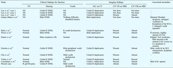
Duplication of the internal auditory canal is a rare condition. To date, only 11 cases have been reported in the literature, including one case of triplication of the internal auditory canal.Reference Lee, Cha, Jeon, Bae, Han and Kim1–Reference Casselman, Offeciers, Govaerts, Kuhweide, Geldof and Somers9 The presence of sensorineural hearing loss on the affected side is universally detected, with absence of the vestibulocochlear nerve on magnetic resonance imaging (MRI) in nearly all cases (Table I). However, facial nerve function and calibre (on MRI) were normal in all reported cases in which these features were mentioned.
Reported cases of internal auditory canal duplication

FN = facial nerve; vestib = vestibular; IAC = internal auditory canal; CT = computed tomography; CNVII and CNVIII = the facial and vestibulocochlear nerves respectively; MRI = magnetic resonance imaging; NS = not specified; unilat = unilateral; R = right; SNHL = sensorineural hearing loss; bilat = bilateral; lat = lateral; SCC = semicircular canal; L = left; EAC = external auditory canal; HL = hearing loss; grd = grade
This report describes an unusual case of unilateral duplication of the internal auditory canal, with sensorineural hearing loss and facial nerve dysfunction, and also presents a review of the literature.
Case report
A 34-year-old man presented to our institution with a three-month history of left-sided, low frequency tinnitus. On further questioning, he claimed to have had right-sided hearing loss since childhood. He denied otorrhoea and otalgia. Occasional episodes of imbalance were reported. There was no family history of sensorineural hearing loss. His perinatal history was unremarkable, with no history of childhood meningitis or chronic otitis media.
Examination of the head and neck revealed a right House–Brackmann grade III facial nerve palsy, which the patient claimed had been present since childhood. Normal tympanic membranes were noted bilaterally. The Weber test lateralised to the left, while the Rinne test showed air conduction to be greater than bone conduction.
Pure tone audiometry revealed a profound sensorineural hearing loss on the right.
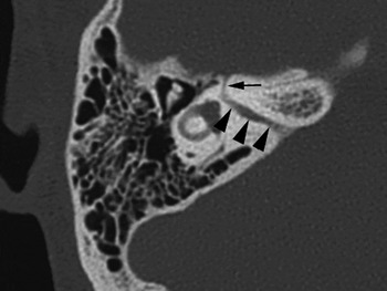
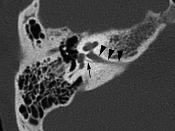
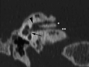
High resolution computed tomography (CT) of the temporal bones demonstrated duplication of the right internal auditory canal. Both canals were narrow: the maximal calibres of the superior and inferior canals measured 1.3 and 1.2 mm, respectively. The superiorly located canal showed continuity with the labyrinthine segment of the facial canal (Figure 1), while the inferiorly located canal had continuity with the cochlear aperture and singular canal (Figure 2). At the most lateral aspect of the canals, a fused portion was seen in which the bony septum which divided the canals more medially was absent (Figure 3). This fused portion occupied approximately one-third of the length of the canals. The rest of the right facial canal, from the labyrinthine segment to the mastoid segment, and including both the first and second genu, was normal in position and course. There was no focal osseous tumour of the internal auditory canal, nor any evidence of a diffuse sclerotic temporal osseous process. The left internal auditory canal was of normal calibre. The cochleae, vestibules and semicircular canals were of normal configuration bilaterally. No enlargement of the vestibular aqueducts was noted.
Axial, high resolution computed tomography scan of the right temporal bone, showing the superior duplicate internal auditory canal (arrowheads), with its lateral end connected to the labyrinthine segment of the ipsilateral facial canal (arrow). Note the narrow calibre of the duplicate canal.

Axial, high resolution computed tomography scan of the right temporal bone, at a lower level than that shown in Figure 1, showing the inferior duplicate internal auditory canal (arrowheads) connecting to the cochlear aperture. Part of the normal modiolus is seen. The singular canal (arrow) is clearly depicted.

Reconstructed coronal, curved, high resolution computed tomography scan of the right temporal bone, aligned along the plane of the duplicate internal auditory canals. The superior (single asterisk) and inferior (double asterisk) canals are separated by a horizontal bony septum, except at the lateral third of their lengths (arrowhead). The inferior canal points towards the cochlear aperture and modiolus (arrow).

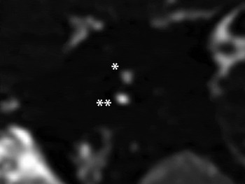
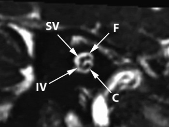
Magnetic resonance imaging, using three-dimensional constructive interference in steady state sequencing, showed no discernible normal cisternal portion of the right facial or vestibulocochlear nerves. Both the superior and inferior canals were vacant (Figure 4). On the left, the normal cisternal portions of the facial and vestibulocochlear nerves were clearly identified, together with normal cochlear, superior and inferior vestibular, and facial nerves, within the left internal auditory canal (Figure 5). The inner ear structures were normal bilaterally.
Parasagittal, oblique, constructive interference in steady state magnetic resonance imaging scan of the right temporal bone, showing a plane perpendicular to that of the duplicate internal auditory canals. The cross-sectional view of the narrow superior (single asterisk) and inferior (double asterisk) canals show them to be empty.

Parasagittal, oblique, constructive interference in steady state magnetic resonance imaging scan of the left temporal bone, showing a plane perpendicular to that of the left internal auditory canal. The normal neural contents of the left internal auditory canal are depicted. SV = superior vestibular nerve; F = facial nerve; IV = inferior vestibular nerve; C = cochlear nerve

The patient was placed on a trial of oral betahistine dihydrochloride. After four months, there was resolution of his left-sided tinnitus.
Discussion
Narrowing of the internal auditory canal is a rare condition, constituting 12 per cent of congenital temporal bone abnormalities.Reference Jackler, Luxford and House10 The normal calibre of the internal auditory canal has classically been described as ranging from 2 to 8 mm, with a mean diameter of 4 mm.Reference Valvassori and Pierce11
Internal auditory canal stenosis may have a congenital or acquired aetiology. Acquired forms of internal auditory canal stenosis are seen in patients with conditions such as osseous osteoma or exostosis causing focal narrowing of the canal.Reference Coakley, Turner and Fagan12, Reference Doan and Powell13 More generalised skeletal conditions, such as osteopetrosis, fibrous dysplasia and Paget's disease, are known to cause narrowing of the internal auditory canal.Reference Morrissey, Talbot and Schleuning14, Reference Roberto, Ettorre and Iurato15 Our patient's CT scan showed no evidence of either an osseous tumour or generalised osseous sclerosis.
In reported cases of internal auditory canal stenosis, normal inner ear structures are rarely found.Reference Nakamura, Koda and Koike16 Other inner ear anomalies are often associated, such as labyrinthine aplasia (also known as Michel aplasia), common cavity, incomplete partition of cochlea, and dysplasias of the semicircular canals or vestibules.Reference Sakina, Goh, Abdullah, Zulfiqar and Saim17, Reference Phelps, Lloyd and Shelton18 Occasionally, internal auditory canal stenosis is seen together with other middle or external ear anomalies, or with systemic syndromes such as Goldenhar or Treacher Collins syndrome.Reference Sakina, Goh, Abdullah, Zulfiqar and Saim17, Reference Shelton, Luxford, Tonokawa, Lo and House19 No other bony anomalies of the inner ear were seen in our patient's cross-sectional imaging.
Stenosis of the internal auditory canal may be associated with ipsilateral sensorineural hearing loss. There are two widely accepted hypotheses for this association.
The first hypothesis states that the embryonic cochlea and vestibule induce the growth of the vestibulocochlear nerve. The bony internal auditory canal develops around the facial and vestibulocochlear nerve by a process of chondrification and ossification of the enveloping mesoderm, in the eighth week of gestation.Reference Yates, Patel, Millman and Gibson20 As such, the internal auditory canal assumes a narrower diameter if there is aplasia or hypoplasia of the vestibulocochlear nerve.
The second hypothesis emphasises the stenotic canal as the primary defect, which inhibits the growth of the vestibulocochlear nerve and results in impaired transmission of an induction signal from the cochlea and vestibule. This hypothesis is considered less likely, due to the normal facial nerve function and morphology usually encountered in these patients.Reference Yates, Patel, Millman and Gibson20
Occasionally, patients with internal auditory canal stenosis possess normal bilateral hearing, or present with sensorineural hearing loss in the radiographically normal, contralateral ear.Reference Rothschild, Wackym, Silvers and Som21 In the former case, the presence of a functionally normal cochlear nerve is presumed despite a stenotic canal, which is often the case with the facial nerve. In the latter case, the hearing loss is probably caused by a lesion within the cochlear nerve or neuroepithelium.
Most cases of internal auditory canal stenosis preserve normal function of the ipsilateral facial nerve.Reference Sakina, Goh, Abdullah, Zulfiqar and Saim17, Reference Baek, Chae and Jung22 To our knowledge, there are only four reports of facial palsy associated with internal auditory canal stenosis.Reference Nakamura, Koda and Koike16, Reference Sakina, Goh, Abdullah, Zulfiqar and Saim17, Reference Curtin and May23, Reference Lin, Huang and Leung24 Curtin and May described a patient whose facial canal had split into two divisions in its labyrinthine segment, with atrophy of the vestibulocochlear nerve.Reference Curtin and May23 Nakamura et al. reported a 37-year-old woman with internal auditory canal stenosis and ipsilateral deafness for 27 years, who presented with an acute onset of ipsilateral facial palsy which responded to corticosteroids.Reference Nakamura, Koda and Koike16 In their series of internal auditory canal stenosis cases, Sakina et al. reported a 10-year-old boy with unilateral sensorineural hearing loss, ipsilateral facial nerve palsy, and hypoplasia of both the facial and vestibulocochlear nerves, found on MRI.Reference Sakina, Goh, Abdullah, Zulfiqar and Saim17 Lin et al. described a five-year-old girl with congenital unilateral facial palsy and sensorineural hearing loss ipsilateral to an internal auditory canal stenosis; MRI examination revealed hypoplastic trigeminal, facial and vestibulocochlear nerves on the side of the stenosis.Reference Lin, Huang and Leung24
A summary of all reported cases of duplicate internal auditory canals, to our best knowledge, is presented in Table I. Eleven of these 12 cases (including our patient) presented with sensorineural hearing loss ipsilateral to the duplicate internal auditory canals. Although facial nerve function was not recorded in four cases, it was reported as normal in seven other patients. Of eight patients who underwent MRI, all demonstrated evidence of normal facial nerve calibres.
To our best knowledge, our patient represents the first reported case of duplication of the internal auditory canal in the presence of dysfunction of both the facial and vestibulocochlear nerves. Three-dimensional constructive interference in steady state MRI showed vacant, duplicate right internal auditory canals. Intriguingly, the patient had a paradoxical grade III facial nerve palsy, despite no discernible MRI evidence of an ipsilateral facial nerve. We had excluded the possibility of an anomalous facial canal. The labyrinthine, tympanic and mastoid segments and first and second genu of the right facial canal were all normal in position and course, apart from the labyrinthine segment being connected to the superior duplicate internal auditory canal. There was no evidence of cochlear malformation or external auditory canal atresia, conditions commonly associated with an anomalous facial canal.Reference Romo and Curtin25, Reference Yu, Han, Gong, Wang, Dai and Zhao26
We had considered the possibility of severe hypoplasia of the facial nerve resulting in a neural calibre below the limits of spatial resolution of the MRI sequence utilised (i.e. constructive interference in steady state): the so-called ‘imperceptible nerve’.Reference Glastonbury, Davidson, Harnsberger, Butler, Kertesz and Shelton27 Our patient's relatively normal intratemporal facial canal suggests an acquired aetiology for his severe facial nerve hypoplasia. Nevertheless, it does seem remarkable that such severe nerve hypoplasia would be compatible with a House–Brackmann grade III palsy.
A possible explanation may be suggested by the phenomenon of aberrant innervation, similar to the pathogenesis of Duane's syndrome.Reference Gutowski28 Pathological studies in isolated cases of Duane's syndrome have shown hypoplasia and/or absence of the abducens nucleus and/or nerve, with lateral rectus innervation from the inferior division of the oculomotor nerve. In Verzijl and colleagues' study of six patients with Mobius syndrome, five showed no MRI evidence of a facial nerve bilaterally (using the constructive interference in steady state sequence), despite all showing residual lower facial muscular contraction on examination.Reference Verzijl, Valk, de Vries and Padberg29 The authors postulated that other cranial nerves, namely the trigeminal, glossopharyngeal or hypoglossal nerves, may aberrantly innervate some facial muscles, in a mechanism possibly akin to that of Duane's syndrome.
We believe that our patient has a rare case of unilateral duplicate internal auditory canals, with vestibulocochlear nerve aplasia and severe facial nerve hypoplasia, coupled with residual facial motor function occurring possibly through a mechanism of aberrant innervation.
In cases of suspected aplasia or hypoplasia of the facial and vestibulocochlear nerves, the delineation of neural anatomy, calibre and course requires high resolution gradient echo MR imaging, and is essential when determining cochlear implant candidacy.Reference Casselman, Offeciers, Govaerts, Kuhweide, Geldof and Somers9 Examples of appropriate MRI sequences reported in the literature include three-dimensional constructive interference in steady state, three-dimensional balanced fast field echo, three-dimensional magnetisation prepared rapid gradient echo, and three-dimensional driven equilibrium radio frequency pulse.Reference Demir, Cakmakci, Erdag and Men5, Reference Cho, Na, Jung and Hong7 These sequences provide submillimetric resolution, three-dimensional capability, and inherent high contrast between neural structures and surrounding cerebrospinal fluid. At present, we have found that the three-dimensional constructive interference in steady state MRI sequence provides the best depiction of the integrity of intracanalicular neural contents. This high resolution MRI sequence has allowed assessment of the calibre of the cochlear nerves, compared with other intracanalicular nerves within the internal auditory canal. Hypoplasia of the cochlear nerve is commonly defined as a calibre less than that of the facial nerve.Reference Glastonbury, Davidson, Harnsberger, Butler, Kertesz and Shelton27 Alternatively, certain authors have employed an auditory nerve to facial nerve ratio of less than one.Reference Madzivire, Camp, Lane, Witte, Robb and Mun30
Cochlear implantation has enabled many patients with congenital sensorineural hearing loss to obtain auditory stimulation and to develop speech, even in cases of severe inner ear malformation.Reference Papsin31 However, most surgeons agree that children with complete cochleovestibular aplasia (Michel aplasia), cochlear nerve aplasia or cochlear nerve canal aplasia are inappropriate candidates for cochlear implantation.Reference Chadha, James, Gordon, Blaser and Papsin32 Until the 1990s (and certainly before widespread use of pre-surgical MRI), a narrow internal auditory canal was considered a contraindication to cochlear implantation, because of the possibility of an absent cochlear nerve.Reference Shelton, Luxford, Tonokawa, Lo and House33 Traditionally, such children have not been considered for cochlear implantation because of questionable post-operative outcomes.
• Internal auditory canal duplication is rare; vestibulocochlear nerve (CN VIII) aplasia is often also present, but the facial nerve (CN VII) is usually normal
• A rare case of ipsilateral duplicate internal auditory canals is presented, with CN VIII aplasia and severe CN VII hypoplasia
• Patients with a narrow or duplicated internal auditory canal and CN VII and/or CN VIII dysfunction should undergo high resolution gradient echo magnetic resonance imaging for assessment of neural integrity
While candidacy for cochlear implantation has expanded in patients with cochlear nerve hypoplasia, there has also been a suggestion that auditory brainstem implantation is the treatment of choice for these patients. A study by Colletti found that auditory brainstem implantation had better outcomes in nontumour patients, compared with those with tumour conditions (e.g. neurofibromatosis).Reference Colletti34 Previously, it had been observed that children with narrowing of the internal auditory canal or cochlear canal had poorer post-implantation performance than any other patient group.Reference Papsin31 In contrast, Colletti and Zoccante have reported that auditory brainstem implantation in patients with narrow internal auditory canals and hypoplastic cochlear nerves shows promising auditory outcomes.Reference Colletti and Zoccante35 We strongly believe that the decision to perform either cochlear implantation or auditory brainstem implantation in a patient with a narrow internal auditory canal should be based both on MRI evidence of a remnant cochlear nerve and on the experience of the surgeon.
Conclusion
We present a rare case of both vestibulocochlear nerve aplasia and severe facial nerve hypoplasia in a patient with duplication of the right internal auditory canal. Although the facial nerve was imperceptible on MRI, its presence was assumed on the basis of residual facial motor function (i.e. House–Brackmann grade III).
In such cases, computed tomography and MRI provide important complementary anatomical detail regarding the integrity of the neural elements within the internal auditory canal, as well as any associated bony anomaly of the inner ear. We recommend that MRI with a three-dimensional high resolution gradient echo sequence (e.g. three-dimensional constructive interference in steady state) should be performed in all patients with an abnormal internal auditory canal and clinical evidence of facial and/or vestibulocochlear nerve dysfunction.