




No CrossRef data available.
Article contents
Ultrastructural and molecular characterization of Bacillidium vesiculoformis n. sp. (Microspora: Mrazekiidae) in the freshwater oligochaete Nais simplex (Oligochaeta: Naididae)
Published online by Cambridge University Press: 13 December 2004
Abstract
The development of a new species, Bacillidium vesiculoformis n. sp. (Microspora, Mrazekiidae), is described from the freshwater oligochaete Nais simplex (Oligochaeta, Naididae). Initial stages of parasite development consist of a monokaryotic merogony within a haemocyte of the intestinal blood sinus. The resulting hypertrophied haemocyte is attached to the chloragocytes of the sinus by fine cytoplasmic extensions with the sinus around the cell becoming greatly enlarged. The meronts within the haemocyte form diplokaryotic sporonts that undergo sporogenesis directly within the cytoplasm of the host cell. The infected cell becomes packed with spores and developmental stages, causing it dramatically to increase in size, eventually rupturing the oligochaete and cell. Sporogony appears to be disporoblastic. Released spores were observed to have an adhesive quality. Transmission studies conducted with mature spores failed to transmit the parasite horizontally although vertical transmission was observed. Phylogenetic analysis of the parasite demonstrated that B. vesiculoformis clustered with microsporidian parasites of bryozoa and two other microsporidians, Janacekia debaiseuxi and an unidentified Bacillidium sp.
Information
- Type
- Research Article
- Information
- Copyright
- © 2004 Cambridge University Press
INTRODUCTION
Microsporidia of the genus Bacillidium are typically parasites of oligochaetes (Larsson & Gotz, 1996). This atypical microsporidian genus currently comprises 7 species, characterized by a large, rod-shaped spore, an exospore composed of 2 distinct components and a polar filament with an anterior manubrium (Larsson, 1994). The manubrium is a unique form of extrusion apparatus that is considered analogous to the polar filament of other microsporidia (Gotz, 1981).
Early studies into species of Bacillidium described the development of the parasites by light microscopy (Janda, 1928; Jirovec, 1936). Details of spore structure could not be ascertained and, therefore, observations were limited to nuclear division, nature of cells infected and effects on the host. Limited transmission studies suggested that the life-cycle was indirect (Janda, 1928). Recent descriptions of Bacillidium spp. have concentrated on ultrastructural examination of sporogony. This has confirmed that they are disporoblastic producing diplokaryotic spores. To date, all stages of development described using electron microscopy have been diplokaryotic and it has been proposed that the Bacillidium are diplokaryotic during both sporogony and merogony in the oligochaete host (Larsson, 1994).
An initial molecular phylogenetic study on an undescribed Bacillidium sp. demonstrated that it did not appear to cluster with any other species of microsporidian, thus reflecting the unique nature of the spore (Nilsen, 1999). Recently, a number of microsporidian genera infecting freshwater bryozoa, have been described (Canning et al. 2002; Morris & Adams, 2002). Phylogenetic analysis of the small subunit ribosomal gene of these parasites has shown that they are clustered around the Bacillidium sequence identified by Nilsen (1999). Canning et al. (2002) proposed the new family Pseudonosematidae for the microsporidia infecting bryozoa, based on the marked differences in spore structure between the bryozoan infecting parasites and Bacillidium spp., with the expectation that the sequencing of further Bacillidium spp. would clarify the relationship between the groups. Here we describe the elucidation of a new species, Bacillidium vesiculoformis n. sp., using light and electron microscopy and discuss its phylogenetic position.
MATERIALS AND METHODS
Collection and identification of infected oligochaetes
Fifty-five oligochaetes were collected from a static 7 litre plastic aquarium, containing bryozoan colonies obtained from Airthrey Loch, University of Stirling, Scotland in July 2002. The bryozoa had been collected from submerged branches in the loch and maintained in the aquarium following the methods of Morris, Morris & Adams (2002), for 3 weeks as part of a larger study.
The oligochaetes were collected directly from the surface of the bryozoan colonies and the aquarium surfaces using a plastic pipette. They were placed into cell wells and examined under an inverted microscope for evidence of microsporidian infection. They were maintained in a 17 °C incubator, the water in the well being replaced weekly with fresh distilled water and a drop of cultured Gloecaspa sp. for food. All oligochaetes were examined daily for evidence of infection over the course of 3 weeks. Observations were noted on the development of the parasites within the infected oligochaetes and the effect of infection on the host.
Collection of samples
Infected oligochaetes were identified using the key of Kathman & Brickhurst (1999). The oligochaetes were transversely bisected, with one half being used for ultrastructural studies and the other half being retained for molecular characterization. Oligochaetes were sampled for study either, when the infection was first noticed, when spores were first noted, or immediately prior to spore release.
Samples taken for transmission electron microscopy were immediately fixed in Karnovsky's fixative for 4 h at 4 °C followed by a rinse in cacodylate buffer (pH 7.2) overnight. They were post-fixed in 1% osmium tetroxide for 1 h, dehydrated through an ethanol series and embedded in Spurr's resin. Ultrathin sections were mounted on formvar-coated grids, stained with lead citrate/uranyl acetate and viewed at 80 kV using a Philips 201 electron microscope. Semi-thin sections (1 μm) were also taken and stained with 1% toluidine blue for examination under light microscopy.
DNA from samples taken for molecular characterization was extracted using an ABGene mini-prep kit. This kit used magnetic separation to purify DNA. Purified DNA was resuspended in 10 μl of nanopure water and stored in 5 μl aliquots at −20 °C before characterization.
Molecular characterization
Amplification and sequencing of small subunit (SSU), internal transcribed spacer (ITS), and partial large subunit (LSU) rDNA from infected tissue was undertaken using a range of microsporidian specific primers, V1f (Weiss et al. 1994), 18sf (Baker et al. 1995), 530f, 228r, 580r (Vossbrinck et al. 1993), HG4f, HG4r (Gatehouse & Malone, 1998). Amplifications were performed as described by Terry et al. (2003). PCR products were cleaned using QIAquick gel extraction kits (Qiagen, Inc. Sussex, UK) and sent for sequencing at the Natural History Museum, London.
A homology search was performed using the FASTA program (European Bioinformatics Institute, Cambridge). The sequence was then aligned with other microsporidian SSU rDNA sequences from GenBank using Bioedit (Hall, 2001), removing regions of ambiguity. The 34 microsporidian sequences used are as follows: Amblyospora connecticus (AF25685); Ameson michaelis (L15741); Antonospora scoticae (AF024655); Bacillidium sp. (AF104087); Brachiola algerae (AF069063); Bryonosema plumatellae (AF484690); Cystosporogenes operophterae (AJ302320); Edhazardia aedis (AF027684); Encephalitozoon cuniculi (L17072); Encephalitozoon hellem (AF118142); Endoreticulatus schubergi (L39109); Enterocytozoon bieneusi (AF024657); Glugea anomala (AF104084); Ichthyosporidium sp. (L39110); Intrapredatorus barri (AY013359); Janacekia debaisieuxi (AJ252950); Kabatana takedai (AF356222); Loma acerinae (AJ252951); Microgemma caulleryi (AY033054); Nosema bombycis (L39111); Nosema granulosis (AJ011833); Nucleospora salmonis (U10883); Oligosporidium occidentalis (AF495379); Ordospora colligata (AF394529); Pleistophora typicalis (AF104080); Pseudonosema cristatellae (AF484694); Schroedera plumatellae (AY135024); Septata intestinalis (L39113); Spraguea lophii (AF033197); Thelohania solenopsae (AF134205); Trichonosema pectinatellae (AF484695); Vairimorpha necatrix (Y00266); Visvesvaria acridophagus (AF024658); Vittaforma corneum (U11046). Sequences were rooted against two zygomycete fungal sequences, Basidiobolus ranarum (D29946) and Mycotypha microspora (AF157148). Parsimony, neighbour-joining, and maximum likelihood analyses were conducted using PAUP*4.0b10 (Swofford, 2002) and the GTR+I+G substitution model (4 gamma classes). A heuristic search was employed with random stepwise addition (10 replicates) and TBR branch swapping. Nodal support of the maximum likelihood analysis was assessed by bootstrap analysis (100 replicates).
Transmission studies
Spores released from a moribund oligochaete were used in experiments to infect either bryozoa or oligochaetes. Six oligochaetes, that did not show signs of infection, were selected from those collected and placed into a fresh 24-cell well plate. These oligochaetes were identified as the same species as the infected specimen. Spores released from the infected oligochaete were then introduced into 3 of the cell wells and the oligochaetes monitored for infection over 14 days using an inverted microscope.
For exposure of bryozoan colonies, statoblasts collected from apparently healthy colonies of Plumatella repens were collected, hatched under inverted Petri dishes (Wood, 1989) and maintained as above. Three colonies on one plate were exposed to infection by pipetting spores directly in the water current created by the tentacular crown (lophophore) of a bryozoan zooid. The colonies were then cultured for 14 days and monitored for evidence of infection using an inverted microscope. The transparent nature of cultured bryozoa allows for relatively rapid diagnosis of developing infections.
RESULTS
Five oligochaetes, collected from the bryozoan tank, were found to have microsporidian infections, representing a prevalence of 9%. These oligochaetes were all identified as Nais simplex Piguet, 1906.
Effect of parasite development upon the host
The first identified stage of infection appeared as red swellings along the length of the intestine (Fig. 1A). The swellings increased in size and number, becoming increasingly opaque, until they started to fill the coelom of the oligochaete (Fig. 1B). At this stage, in live oligochaetes, cylindrical spores could be observed within the swellings. The swellings continued to increase in size causing the body wall of the oligochaete to distend (Fig. 1C). Five days after the initial infection was identified, a portion of the oligochaete ruptured releasing spores, developmental stages and host tissue, into the cell well. The remaining portion of the oligochaete, although alive, was incapable of crawling and survived for several hours before rupturing again for a final time. The released spores were cylindrical 12·2 μm long and 1·6 μm in diameter (Fig. 1D). One oligochaete in the early stage of infection was noted to develop eye-spots midway down its body. The infection persisted in this oligochaete as it underwent binary division. Both resultant individuals were infected with the microsporidian.

Fig. 1. (A) Swellings (caused by infected haemocytes) observed along the line of the intestine during the initial stages of observable infection. Swellings indicated by arrows. (B) Enlargement of infected haemocytes and their increasing opacity within the oligochaete. (C) Distension of body wall of oligochaete by infected cells. (D) Bacilliform spores released from oligochaete. (E) Section through an oligochaete with the early stages of infection. Two infected haemocytes (H) can be observed within the enlarged peri-intestinal sinus (S). Cytoplasmic extensions from an infected haemocyte appear to be interact with the chloragocytes surrounding the sinus (arrow). The intestinal lumen is indicated with an (I). (F) Monokaryotic meronts (arrows) surrounding lobed nucleus (N) of infected cell. (G) Diplokaryotic sporont within hypertrophic haemocyte.
Released spores were difficult to dislodge from the cell well using a pipette, appearing to have an adhesive quality. It was noted that ciliates, introduced into the well with the oligochaete, would rapidly ingest the spores. This appeared to be in preference to the remains of the worm in the well. Even though up to 5 spores were noted within a single ciliate no adverse affects or infection was observed. The ciliates presumably ejected the spores after a period of time as these eventually became evenly distributed over the base of the cell well.
Ultrastructural examination of microsporidian development
The earliest stage of development examined was in an oligochaete with characteristic red swellings down the length of its intestine, but no visible spores. Ultrastructural examination of this oligochaete revealed there to be hypertrophied haemocytes within the peri-intestinal blood sinus (Fig. 1E). These haemocytes contained numerous monokaryotic meronts, which resided in direct contact with the host cell cytoplasm, and consisted of a round nucleus that occupied most of the cell volume (Fig. 1F). Division of the meronts was not noted, but occasionally parasites containing large nuclei with flocculent chromatin, suggestive of nuclear division, were observed. The host cell nucleus was notably enlarged and lobed, while the cell surface possessed many cytoplasmic filaments that appeared to interact with the chloragocytes enclosing the sinus, although direct contact with these cells was not observed. The sinus was markedly enlarged, with the infected cell appearing to be suspended by the filaments in the centre of it.
Sections were examined from oligochaetes when spores were first noted in the infected haemocytes (opaque swellings) and later when the body wall of the oligochaete was distended by spore masses. From these sections, a partial developmental sequence was deduced. The earliest sporogonic stages noted were diplokaryotic sporonts but the transformation of meronts to sporonts was not observed. Initially the sporonts were condensed and contained a well-defined diplokaryon occupying the majority of the cell (Fig. 1G). One sporont was observed apparently in the final processes of dividing by binary division. Sporogenesis occurred directly in the cytoplasm of the host cell and was asynchronous, with sporonts, sporoblasts and mature spores all within the same haemocyte. The initiation of sporogenesis was signalled by the cytoplasm of the sporonts extending, producing an elongate sporoblast, with the diplokaryon positioned at one end (Fig. 2A).

Fig. 2. (A) First stage of sporogenesis with the sporoblast elongating. (B) Transverse section through early sporoblast. Electron-dense material is forming on the surface of the sporoblast with an electron-lucent area immediately beneath it. Arrowheads indicate these two layers. The forming manubrium is composed of 3 layers, an electron-dense outer layer surrounding a granular matrix with an electron-dense core. (C) Transverse section through developing sporoblast. The manubrium is differentiating into layers. The developing exospore and endospore layers now completely encircle the sporoblast with spherical/granular material clearly associated with the exospore. (D) Formation of anchoring apparatus. The exospore and endospore layers of the spore wall are clearly identifiable. The developing anchoring disk (A) caps the manubrium and is surrounded by the polar sac (PS). The manubrium is surrounded by an additional lucent layer (arrow). (E) Transverse section through mature spore. The manubrium is composed of 7 layers with a wide endospore surrounded by a relatively thin exospore. (F) Section through surface of spore wall. The exospore is embedded with granular material. (G) Section through anterior of mature spore. Anchoring disc (A) is at the apex of the manubrium and extends into the polar sac (PS). The electron-dense anterior polaroplast (AP) is followed by a globular posterior polaroplast (PP). (H) Mature spore of Bacillidium vesiculoformis. Structures labelled are diplokaryon (D), posterior dense bodies (B), manubrium (M) and the polar filament (arrow).
During sporogenesis the development of the spore wall and the development of the manubrium occurred in concert. This was clearly observed by transverse sections through sporoblasts by which the development of the spore could be charted. After elongation of the sporoblast, areas of cytoplasm associated with the plasma membrane became increasingly electron-lucent with the surface of the sporoblast becoming thickened and electron-dense (Fig. 2B). The manubrium at this time appeared to consist of an electron-dense outer layer surrounding a granular matrix with an undifferentiated dense core. The changes on the surface of the plasma membrane continued, extending around the sporoblast, with the electron-dense layer becoming recognizable as the developing exospore and the electron-lucent layer beneath it the endospore. Spheroids of material resembling granules measuring approximately 25 nm in diameter were associated with the developing exospore but their origin could not be determined. The manubrium continued to differentiate with the granular layer becoming homogenous and a further electron lucent layer forming around the structure (Fig. 2C, D). The earliest observed sections demonstrating the formation of the anchoring apparatus occurred when the manubrium extended the entire length of the cell, and the developing endospore and exospore were clearly differentiated (Fig. 2D). The developing apparatus, at the anterior of the sporoblast, consisted of a dense anchoring disc, covering the core of the manubrium and capped by a moderately dense polar sac that extended around the anchoring disc and the apex of the manubrium.
As the endospore expanded in diameter, the manubrium continued to differentiate into more layers, and the developing spores became more regular in transverse section. With the development of the endospore, fixation of the spores became increasingly poor. The contents of the vast majority of mature spores were not fixed.
Mature spores were roughly cylindrical with a manubrium composed of 7 layers, an endospore of 100 nm and an exospore of 25 nm (Fig. 2E). The layers of the manubrium alternating electron lucent (layer 1), dense (layer 2), lucent (layer 3), moderate (layer 4), dense (layer 5), light (layer 6) with a moderately dense core (layer 7). The spore surface was covered in dense spherical granules that appeared to be embedded in the exospore (Fig. 2F). In the mature spore the polar sac was a comparatively flat, crescent structure that was composed of dense material (Fig. 2G). The anchoring apparatus appearing as a moderately lucent area beneath the sac, capping the outer layers of the manubrium and appearing continuous with layer 4 of this structure. The apparatus appeared to extend into the polar sac region where this structure became electron lucent. The anterior part of the polaroplast was an electron dense, cup like body that extended back from the polar sac into the spore. It was 1/10 the length of the spore and was lined with two 5 nm thick unit membranes although the internal organization of this area of the polaroplast could not be discerned. The posterior polaroplast did not have a clearly definable structure but appeared to contain electron-dense globular material.
The manubrium extended nearly the entire length of the spore, narrowing to a short polar filament at the posterior. The diplokaryon could be distinguished, with the nuclei lying in tandem along the side of the spore while in the posterior of the spore there were spherical dense bodies of various sizes (Fig. 2H).
The developing spores were initially contained within the cytoplasm of the host cell. As the infection progressed and the spores matured so the cytoplasm became reduced and vacuous, appearing to be replaced by an electron-lucent matrix. The hypertrophied cell at this stage was still contained within the peri-intestinal sinus, being encircled by host cells of uncertain characterization, presumably leucocytes, underlying the chloragocytes of the sinus (Fig. 3A). As the cytoplasm of the infected cell became replaced by the electron-lucent matrix so it increased in size. The body wall of the oligochaete became reduced at points and the infected cell was noted to almost breach the exterior of the worm (Fig. 3B).

Fig. 3. (A) Haemocyte with reduced cytoplasm containing mature spores (S) contained with the peri-intestinal sinus (B) surrounded by a layer of host cells (*). The surrounding chloragocytes of the sinus are labelled with an arrow. Exterior of oligochaete indicated with (E). (B) Enlarged haemocyte containing mature spores (S) breaking through the cuticle of the oligochaete. Chloragocytes surrounding the haematocyte indicated with an arrow. Exterior of oligochaete indicated with (E).
Phylogenetic analysis
1580 bp of sequence (GC content 46·5%) representing the complete SSU, ITS, and partial LSU rDNA of the parasite was successfully amplified and the sequence has been deposited in GenBank, Accession No. AJ581995. The FASTA search revealed that the sequence had the closest homology to the bryozoan parasites, Pseudonosema cristatellae and Trichonosema pectinatellae, and the oligochaete parasite, Bacillidium sp. Parsimony, neighbour-joining and maximum likelihood analyses all gave the same overall tree topography placing the parasite within the clade containing Bacillidium sp., Schroedera plumatellae and Pseudonosema cristatellae (Fig. 4). Therefore, based on ultrastructural characteristics, host type and molecular data, we propose to name this parasite Bacillidium vesiculoformis.

Fig. 4. Phylogenetic tree of microsporidia based on SSU rDNA using maximum likelihood. Bacillidium vesiculoformis is placed within the poorly resolved clade containing both bryozoan (e.g. Pseudonosema cristatellae) and oligochaete (Bacillidium sp.) parasites (highlighted box). The boot strap values (100 replicates) on the tree represent the percentage of bootstrap replicates that gave that topology. Neighbour-joining and parsimony analyses resulted in identical placement of B. vesiculoformis within the tree (data not shown).
Systematic Position
Phylum Microspora, Class Dihaplophasea, Order Dissociodihaplophasida, Family Mrazekiidae
Description of the species
- Name: Bacillidium vesiculoformis n. sp.
- Type host: Nais simplex Piguet, 1906 (Oligochaeta: Naididae)
- Type locality: Southern end of Airthrey Loch, University of Stirling campus. National grid reference NS 804963.
- Infected cell type: Haemocyte
- Type material: Hapantotype material has been deposited in the collection of the Natural History Museum, London. This material comprises embedded blocks of oligochaetes containing early stages of parasite development (registration number 2004[ratio ]6[ratio ]8[ratio ]2) and later developmental stages including sporonts and spores (registration number 2004[ratio ]6[ratio ]8[ratio ]1).
- Etymology: The specific name is derived from the latin for ‘bladder forming’ in reference to the enlargement of the blood sinus around the infected haemocytes.
- Description of spores: Diplokaryotic spores, bacilliform, 12·2 μm±0·4 μm in length, 1·6 μm±0·2 μm (n=20) in width, with a manubrium composed of 7 layers that extends nearly the entire length of the spore, tapering at the posterior to a polar filament. Elongate diplokaryon positioned midway along spore. Electron-dense anterior polaroplast occupying one tenth of the spore length, while the posterior polaroplast appeared globular of uncertain length. Electron-lucent endospore, 100 nm in diameter, surrounded by dense exospore, 25 nm in diameter, embedded with granular material on its surface.
Development
Monokaryotic merogony presumably by binary division followed by diplokaryotic, disporoblastic (?) sporogony. Sporogenesis occurs in direct contact with host cell cytoplasm.
Transmission studies
Neither transmission study induced an observable infection. However, while the oligochaetes were observed to graze on the spores introduced into their well, ingestion of the spores into the bryozoan colonies was more difficult to assess. The majority of spores generally sank quickly out of the water current generated by the lophophore. However, occasional spores were noted to remain in the current to be transported towards the lophophore of the bryozoan's zooid. However, as soon as one spore touched a tentacle the lophophore would immediately retract and the filter-feeding stop. As such, although contact between spores and bryozoa was observed, actual ingestion of the spores was not ascertained. At the end of the experiment, no colonies appeared to be infected.
DISCUSSION
This extensive ultrastructural study, at various time-points through infection, has enabled partial elucidation of the life-cycle of this parasite in its oligochaete host. To date, ultrastructural observations on the development of Bacillidium spp. have suggested that they are diplokaryotic throughout their development (Larsson, 1994). However, as Bacillidium infections are rarely encountered, these observations have all been conducted after the onset of sporogony when spores are clearly observable within hypertrophied cells. In this study, the early development of B. vesiculoformis was seen to have a distinct phase of monokaryotic merogony that preceded the diplokaryotic sporogony. It is unknown whether monokaryotic merogony occurs in other species of Bacillidium, although light microscopical studies have suggested the presence of these stages during the early development of B. criodrili (Jirovec, 1936). A relatively large number of infected cells were noted in every infected worm examined. However, no specialized auto-infective spores were noted. Therefore, it is likely that the formation of the hypertrophied cells either represented exposure to multiple spores, as would be expected if the oligochaete ingested another host, fragmentation of an existing infected cell or meront infection of haemocytes.
The structure of the spore was typical for Bacillidium, in that it was bacilliform, diplokaryotic, possessed an exospore with a distinct surface layer and a well-developed manubrium. Most species of Bacillidium have been reported within the coelomic lymphocytes of oligochaetes. However, B. haematobium has been recorded as developing within the haemolymph (Jirovec, 1936). As all known microsporidia are obligate parasites that require a host cell it is likely that B. haematobium developed within a blood cell in the blood vessel. Ultrastructural data for B. haematobium does not presently exist making comparisons with the B. vesiculoformis impossible. However, as the spore length of B. haematobium is significantly larger than that described for B. vesiculoformis (16–17 μm compared to 12·4 μm) and the host oligochaete species is different we consider that B. vesiculoformis represents a new species.
The major morphological difference between B. vesiculoformis and B. criodrili, the type species for the genus was the absence of an exospore-derived sac. The absence of a membranous sac derived from the exospore may be a significant feature of B. vesiculoformis that clearly distinguishes it from B. criodrili. The granular material embedded in the exospore appears to have more in common with B. filiferum in that this species has projections emanating from the exospore rather than a membranous sac (Larsson, 1989). For B. filiferum the projections are considered to be composed of the same material as the exospore, while the granular material of B. vesiculoformis appears to be of a distinct composition. The projections and material associated with the exospore of Bacillidium spp. are likely to have a biological function, and for B. vesiculoformis this material may contribute to the adherent nature of the fresh spores.
Phylogenetic data have previously indicated that a Bacillidium sp., Janacekia debaiseuxi and microsporidia infecting bryozoa are closely related, although marked differences in spore structure and development exist (Canning et al. 2002; Morris & Adams, 2002). The position of B. vesiculoformis further suggests that there is a phylogenetic relationship between members of the genus Bacillidium and those microsporidia infecting bryozoa, although, bootstrap support within the clade is poor. As such, the relationships between these parasites remains unclear and will only be resolved with the entry of additional closely related sequences into the analyses or by the elucidation of their life-cycles.
Bacillidium spp. are thought to have indirect life-cycles as previous direct transmission experiments have failed (Janda, 1928). In this study we observed the vertical transmission of B. vesiculoformis, a route that may be important in sustaining this parasite in its oligochaete host. However, this is unlikely to be the only route of propagation for the parasite. The rupturing of the host and subsequent release of spores suggests that horizontal transmission also features within the parasite life-cycle. With the failure to induce the infection in oligochaetes directly, we conclude that another host is involved in the life-cycle of B. vesiculoformis. Although the phylogenetic association of bryozoa infecting microsporidia and B. vesiculoformis suggests that the bryozoa could be alternate hosts, transmission experiments failed to transmit the parasite. This may be due to the wrong bryozoan species being exposed to the spores. However, as the spores adhere to substrates, it is not clear how they would directly infect bryozoa that filter-feed seston. If bryozoa are involved in the life-cycle, it is possible that a further host is involved in the transmission of B. vesiculoformis. Such a host is presently unknown but the observation of ciliates ingesting the spores suggests that they may act as a paratenic host for the parasite or act as an aid for spore dispersal.
The authors would like to thank DEFRA project grant FC1111 and NERC grant no NER/A/S/1999/60040 for funding these studies. The authors would also like to thank Mr L. Brown for assistance in preparation of EM specimens, Professor J. I. R. Larsson, University of Lund, for clarification on points of Bacillidium biology and the Nuffield foundation for funding Ms K. B. Ferguson through a Nuffield science bursary.
References
REFERENCES
BAKER, M. D.,
VOSSBRINCK, C. R.,
DIDIER, E. S.,
MADDOX, J. V. &
SHADDUCK, J. A. (
1995).
Small subunit ribosomal DNA phylogeny of various microsporidia with emphasis on AIDS related forms.
Journal of Eukaryotic Microbiology
42,
564–570.CrossRefGoogle Scholar
CANNING, E. U.,
REFARDT, D.,
VOSSBRINCK, C. R.,
OKAMURA, B. &
CURRY, A. (
2002).
New diplokaryotic microsporidia (Phylum Microsporidia) from freshwater bryozoans (Bryozoa, Phylactolaemata).
European Journal of Protistology
38,
247–265.CrossRefGoogle Scholar
GATEHOUSE, H. S. &
MALONE, L. A. (
1998).
The ribosomal RNA gene region of Nosema apis (Microspora): DNA sequence for small and large subunit rRNA genes and evidence of a large tandem repeat unit size.
Journal of Invertebrate Pathology
71,
97–105.CrossRefGoogle Scholar
GOTZ, P. (
1981).
Homology of the manubrium of Mrazekia brevicauda and the polar filament of other Microsporidia.
Zeitschrift für Parasitenkunde
64,
321–333.Google Scholar
HALL, T. (
2001).
Bioedit: Biological sequence alignment editor for Windows 95/98/NT Edition 5.0.9.
http://www.mbio.ncsu.edu/BioEdit/bioedit.html.
JANDA, V. (
1928).
Über Microorganismen aus der Leibshöhle von Criodrilus lacuum Hoffm. und eigenartige Neubildungen in der Körperwand dieses Tieres.
Archive für Protistenkunde
63,
84–93.Google Scholar
JIROVEC, O. (
1936).
Zur kenntnis von Oligochäten parasitierenden Microspodien aus der Familie Mrázekidae.
Archive für Protistenkunde
87,
314–344.Google Scholar
KATHMAN, R. D. &
BRINKHURST, R. O. (
1999).
Guide to the Freshwater Oligochaetes of North America.
Aquatic Resources Center, College Grove,
Tennessee, USA.
LARSSON, J. I. R. (
1989).
The light and electron microscopic cytology of Bacillidium filiferum sp. nov. (Microspora, Bacillidiidae).
Archive für Protistenkunde
137,
346–355.Google Scholar
LARSSON, J. I. R. (
1994).
Characteristics of the genus Bacillidium Janda, 1928 (Microspora, Mrazekiidae)- reinvestigation of the type species B. criodrili and improved diagnosis of the genus.
European Journal of Protistology
30,
85–96.Google Scholar
LARSSON, J. I. R. &
GOTZ, P. (
1996).
Ultrastructural evidence for the systematic position of Jirovecia brevicauda (Leger & Hesse, 1916) Weisser, 1985 (Microspora, Mrazekiidae).
European Journal of Protistology
32,
366–371.CrossRefGoogle Scholar
MORRIS, D. J. &
ADAMS, A. (
2002).
Development of Schroedera plumatellae gen.n., sp. n. (Microsporidia) in Plumatella fungosa (Bryozoa: Phylactolaemata).
Acta Protozoologica
41,
383–396.Google Scholar
MORRIS, D. J.,
MORRIS, D. C. &
ADAMS, A. (
2002).
Development and release of a malacosporean (Myxozoa) from Plumatella repens (Bryozoa: Phylactolaemata).
Folia Parasitologica
49,
25–34.CrossRefGoogle Scholar
NILSEN, F. (
1999).
Small subunit rDNA phylogeny of Bacillidium sp. (Microspora, Mrazekiidae) infecting oligochaets.
Parasitology
118,
553–558.Google Scholar
SWOFFORD, D. L. (
2002).
PAUP*. Phylogenetic Analysis Using Parsimony (*and other methods), v. 4.0b10.
Sinauer Associates,
Sunderland, Mass.
TERRY, R. S.,
MacNEIL, C.,
DICK, J. T. A.,
SMITH, J. E. &
DUNN, A. M. (
2003).
Resolution of a taxonomic condundrum: an ultrastructural and molecular description of the life cycle of Pleistophora mulleri (Pfeiffer 1895; Georgevitch 1929).
Journal of Eukaryotic Microbiology
50,
266–273.CrossRefGoogle Scholar
VOSSBRINCK, C. R.,
BAKER, M. D.,
DIDIER, E. S.,
DEBRUNNER-VOSSBRINCK, B. A. &
SHADDUCK, J. A. (
1993).
Ribosomal DNA sequences of Encephalitozoon hellem and Encephalitozoon cuniculi: species identification and phylogenetic construction.
Journal of Eukaryotic Microbiology
40,
354–362.CrossRefGoogle Scholar
WEISS, L. M.,
ZHU, X.,
CALI, A.,
TANOWITZ, H. B. &
WITTNER, M. (
1994).
Utility of microsporidian rRNA in diagnosis and phylogeny: a review.
Folia Parasitologica
41,
81–90.Google Scholar
WOOD, T. S. (
1989)
Ectoproct bryozoans of Ohio.
Ohio Biological Survey Bulletin
8,
1–70.Google Scholar

Fig. 1. (A) Swellings (caused by infected haemocytes) observed along the line of the intestine during the initial stages of observable infection. Swellings indicated by arrows. (B) Enlargement of infected haemocytes and their increasing opacity within the oligochaete. (C) Distension of body wall of oligochaete by infected cells. (D) Bacilliform spores released from oligochaete. (E) Section through an oligochaete with the early stages of infection. Two infected haemocytes (H) can be observed within the enlarged peri-intestinal sinus (S). Cytoplasmic extensions from an infected haemocyte appear to be interact with the chloragocytes surrounding the sinus (arrow). The intestinal lumen is indicated with an (I). (F) Monokaryotic meronts (arrows) surrounding lobed nucleus (N) of infected cell. (G) Diplokaryotic sporont within hypertrophic haemocyte.
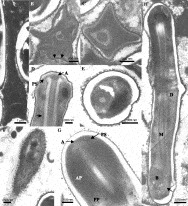
Fig. 2. (A) First stage of sporogenesis with the sporoblast elongating. (B) Transverse section through early sporoblast. Electron-dense material is forming on the surface of the sporoblast with an electron-lucent area immediately beneath it. Arrowheads indicate these two layers. The forming manubrium is composed of 3 layers, an electron-dense outer layer surrounding a granular matrix with an electron-dense core. (C) Transverse section through developing sporoblast. The manubrium is differentiating into layers. The developing exospore and endospore layers now completely encircle the sporoblast with spherical/granular material clearly associated with the exospore. (D) Formation of anchoring apparatus. The exospore and endospore layers of the spore wall are clearly identifiable. The developing anchoring disk (A) caps the manubrium and is surrounded by the polar sac (PS). The manubrium is surrounded by an additional lucent layer (arrow). (E) Transverse section through mature spore. The manubrium is composed of 7 layers with a wide endospore surrounded by a relatively thin exospore. (F) Section through surface of spore wall. The exospore is embedded with granular material. (G) Section through anterior of mature spore. Anchoring disc (A) is at the apex of the manubrium and extends into the polar sac (PS). The electron-dense anterior polaroplast (AP) is followed by a globular posterior polaroplast (PP). (H) Mature spore of Bacillidium vesiculoformis. Structures labelled are diplokaryon (D), posterior dense bodies (B), manubrium (M) and the polar filament (arrow).
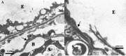
Fig. 3. (A) Haemocyte with reduced cytoplasm containing mature spores (S) contained with the peri-intestinal sinus (B) surrounded by a layer of host cells (*). The surrounding chloragocytes of the sinus are labelled with an arrow. Exterior of oligochaete indicated with (E). (B) Enlarged haemocyte containing mature spores (S) breaking through the cuticle of the oligochaete. Chloragocytes surrounding the haematocyte indicated with an arrow. Exterior of oligochaete indicated with (E).
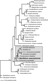
Fig. 4. Phylogenetic tree of microsporidia based on SSU rDNA using maximum likelihood. Bacillidium vesiculoformis is placed within the poorly resolved clade containing both bryozoan (e.g. Pseudonosema cristatellae) and oligochaete (Bacillidium sp.) parasites (highlighted box). The boot strap values (100 replicates) on the tree represent the percentage of bootstrap replicates that gave that topology. Neighbour-joining and parsimony analyses resulted in identical placement of B. vesiculoformis within the tree (data not shown).
 You have
Access
- 19
- Cited by