Introduction
Incudostapedial erosion following chronic otitis media accounts for up to 80 per cent of ossicular chain discontinuities. Since ossiculoplasty was first introduced in the 1950s, a number of materials have been used in attempts to bridge the incudostapedial gap and restore hearing.Reference Wullstein1 These materials include autografts using the patient's own ossicles or cortical bone, and prostheses made from biocompatible materials such as Teflon®, Plastipore™, titanium and gold.
In 1990, Geyer and Helm first reported the use of cement in middle-ear surgery.Reference Geyer and Helms2 Biocements have enabled the natural reconstruction of ossicles in an attempt to preserve the natural cantilever effect of the ossicular chain.Reference Maassen and Zenner3 The aim of this study was to determine the effectiveness of cement in rebridging isolated erosion of the incudostapedial joint.
Methods
A preliminary database search revealed that no previous reviews had been published on bone cements. Both authors performed a thorough search of the Embase, Medline and Pubmed databases from January 1998 to December 2012 using keywords and subject headings including the terms ‘ossiculoplasty’, ‘cement’, ‘incus’, ‘erosion’, ‘incudostapedial’ and ‘rebridging’. Inclusion criteria were papers published in English, case series or comparative studies with more than 10 patients, isolated incudostapedial erosion through chronic ear disease, mobile stapes and malleus, minimal data on air-bone gaps less than 20 dB (or less than 10 dB), and follow up for at least 1 year post-surgery. Exclusion criteria were traumatic dislocations, congenital malformations leading to fixation, cement used to secure prosthesis or bridge gaps between the malleus and stapes, and case studies. Additional papers were selected from the reference lists of shortlisted papers if they met inclusion criteria.
Statistical analysis
Statistical analysis was difficult to perform for a number of reasons. Only one paper described a prospective randomised study; the remaining studies were retrospective comparative studies and case series. In addition, inclusion criteria and follow-up periods varied among studies.
Results
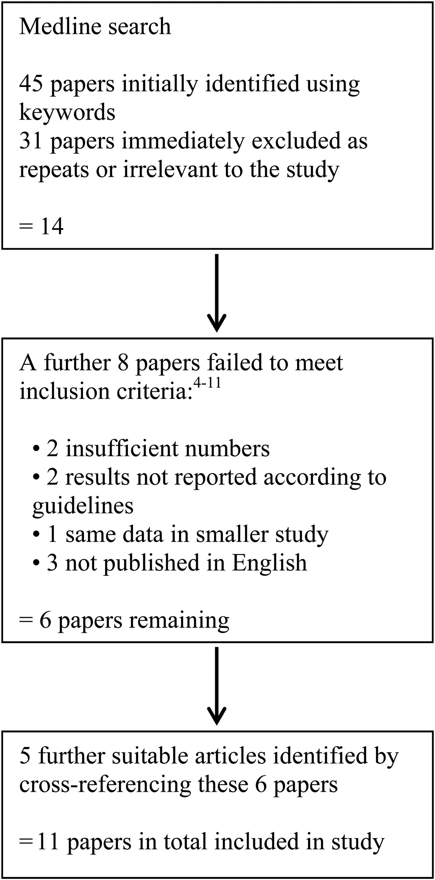
From the Medline search performed in December 2012, 6 papers were selected from a possible 45. Five further papers were selected from the reference lists of shortlisted papers. An identical search of Embase and Pubmed identified no new papers. Figure 1 shows a summary of the study selection process.
Flow chart demonstrating selection criteria for the study

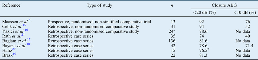
Ideally, we would have performed a formal meta-analysis of these studies. However, patient heterogeneity would have limited the conclusions; therefore, we performed a systematic review of the data. In a total of 110 patients in 2 retrospective comparative studies and 1 case series, rebridging ossiculoplasty was performed using hydroxyapatite cement.Reference Elsheikh, Elsherief and Elsherief12–Reference Babu and Seidman14 Closure of air-bone gaps of less than 20 dB and less than 10 dB were achieved in 80–94.4 per cent and 29–75 per cent of cases, respectively (Table I). Glass ionomer cement was used to perform rebridging ossiculoplasty in 318 patients in 3 comparative studiesReference Maassen and Zenner3, Reference Celik, Aslan Felek, Islam, Demirci, Samim and Oztuna15, Reference Yazici, Uzunkulaoglu, Emir, Kizilkaya, Dogan and Samim16 and 5 case series.Reference Baglam, Karatas, Durucu, Kilic, Ozer and Mumbuc17–Reference Rath, Bauer, Pytel, Vona, Szanyi and Lujber21 Closure of air-bone gaps less than 20 dB and less than 10 dB were achieved in 74–94 per cent and 40–76 per cent of cases, respectively. However, closure to less than 10 dB was only achieved in 121 patients (Table II).Reference Maassen and Zenner3, Reference Celik, Aslan Felek, Islam, Demirci, Samim and Oztuna15, Reference Bayazit, Ozer, Kanlikama, Durmaz and Yilmaz18, Reference Rath, Bauer, Pytel, Vona, Szanyi and Lujber21
Studies using hydroxyapatite cement to restore isolated incudostapedial erosion
ABG = air-bone gap
Studies using glass ionomer cement to restore isolated incudostapedial erosion
*Four patients had a gap between the malleus and stapes; †reported in paper as 86.7 per cent, but that was when two failures due to fractured cement were excluded; if included, the success rate dropped to 76.5 per cent. ABG = air-bone gap
Discussion
Since the introduction of ossiculoplasty in the late 1950s, numerous materials have been introduced in an attempt to restore hearing.Reference Wullstein1 These have included autogenous materials such as the patient's own incus and cortical bone. Although freely available, bone refashioning is time consuming, can result in necrosis if overheated and can displace ankylosed bone with surrounding bone. There is also a possibility of disease re-implantation if the squame is not fully removed. Alternatives such as partial ossicular replacement prostheses made of biocompatible materials are expensive, may require a large inventory and can be displaced and extruded.
Bone cements such as hydroxyapatite and glass ionomer cement have been used since the late 1980s as luting substances in dentistry.Reference LeGeros, Kijkowska and LeGeros22 They are cheap, easy to mix and start to set within a few minutes to form a putty-like substance that is relatively straightforward to work with. They are adhesive (provided all mucosa has been meticulously removed and surfaces are dry), restore the natural continuity of the ossicular chain, and instigate osseointegration and re-epithelialisation.
The disadvantages of both types of cement include fracturing leading to deterioration in hearing. With glass ionomer cement, foreign body reaction and neurotoxicity caused by the release of aluminium ions during setting has been reported. For this reason, glass ionomer cement is licensed for use in the middle ear only when no contact with the dura can be guaranteed. It has also been suggested that the facial nerve and round and oval windows can be protected with a topical haemostatic agent such as Spongostan™ to prevent potential paralysis or sensorineural hearing loss.
This systematic review identified only one prospective, randomised study: the others were retrospective comparative studies and case series. The severity of middle-ear disease resulting in incudostapedial joint erosion and the degree of erosion varied between papers. Although all studies used recognised guidelines for reporting outcomes, their follow-up periods varied. Such heterogeneity would limit the value of a meta-analysis of these data.
Nevertheless, in two retrospective comparative studies and one case series, 110 patients with chronic otitis media underwent closure of an isolated erosion of the incudostapedial joint using hydroxyapatite cement (Table II).Reference Elsheikh, Elsherief and Elsherief12–Reference Babu and Seidman14 Closure of air-bone gaps of less than 20 dB and less than 10 dB was achieved in 80–94.4 per cent and 29–75 per cent of cases, respectively. This was not significantly better than incus interposition in Somers and colleague's study,Reference Somers, Van Rompaey, Claes, Salembier, van Dinther and Andrzej13 but was significantly better (p < 0.001) than the partial ossicular prosthesis used by Elsheikh and colleagues.Reference Elsheikh, Elsherief and Elsherief12
Elsheikh and colleagues classified the gaps into two categories: those less than 2 mm and those more than 2 mm.Reference Elsheikh, Elsherief and Elsherief12 For defects greater than 2 mm, closure of the air-bone gap to less than 20 dB after one year occurred in 82 per cent of patients (and 29 per cent for air-bone gap less than 10 dB). For defects of less than 2 mm, closure of the air-bone gap to less than 20 dB occurred in 92 per cent of patients (75 per cent for air-bone gap less than 10 dB). However, there was no statistically significant difference between these groups (p > 0.5). There were no complications with the patient group with defects less than 2 mm; however, three patients in the group with defects greater than 2 mm (9 per cent) went on to develop recurrent conductive hearing loss (no reason given). Somers and colleagues did not quantify the size of the erosion, but did use 5-0 Vicryl wire (polygalactin 910) suture as a supporting framework for larger gaps.Reference Somers, Van Rompaey, Claes, Salembier, van Dinther and Andrzej13 No complications were described in the study.Reference Somers, Van Rompaey, Claes, Salembier, van Dinther and Andrzej13 Babu and colleagues classified defects into two categories: 1–2 mm and 2–3 mm.Reference Babu and Seidman14 In this study, only one patient with a defect greater than 3 mm had closure of an air-bone gap greater than 20 dB; however, this patient experienced a subjective improvement in hearing.
In 1 prospective and 2 retrospective comparative trials, a total of 68 patients underwent reconstruction of an isolated erosion of the incudostapedial joint using glass ionomer cement.Reference Maassen and Zenner3, Reference Celik, Aslan Felek, Islam, Demirci, Samim and Oztuna15, Reference Yazici, Uzunkulaoglu, Emir, Kizilkaya, Dogan and Samim16 Closure of the air-bone gap to less than 20 dB or to less than 10 dB was achieved in 78.6–94 per cent or 52–76 per cent of patients, respectively (Maassen and colleaguesReference Maassen and Zenner3 and Celik and colleaguesReference Celik, Aslan Felek, Islam, Demirci, Samim and Oztuna15 only). Closure of the air-bone gap was significantly better than that achieved by incus interposition (Maassen et al. p < 0.028; Yazici et al. p < 0.0001), but Maassen and colleagues found no difference in outcome between cement and Plester's gold-titanium-angled prosthesis.Reference Maassen and Zenner3, Reference Yazici, Uzunkulaoglu, Emir, Kizilkaya, Dogan and Samim16 Similarly, Celik and colleagues found no significant difference in closure of the air-bone gap using cortical bone.Reference Celik, Aslan Felek, Islam, Demirci, Samim and Oztuna15 Maassen and colleagues found that hearing results after creation of a neo-incudal joint using a droplet of saline on the stapes head were better than those after creation of a stiff joint.Reference Maassen and Zenner3 Notably, four previous cases with isolated incudostapedial joint erosion that were not included in the study had air-bone gaps greater than 20 dB post-surgery.
Neither Maassen and colleagues nor Yazici and colleagues quantified the size of the erosions, although in four patients from the latter study a large gap in the incus was rebridged between the malleus and stapes.Reference Maassen and Zenner3, Reference Yazici, Uzunkulaoglu, Emir, Kizilkaya, Dogan and Samim16 Air-bone gaps of less than 20 dB were present in 92 per cent of patients in Maassen and colleague's study and 78.6 per cent in Yazici and colleague's study.Reference Maassen and Zenner3, Reference Yazici, Uzunkulaoglu, Emir, Kizilkaya, Dogan and Samim16 In Celik and colleague's study, all patients had gaps of less than 2 mm, with air-bone gap of less than 20 dB in 94 per cent of patients. No complications were reported in all three studies.Reference Maassen and Zenner3, Reference Celik, Aslan Felek, Islam, Demirci, Samim and Oztuna15, Reference Yazici, Uzunkulaoglu, Emir, Kizilkaya, Dogan and Samim16
In a further 5 case series using glass ionomer cement (total of 250 patients) closure of the air-bone gap to less than 20 dB was achieved in 60–86.7 per cent.Reference Baglam, Karatas, Durucu, Kilic, Ozer and Mumbuc17–Reference Rath, Bauer, Pytel, Vona, Szanyi and Lujber21 Only 2 studies involving a total of 77 patients reported closure of the air-bone gap to less than 10 dB in between 40 and 71.4 per cent.Reference Bayazit, Ozer, Kanlikama, Durmaz and Yilmaz18, Reference Rath, Bauer, Pytel, Vona, Szanyi and Lujber21 Within this patient group, the erosion size was reported by Brask alone, as 1–2 mm.Reference Bayazit, Ozer, Kanlikama, Durmaz and Yilmaz18 Air-bone gap closure was less than 20 dB in 81.3 per cent.Reference Brask19
• Biocompatible bone cements are cheap and easy to use, with relatively few complications
• They can restore the natural continuity of the ossicular chain to maintain its cantilever effect
• Closure of air-bone gaps to less than 20 dB range is achieved in 74–94.4 per cent of patients (29–76 per cent for air-bone gaps less than 10 dB)
• Results are comparable with other methods used to restore isolated incus erosion
• Outcomes are better for gaps of less than 2 mm
The procedure failed in three patients as a result of fractured cement,Reference Hafiz20, Reference Rath, Bauer, Pytel, Vona, Szanyi and Lujber21 and transient grade 2 facial weakness resulted from haemorrhage and glass ionomer cement spreading to the middle-ear cleft.Reference Bayazit, Ozer, Kanlikama, Durmaz and Yilmaz18 No other complications were reported.
Conclusion
Our study indicates that cement is effective in restoring conductive hearing loss as a result of isolated incudostapedial erosion. The success rate for closure to less than 20 dB varies between 74 and 94.4 per cent, with success rates appearing to be higher for gaps equal to or less than 2 mm (81.3–94 per cent). It may therefore be helpful to form a neo-incudostapedial joint using saline to mimic the synovial joint between the incus and stapes.
Very few complications have been documented when ossicles have been prepared correctly and cement has been used according to the manufacturer's instructions, with precautions taken to protect neural tissue.
In the future, larger comparative prospective studies should include careful documentation of the length of eroded incus and types of reformation of the incudostapedial joint, with standardised reporting of results.