Introduction
Taenia taeniaeformis is a cestode of cosmopolitan distribution and parasitizes the small intestine of felids (Felis catus, F. rufus, F. silvestris) and dogs, with some spillover into foxes (Vulpes vulpes), coyote/wolf hybrids (Canis latrans × C. rufus) and occasionally birds and humans (Sterba et al., Reference Sterba, Blazek and Barus1977; Jones & Pybus, Reference Jones, Pybus, Samuel, Pybus and Kocan2001). Its infective form Cysticercus fasciolaris, also named Taenia crassicollis, Hydatigen fasciolaris, Strobilocercus fasciolaris and bladder worms, is commonly found encysted in the livers of rodents (Georgi & Georgi, Reference Georgi, Georgi, Georgi and Georgi1990; Hanes, Reference Hanes1995; Jones & Pybus, Reference Jones, Pybus, Samuel, Pybus and Kocan2001). The partially developed eggs (onchospheres) are released from the mature proglotids of adult worms and pass out with the faeces. When a viable onchosphere is ingested by a susceptible intermediate host, the egg membrane is removed during passage through the stomach or intestine and the larva migrates to the liver, usually to the surface, where it continues its development inside a cyst formed by the host connective tissue (Bowman et al., Reference Bowman, Lynn and Eberhard2003).
In intermediate hosts, such as rats and mice, the occurrence of T. taeniaeformis larvae can vary by locality (Kataranovski et al., Reference Kataranovski, Zolotarevski, Belij, Mirkov, Stošić, Popov and Kataranovski2010). In Brazil, Bonfim (Reference Bonfim2001), examined Rattus novergicus for C. fasciolaris, and recorded that 8% of animals were infected. In Egypt, Wanas et al. (Reference Wanas, Shehera and Rashed1993) found a wide range of C. fasciolaris prevalence (25–41%) in rodents (Rattus rattus, Mus musculus and Gerbillus gerbillus); however, in the USA low prevalences (6–18%) have been registered in rodents (McBee, Reference McBee1977; Lochmiller et al., Reference Lochmiller, Jones, Whelan and Kirkpatrick1982; Theis & Schwab, Reference Theis and Schwab1992).
In the Yucatan, there is no report of the presence of C. fasciolaris in animal populations. The aim of this study was to evaluate the occurrence of the larval cestode C. fasciolaris in rodent populations from the Cuxtal ecological reserve, Yucatan, Mexico.
Materials and methods
The study was carried out in the village of Molas located south-east of Merida, Yucatan, Mexico, which is located between 20°49′ and 20°48′ N and 89°37′ and 89°38′ W. Molas has a population of 1859 inhabitants, is located within the ecological reserve ‘Cuxtal’, and is surrounded by tropical forest.
In the village 40 households were randomly selected and sampled every month for three consecutive nights in two periods. The first period corresponded to the rainy season (October–December 2009) and the second to the dry season (February–April 2010). In each household, 12 Sherman live traps (7.5 × 23 × 9 cm, H.B. Sherman Traps Inc., Tallahassee, Florida, USA) were either placed both inside and outside in backyards, or only in backyards, depending on the authorization of the owners. The trapping effort for each period was 4320 nights/trap. All traps were baited with a mixture of vanilla and oats. Traps were inspected in the morning and collected animals were transported to the laboratory. For each animal examined, the trapping locality, conventional mammalian somatic measurements and sex were recorded. Animals were separated into two age–weight groups: juveniles ( < 9 g female, < 10 g male) and adults ( ≥ 9 g female, ≥ 10 g male) (Krebs et al., Reference Krebs, Kenney and Singleton1995).
All rodents were euthanized by cervical dislocation and necropsied for inspection of the liver for the presence of C. fasciolaris (larval stage). Larval capsules were opened through a small slit to release the parasites. Cestode parasites were recovered alive, counted and identified under a stereoscopic microscope. The collected cestodes were kept in 95% ethanol (Rodriguez-Vivas & Cob-Galera, Reference Rodríguez-Vivas and Cob-Galera2005) and their identification was based on Bowman et al. (Reference Bowman, Lynn and Eberhard2003).
Prevalence of C. fasciolaris infection in rodents was calculated. A primary screening of C. fasciolaris infection in mice was performed using 2 × 2 contingency tables of exposure variables (place of trap, season, sex of rodent and age of rodent). All variables with P < 0.20 were analysed by a logistic-binomial regression model using SPSS (version 13.0 K; SPSS Inc., Chicago, Illinois, USA), which provides exact regression estimates, 95% confidence intervals (95% CIs), odds ratio (OR: a measure of association which quantifies the relationship between the exposure variables and outcomes) and P values (regression coefficient).
Results
During the study 411 rodents were trapped, of which 322 were M. musculus (78.3%), 57 R. rattus (13.9%), 27 Peromyscus yucatanicus (6.6%), 3 Ototylomys phyllotis (0.7%), 1 Heteromys gaumeri (0.2%) and 1 Reithrodontomys gracilis (0.2%). From the 322 M. musculus trapped, 122 were females and 200 males, 67 were juveniles and 255 adults. One hundred and eleven individuals were trapped inside households and 211 in the backyards.
During necropsy, cestode cysts were found in the liver of rodents, and identified. All cysts were classified as the larval stage of T. taeniaeformis (Cysticercus fasciolaris, Rudolphi, 1808). Cysticercus cysts were white and barely involved the hepatic parenchyma. From the positive rodents, 26 (83.9%), 4 (12.9%) and 1 (3.2%) individuals had one, two and three cysts, respectively. Each cyst was characterized by a variably thick fibrous capsule surrounding one loosely coiled white C. fasciolaris with flocculent white to opalescent fluid. Different sizes of C. fasciolaris were removed from liver cysts (11 cm long prior to fixation, ranging from 3 to 17 cm). Most of the larvae were characterized by an extruded scolex with a long neck (strobila), pseudosegmentations along the length, and a relatively small terminal bladder. Cysticercus fasciolaris is unique in that the scolex is not invaginated into the bladder but attached to it by the strobila. The overall prevalence of rodents with C. fasciolaris cysts was 7.5% (31/411). Cysticercus fasciolaris cysts were observed in 29 M. musculus (9%) and two R. rattus (3.5%). Peromyscus yucatanicus, O. phyllotis , H. gaumeri and R. gracilis did not show cysts in their livers.
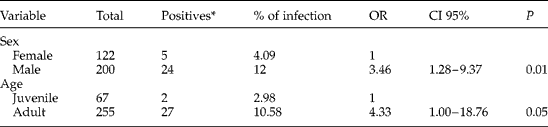
Out of the 322 M. musculus trapped, 4.09% (5/122) of females and 12% (24/200) males were positive for C. fasciolaris. Also, 2.9% (2/67) of juveniles and 10.5% (27/255) of adults were positive. Similarly, 9.9% (11/111) of rodents trapped inside houses and 8.5% (18/211) trapped in backyards were positive.
The χ2 analysis (univariate analysis) of the variables place of trapping and season had P values >0.2. The variables sex and age, with P value < 0.2, were included in the logistic regression (multivariate analysis). Table 1 shows the logistic regression analysis where sex and age variables were significant (P ≤ 0.05). Mus musculus males were 3.46 (OR value) times more likely to have C. fasciolaris than females. Also, M. musculus adults were 4.33 (OR value) times more likely to have the cestode compared with juveniles.
Results of logistic regression analysis of Mus musculus positive for liver cysts of Cysticercus fasciolaris in the community of Molas, Yucatan, Mexico.
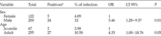
* Positive with one, two or three C. fasciolaris cysts; CI, confidence interval at 95%; OR, odds ratio; P probability value.
Discussion
Prevalence of T. taeniaeformis is considered higher in felids than in canids, and prevalence of C. fasciolaris differs in different rodent hosts, but generally is highest in rats, cotton rats and muskrats (Jones & Pybus, Reference Jones, Pybus, Samuel, Pybus and Kocan2001). However, Brandt & Sewell (Reference Brandt and Sewell1981) showed that T. taeniaeformis strains from Scotland, Belgium and Iraq were more infective for mice than rats, whereas the situation was reversed with a Malaysian strain. In the present study, mice (M. musculus) had a higher prevalence of infection (9.0%) when compared with rats (R. rattus) (3.5%). It has been reported that prevalence of the parasite in mice varies by locality and country (Kumar et al., Reference Kumar, Reddy, Aparna, Srinivas, Nagarajan, Venkatesan, Sreekumar and Sesikaran2006). The prevalence revealed by our study was similar to that found in rodents in the USA (6–18%) (McBee, Reference McBee1977; Lochmiller et al., Reference Lochmiller, Jones, Whelan and Kirkpatrick1982; Theis & Schwab, Reference Theis and Schwab1992); however, it was low when compared with reports from Egypt (25–41%) (Wanas et al., Reference Wanas, Shehera and Rashed1993) and Malaysia (95%) (Al-Salihi et al., Reference Al-Salihi, Sheikh and Saied2009).
Rodents are infected by food contaminated with cat faeces harbouring eggs of T. taeniaeformis. Two cycles can occur: an urban cycle that involves the domestic cat and house and field rodents; and a sylvatic cycle that involves wild felines and wild rodents (Taylor et al., Reference Taylor, Coop and Wal2007). Although in the present study no wild species of rodents were infected with C. fasciolaris, its presence in backyards of households in Molas could mean a potential risk of acquiring this infection, primarily by P. yucatanicus (6.56%), which was the most trapped wild species and endemic to the Yucatan peninsula.
Adult cestodes in the intestine of dogs and cats rarely cause serious disease, and clinical signs, if present, may depend on the degree of infection, age, condition and breed of the host (Kahn & Line, Reference Kahn and Line2005). The intermediate host shows more signs of illness than the definitive host (Hanes, Reference Hanes1995; Karimi et al., Reference Karimi, Chalechale, Bahiraie and Azadbakht2009). The majority of rodents with C. fasciolaris in this study had only one cyst. This finding is in agreement with those of Al-Salihi et al. (Reference Al-Salihi, Sheikh and Saied2009) who found that primary infection confers a strong immunity to rodents and it is unusual to find more than a few cysts in animals infected under natural conditions. However, Iwaki et al. (Reference Iwaki, Nonaka, Okamoto, Oku and Kamiya1994) suggest that the difference in infectivity (number of cysts in the liver) may be the result of genetic mutation of the cestode and is not host induced. Further studies would be required to test this hypothesis.
A study conducted by Mikesic & Drickamer (Reference Mikesic and Drickamer1992) demonstrated that the home-range size of mice (M. musculus) varies significantly between the sexes and increases with age. Home-range sizes of juveniles of the two sexes are nearly equal, but males increase home-range area after maturity. Most investigators examining movements of house mice report differences of home-range sizes between the sexes (e.g. Fitzgerald et al., Reference Fitzgerald, Karl and Moller1981; Mikesic & Drickamer, Reference Mikesic and Drickamer1992), with males almost invariably occupying larger home ranges than females. In polygynous mating systems, males presumably occupy large home ranges to increase their access to females. In our study, the large home ranges of adult males might explain the higher probability of infection with C. fasciolaris (males: OR 3.46, and adult: OR 4.33). These findings are in agreement with those of Lochmiller et al. (Reference Lochmiller, Jones, Whelan and Kirkpatrick1982), who found that prevalence of C. fasciolaris in Microtus pinetorum was greater in adults (14.3%) than in juveniles (2.7%), and Theis & Schwab (Reference Theis and Schwab1992), who found that prevalence was significantly different in young (1.2%) and adult (4.2%) Peromyscus maniculatus, but there were no significant differences in prevalence or intensity related to host sex. Theis & Schwab (Reference Theis and Schwab1992) suggested that differences may relate to different passive immunity in juveniles. Natural passive transfer of resistance to cestodes from mothers to their offspring has been shown to occur in both laboratory and domestic animals (Musoke et al., Reference Musoke, Williams, Leid and Williams1975). As examples, Rickard & Arundel (Reference Rickard and Arundel1974) showed that lambs were protected against Taenia ovis after suckling ewes which were either naturally or artificially immunized against this parasite. A study conducted by Musoke et al. (Reference Musoke, Williams, Leid and Williams1975) demonstrated that young rats born of mothers infected with T. taeniaeformis are immune to challenge infection and antibodies are primarily responsible for this protection. Also, Sheelagh & Soulsby (Reference Sheelagh and Soulsby1978) found that neonatal mice were protected against infection with T. taeniaeformis when fed purified colostral IgA and serum IgG from immune donors.
Adults of T. taeniaeformis have been recovered from the intestine of humans (Morishita & Sawada, Reference Morishita and Sawada1966; Bowman et al., Reference Bowman, Lynn and Eberhard2003) and C. fasciolaris infection in the liver of humans has been reported (Sterba & Barus, Reference Sterba and Barus1976; Sterba et al., Reference Sterba, Blazek and Barus1977); however, this cestode is considered to pose a low health risk.
From our study we can conclude that in the Yucatan, Mexico, the prevalence of C. fasciolaris is higher in M. musculus, and adult males had a higher probability of infection. Peromyscus yucatanicus is sylvatic but tends to visit households (Young & Jones, Reference Young and Jones1983), so, although no parasites were found, the presence of this rodent could pose a risk as it may be, or become, a reservoir of infection.
Acknowledgements
This project was founded by Promep-Mexico (Red epidemiológica de enfermedades zoonóticas y transmitidas por vectores de importancia en salud pública). We would like to thank Dr Jessica Malakovski and Susana Mendez for the critical review of this paper.