Introduction
Bilateral conductive hearing loss is common. It may be congenital in the form of bilateral microtia, or acquired as in chronic suppurative ear disease.Reference Tran, Vrabec, Manolidis and Coker1 Chronic suppurative ear disease can result in damage to the ossicular chain, including necrosis, discontinuity and fixation.Reference Carter and Hoff2 Where ossicular reconstruction is unsuccessful or not possible, bilateral conductive hearing loss ensues. Rehabilitating the hearing under such circumstances can be challenging, and could involve ossiculoplasty, conventional air conduction hearing aids or bone conduction hearing aids.Reference Baumgartner, Hamzavi and Boheim3 Bone conduction implants have also shown promise in this area.Reference Bravo-Torres, Der-Mussa and Fuentes-López4 Ensuring access to sound by stimulating both cochleae aids sound localisation and hearing in background noise.Reference Hassepass, Bulla, Aschendorff, Maier, Traser and Steinmetz5
The Bonebridge implant system (Med-El, Innsbruck, Austria) is a bone conduction hearing implant indicated in the treatment of conductive hearing loss.Reference Zernotti and Sarasty6 A retrospective study of 24 consecutive Bonebridge patients demonstrated an average functional hearing gain of 28.8 dB, and a statistically significant increase in monosyllabic word scores at 65 dB in quiet conditions from 4.6 to 53.7 percentage points.Reference Riss, Arnoldner, Baumgartner, Blineder, Flak and Bachner7 Similarly, a multi-centre prospective study of 12 German-speaking adults with conductive or mixed hearing loss demonstrated an average improvement of speech perception, as measured by word recognition scores and a speech recognition threshold at 50 per cent, of 78.8 and 25 dB HL, respectively, at 3 months after implantation.Reference Sprinzl, Lenarz and Ernst8 However, there are few reports of implantation in the paediatric population and no reports of bilateral simultaneous asymmetrical implantation of the Bonebridge in the paediatric population.Reference Baumgartner, Hamzavi and Boheim3
We present the first published case of simultaneous asymmetrical Bonebridge implantation in a child, with the right transducer implanted in the sinodural angle and the left transducer in the middle cranial fossa.
Case presentation
An 11-year-old girl presented with bilateral conductive hearing loss secondary to chronic middle-ear disease. There was a history of tympanosclerosis, fluctuating middle-ear effusions and ossicular fixation.
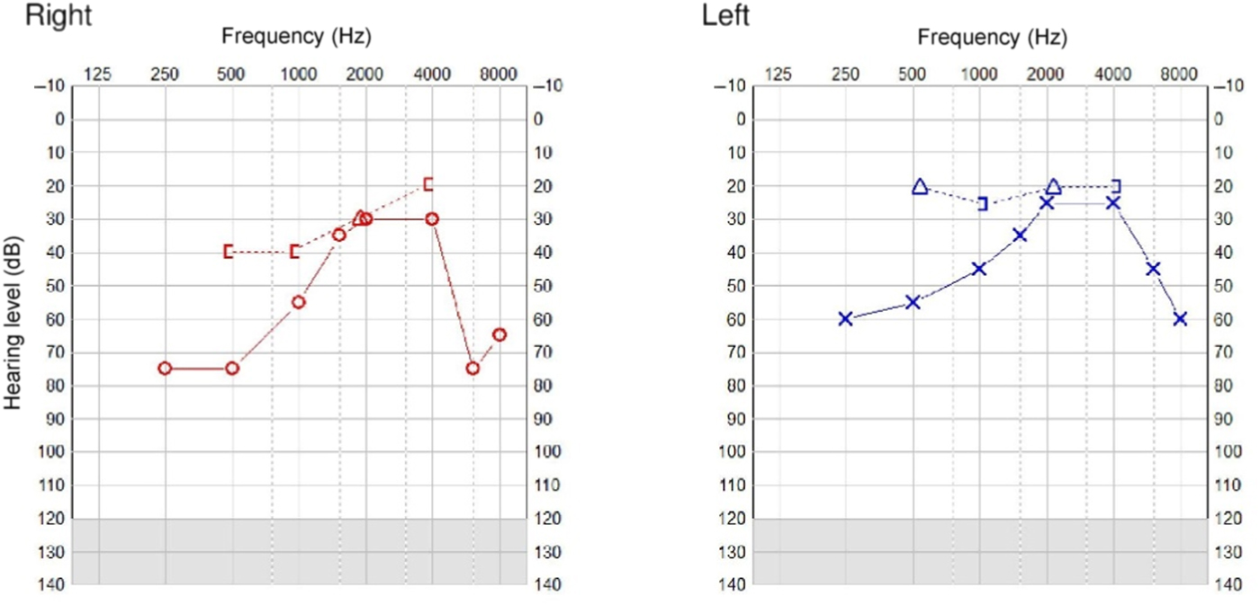
Pure tone audiology showed a bilateral moderate to severe conductive hearing loss and a maximal air–bone gap of 25 dB bilaterally, as shown in Figure 1. Her unaided speech testing score using the Manchester Junior Word List was 60 per cent at 55 dB HL without lip-reading. Her aided speech testing score without lip-reading was 100 per cent at 55 dB HL, but the patient reported poor compliance with air conduction hearing aids because of discomfort.
Pre-operative unaided pure tone audiograms. [ = bone conduction mastoid masked (right); Δ = air conduction masked (right);○ = air conduction unmasked (right); ] = bone conduction mastoid masked (left); × = air conduction unmasked (left)

Temporal bone computed tomography showed bilateral prominent sigmoid sinuses, with the left side being dominant. Pre-operative planning was performed using the BB FastView software provided by Med-El. There was sufficient space for the Bonebridge transducer in the right sinodural angle, but insufficient space in the left sinodural angle or retrosigmoid region because of the size of the sigmoid sinus and small temporal bone. The only region in which there was sufficient bone to accommodate the transducer was in the left middle cranial fossa.
The surgery was performed under general anaesthesia via bilateral post-auricular incisions. The procedure was uneventful.
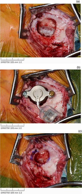
Specifically, a superiorly based periosteal flap was created and the appropriate beds were drilled to house the transducer (Figure 2). On the right side, where the bone conduction floating mass transducer was placed in the sinodural angle, the sigmoid sinus was prominent as expected and was visualised during the surgery. The sinus was skeletonised, with an ‘eggshell’ of bone left on the great vessel. The transducer was positioned, aided with 3 mm and 4 mm lifts to avoid compression of the sinus (Figure 2a,b). On the left side, the bed for the bone conduction floating mass transducer was drilled, with exposure of dura. The dura was preserved and not breeched. Two 2 mm lifts were used to avoid dural compression. Despite the different anatomical positions of the bone conduction floating mass transducer, the subcutaneous magnets were positioned symmetrically in light of the flexible connector between the bone conduction floating mass transducer and the demodulator, which allows up to 90 degrees of bend.
Intra-operative photographs of: (a) right sinodural angle, with skeletonised sigmoid sinus visible; (b) right-sided transducer in situ, with lifts to avoid compression of the sigmoid sinus; and (c) drilled left middle cranial fossa transducer bed.

The patient was discharged home the same day. There were no peri-operative complications.
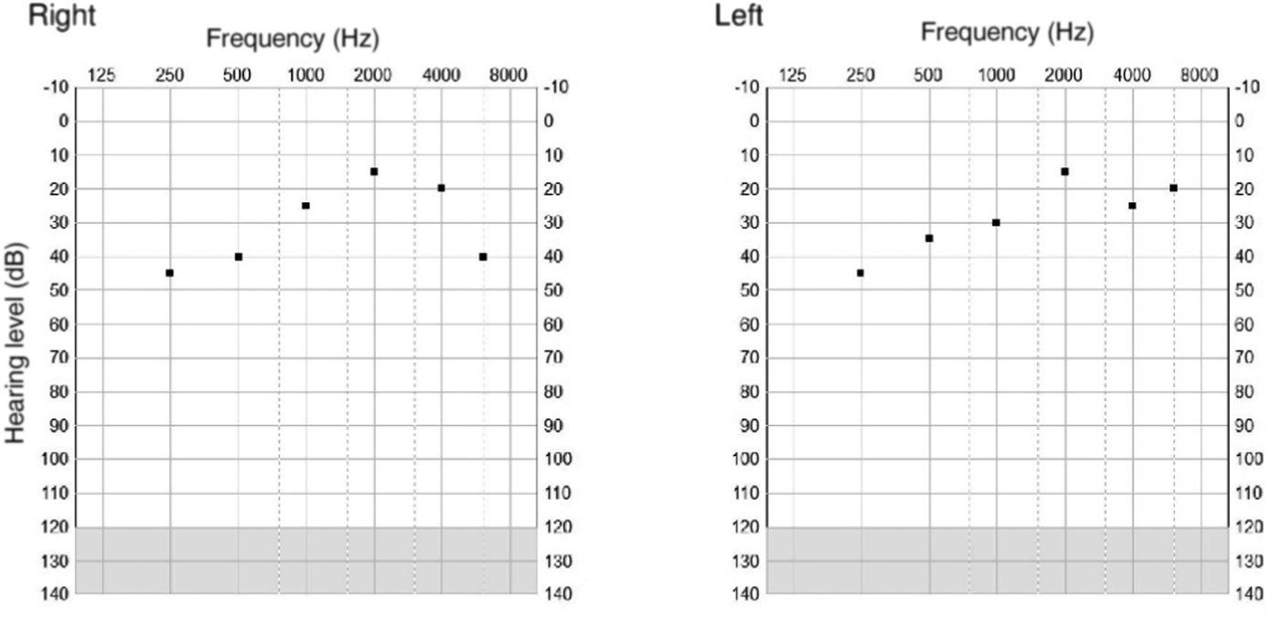
The device was activated five weeks after the surgery with no complications. Aided sound-field testing conducted nine months after surgery demonstrated hearing thresholds of 15–45 dB bilaterally, as shown in Figure 3. The speech testing score was 100 per cent at 55 dB HL.
Aided sound-field testing conducted nine months after surgery.

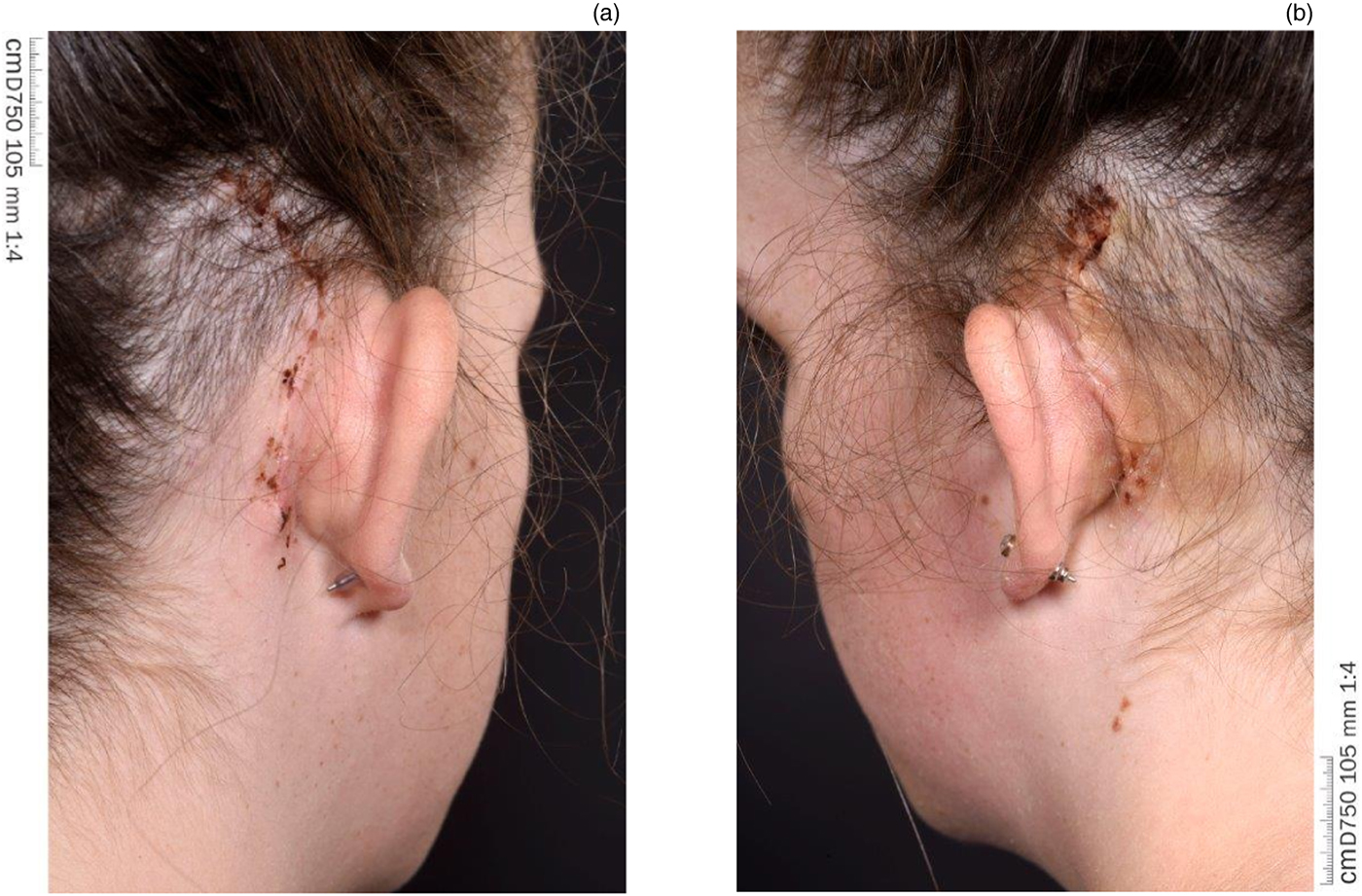
There was a good cosmetic outcome, with minimal projection of the internal component (Figure 4).
Post-operative clinical photographs taken four weeks after surgery, showing the post-auricular surgical wounds on the (a) right and (b) left sides.

The patient was delighted with the improved sound quality compared with her pre-operative air conduction hearing aids. In addition, she finds the external Bonebridge device more comfortable to wear, and compliance has improved significantly.
Discussion
Bonebridge implantation is becoming increasingly popular given the transcutaneous nature of its activity. The implant avoids the well-documented skin complications associated with conventional percutaneous systems.Reference Ricci, Volpe and Faralli9
In order to successfully implant the device, there needs to be sufficient temporal bone into which the transducer should be housed.Reference Rahne, Schilde, Seiwerth, Radetzki, Stoevesandt and Plontke10 This can be challenging in children who have smaller temporal bones and in those who have had previous mastoid surgery. Med-El recommend that the transducer is secured within the sinodural angle or within the retrosigmoid region.
In the case presented here, based on the surgical planning using the company supplied software, it was only deemed possible to position the right transducer in the sinodural angle and the left transducer in the middle cranial fossa.
In this case, other surgical bone conduction implants were discussed. The percutaneous bone-anchored hearing aids (BAHA; Cochlear, Sydney, Australia) were not deemed acceptable to the patient. In addition, round window vibroplasty was not recommended because of fluctuating middle-ear effusions and recurrent episodes of acute otitis media.
Post-implantation speech testing demonstrated an improvement in speech score using the Manchester Junior Word List that was comparable to wearing bilateral BAHA softbands (Cochlear). She attained a score of 100 per cent at 55 dB HL. The patient reported an improvement in sound quality compared with her air conduction hearing aids, although this difference was not measurable. Most significantly, she finds the Bonebridge more comfortable to wear and therefore compliance has improved.
Conclusion
Bilateral asymmetrical simultaneous Bonebridge implantation is safe and can be successful in the paediatric population. Appropriate pre-operative planning is necessary to ensure that sufficient temporal bone is present to house the transducer. Positioning the transducer in different anatomical sites appears not to be detrimental to the overall hearing outcome. If possible, care should be taken to ensure symmetry of the magnet, by utilising the flexibility of the region between the transducer and demodulator, which may be manipulated up to 90 degrees.
Competing interests
None declared