Introduction
Lice in the genus Mulcticola infest members of the Caprimulgidae (Aves: Caprimulgiformes), the nighthawks and nightjars (Price et al. Reference Price, Hellenthal, Palma, Price, Hellenthal, Palma, Johnson and Clayton2003). This group of chewing lice includes 18 species, infesting 24 host species, in 25 countries (Valim and Kuabara Reference Valim and Kuabara2015). The genus has attracted recent attention taxonomically (Valim and Kuabara Reference Valim and Kuabara2015) and ecologically (Galloway Reference Galloway2006; Galloway and Lamb Reference Galloway and Lamb2015). Valim and Kuabara (Reference Valim and Kuabara2015) reviewed the neotropical species, described five new species, and provided a catalogue for all Mulcticola species, while Galloway (Reference Galloway2006) and Galloway and Lamb (Reference Galloway and Lamb2015) calculated infestation parameters (prevalence and intensity) and population variability for one species, Mulcticola macrocephalus (Kellogg) (Phthiraptera, Ischnocera: Philopteridae), infesting the common nighthawk, Chordeiles minor (Forster) in Manitoba, Canada. In the course of the ecological studies in Manitoba, large numbers of lice were collected, including all three immature stages. This species of chewing louse was originally described by Kellogg (Reference Kellogg1896), but many descriptive details for M. macrocephalus were not provided at that time. The abundance of specimens collected from common nighthawks in Manitoba offered the opportunity to redescribe the adults of M. macrocephalus, as well as the three juvenile instars.
Material and methods
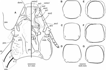
Lice were collected from common nighthawks that had been submitted to two wildlife rehabilitation hospitals in Manitoba, the Wildlife Haven (previously Manitoba Wildlife Rehabilitation Organization) and the Prairie Wildlife Rehabilitation Centre. Nighthawks that died or were euthanised were individually frozen at –20 °C for at least 48 hours to kill all ectoparasites. Birds were washed three times to remove ectoparasites, and samples were passed through a 90-μ sieve. Lice were handpicked from samples and preserved in 70% or 95% ethanol. All specimens from the current descriptive study are deposited in the Museu de Zoologia da Universidade de São Paulo (MZUSP), Brazil, with a number associated as a unique identifier from the SophiA® database for the Phthiraptera collection. Additional specimens are deposited in the J.B. Wallis/R.E. Roughley Museum of Entomology, Department of Entomology, University of Manitoba, Winnipeg, Manitoba, Canada. Lice were permanently slide mounted in Canada balsam, following the technique in Palma (Reference Palma1978). The terminology of body features and chaetotaxy follows that used by Valim and Kuabara (Reference Valim and Kuabara2015; see also Fig. 4).
All measurements are in millimetres and identified by the following abbreviations: ADHPL – anterior dorsal head plate length; ADHPW – anterior dorsal head plate width; AW – abdominal width (at the level of segment V); GL – male genitalia length; HL – head length (at midline and including the hyaline margin); PAW – preantennal width (at the level of preconal setae); PL – paramere length; PTW – pterothorax width (at its posterior level); PW – prothorax width; TL – total length; TW – temporal width (at the level of marginal temporal setae 2 – mts2). The cephalic index measure is calculated by the head length divided by head width.
Systematics
Phthiraptera Haeckel, Reference Haeckel1896
Ischnocera Kellogg, Reference Kellogg1896
Philopteridae Burmeister, Reference Burmeister1838 (sensu lato)
Mulcticola Clay et Meinertzhagen, Reference Clay and Meinertzhagen1938, p. 279
Mulcticola macrocephalus (Kellogg, Reference Kellogg1896) (See Figs. 1–4)
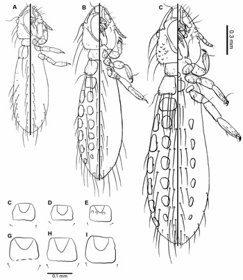
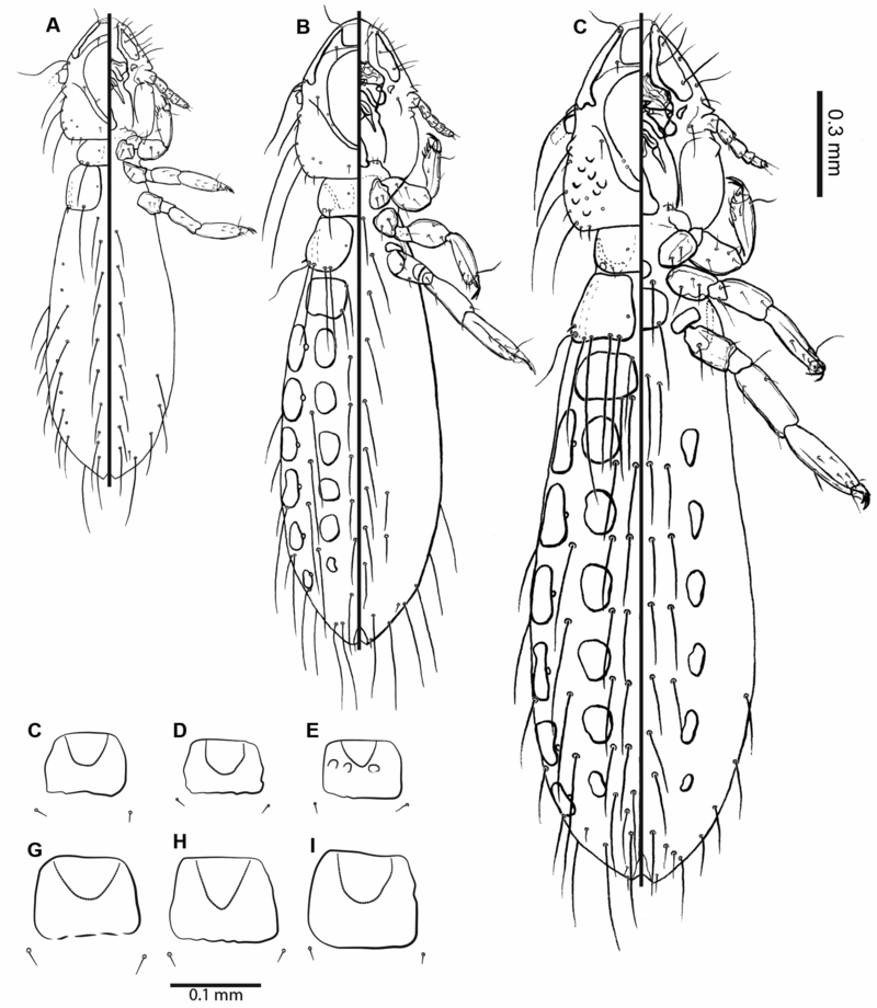
Nymphal habitus of Mulcticola macrocephalus, in dorsoventral views: A, nymph I; B, nymph II; C–E, anterior dorsal head plate from three specimens of nymph II; F, nymph III; G–I, anterior dorsal head plate from three specimens of nymph III.
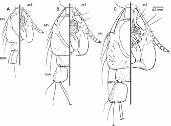
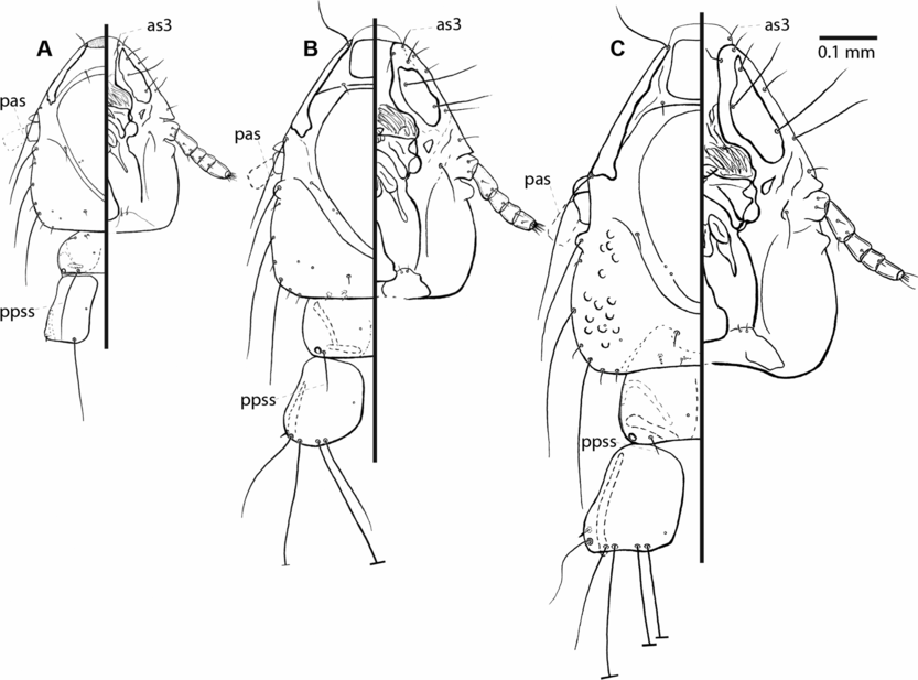
Head in dorsoventral views and thorax in dorsal view of Mulcticola macrocephalus: A, nymph I; B, nymph II; C, nymph III. as, anterior seta; pas, preantennal seta; ppss, prothoracic postspiracular seta.
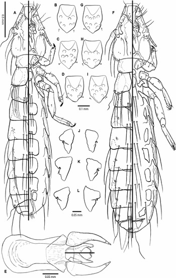
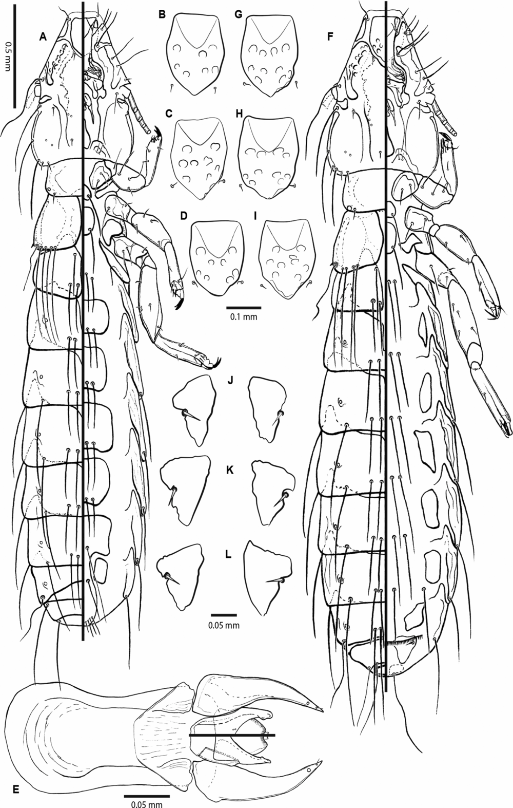
Adults of Mulcticola macrocephalus (Kellogg, Reference Kellogg1896) in dorsoventral views: A, male habitus; B–D, anterior dorsal and ventral head plates of males from three specimens; E, male genitalia with mesosome; F, female habitus; G–I, anterior dorsal and ventral head plates of females; J–L, female subvulvar plates from three specimens.
Mulcticola macrocephalus (Kellogg, Reference Kellogg1896): A, male head in dorsoventral views; B–D, female metasternal plates from three specimens; variation of three specimens; E–G, male metasternal plates from three specimens, Mulcticola nacunda Carriker, Reference Carriker1945 (dotted lines). ads, anterior dorsal seta; as, anterior seta; avs, anterior ventral seta; dsms, dorsal submarginal seta; mds, mandibular seta; mts, marginal temporal seta; os, ocular seta; pas, preantennal seta; pcs, preconal seta; pns, postnodal seta; pos, preocular seta; pts, postemporal seta; vsms, ventral submarginal seta.

Lipeurus macrocephalus Kellogg Reference Kellogg1896, p. 504, type host Chordeiles virginianus henryi (=C. minor henryi).
Nirmus tyrannus: Osborn Reference Osborn1896, p. 228, ex Tyrannus sp. (= error) (junior synonym, see Emerson Reference Emerson1960, p. 162).
Mulcticola macrocephala: Carriker Reference Carriker1947, p. 136 (new combination and unjustified emendation).
Mulcticola macrocephalus: Hopkins and Clay Reference Hopkins and Clay1952, p. 227 (checklist); Carriker Reference Carriker1957, p. 103 (lectotype designation); Emerson and Elbel Reference Emerson and Elbel1957, p. 423 (illustrations of anterior dorsal head plate and male genitalia); Emerson Reference Emerson1972, p. 96 (list of North American species); Price et al. Reference Price, Hellenthal, Palma, Price, Hellenthal, Palma, Johnson and Clayton2003, p. 198 (checklist); Galloway Reference Galloway2006, p. 7 (biological and parasitological indices); Valim and Kuabara Reference Valim and Kuabara2015, p. 12 (catalogue of species); Galloway and Lamb Reference Galloway and Lamb2015, p. 725 (ecology).
Type locality: Palo Alto, California, United States of America
Descriptions of preimaginal stages
Nymph I. General habitus as in Fig. 1A; details of head and thoracic chaetotaxy as in Fig. 2A; cephalic ratio 1.3–1.4, with marginal carina medially interrupted, region smooth and distinctly sclerotised, without an anterior dorsal head plate. Without visible attachment sites for cibarial muscles on the head capsule. The as3 present, hyaline margin completely absent; pas, os, mts1 and mts3 very long; ads set on sclerotised part anterior to ecdysial line. Prothorax with postspiracular setae very long, reaching posterior margin of pterothorax; chaetotaxy of pterothorax, each side: one short, spine-like posterolateral seta plus one long posterior seta. Meso- and metasternal plates without sclerotisation or setae. Abdomen membranous, without sclerotised plates. Tergal chaetotaxy (each side): II with one anterior seta; II–VII, IX with one posteromedial seta; VIII with one short and one long seta; IV–VIII with postspiracular setae. Sternal chaetotaxy (each side): II–VIII with one medial seta; IX with one long and one short seta. Pleural chaetotaxy (each side): IV–V one seta; VI–VII two setae; IX–X one seta. Measurements (n = 9): HL 0.33–0.35; PAW 0.21–0.24; TW 0.25–0.27; PW 0.16–0.20; PTW 0.19–0.25; AW 0.23–0.33; TL 1.00–1.29.
Nymph II. General habitus as in Fig. 1B; details of head and thoracic chaetotaxy as in Fig. 2B; cephalic ratio 1.3, with marginal carina medially interrupted and developed anterior dorsal head plate posteriorly still fused with the head surface. Three variations of the anterior dorsal head plate are presented in Fig. 1C–E. Attachment sites for cibarial muscles indistinct, aspect of anterior dorsal head plate smooth. The as3 present and set on hyaline margin; pas, os, mts1 and mts3 very long; ads set on sclerotised part of head posterior to the anterior dorsal head plate. Prothorax (each side) with postspiracular setae intermediate between very long in nymph I and very short in nymph III; chaetotaxy of pterothorax, each side: one spine-like ventral seta, one trichoid lateral seta, one long posterolateral seta, plus two long posteromedial setae. Chaetotaxy of pterothorax similar to that of imago, less one long seta on posterolateral margin of metathorax. Meso- and metasternal plates without sclerotisation but with one pair of medium-long setae on each segment. Abdomen membranous but with sclerotised pleural and tergal plates. Segment II with one subquadrate tergopleural plate; segments III–VIII with distinct, square-shaped tergal plates fused with pleural ones. Tergal chaetotaxy (each side): II with one anterior seta; II–VIII with one posteromedial seta; IV–VII with postspiracular setae; IX with two long setae. Sternal chaetotaxy (each side): II–VIII with one medial seta; VI–VII with additional pair of setae; IX with two long setae. Pleural chaetotaxy (each side): IV–V one seta; VI–VIII two setae; IX–X one seta. Measurements (n = 6): HL 0.39–0.46; PAW 0.27–0.32; TW 0.30–0.36; ADHPW 0.08–0.09; PW 0.20–0.26; PTW 0.29–0.36; AW 0.34–0.46; TL 1.46–1.79.
Nymph III. General habitus as in Fig. 1F; details of head and thoracic chaetotaxy as in Fig. 2C; cephalic index 1.3, with marginal carina medially interrupted and developed anterior dorsal head plate, posteriorly fused with the head surface. Three variations of the anterior dorsal head plate are presented in Fig. 1G–I. Attachment sites of cibarial muscles clearly discernible on the anterior dorsal head plate. The as3 present and set on hyaline margin; pas, os, mts1 and mts3 very long; ads set on the soft integument of preantennal suture, posterior to the anterior dorsal head plate. Prothorax with postspiracular setae very short, as in imago; chaetotaxy of pterothorax as in imago, with four long posterior setae plus spine-like and trichoid setae. Meso- and metasternal plates with sclerotisation and with one pair of medium-long setae on each segment. Abdomen membranous but with sclerotised pleural, tergal, and sternal plates. Tergal plates as described for nymph II, sternal plates tear-shaped and present on segments III–VIII. Tergal chaetotaxy (each side): II with one anterior seta; II–IX with two posteromedial setae; VIII with one additional seta; IV–VII with postspiracular setae. Sternal chaetotaxy (each side): II–VI with two medial setae; VII–VIII with one seta each; X with one long, two short, plus one long setae. Pleural chaetotaxy (each side): IV–V one seta; VI–VII two setae; VIII three (rarely two) setae; IX–X two setae. Measurements (n = 7): HL 0.54–0.58; PAW 0.37–0.42; TW 0.42–0.46; ADHPW 0.10–0.11; PW 0.29–0.32; PTW 0.39–0.43; AW 0.44–0.61; TL 1.94–2.37.
Redescription of the imago
Head. Triangular, sub-truncate anteriorly, longer than wide, cephalic index 1.2 in both sexes, rarely 1.3 (Fig. 4A). The short clypeal region, narrow hyaline margin, rounded sides, and slightly rounded to straight in front. Anterior dorsal and ventral head plates of characteristic shape, with distinct attachment sites for cibarial muscles on dorsal plate (Fig. 4A); the ratio of anterior head plate 1.3, rarely 1.2 in some females. Coni triangular in shape, short, not reaching the posterior margin of scapus. Postnodal seta short. Eye small, rounded, and prominent. Temples slightly extended, marginal temporal carina narrow. Posterior margin of occipital edge concave, with pts and sensilla s1–s4 present on each side.
Thorax. Entire thorax is shorter than the head. Prothorax trapezoidal, pterothorax also as a larger trapezium, about the same width as the head. Meso- and metasternal plates distinctly sclerotised, each with one pair of medium-long setae, latter plate of diagnostic shape, nearly square (Fig. 4B–G).
Abdomen. Elongated, with the widest segment at V (Fig. 3A, F). The tergal plate of segment II laterally fused with pleura and entirely divided medially, with four posterior setae, plus one pair of anterior setae. Segments III–VII medially entire and fused to pleura laterally, forming a tergo-pleurite. Postspiracular setae set dorsally each side on IV–VII, segment VIII with trichoid setae set posterolaterally; sensilla accessory to postspiracular setae on IV–V. Posterior setae on tergo-pleurites III–VII 4 (rarely 5) and VIII 4 (rarely 2–3); anterior margin of tergal plates III–V (6) or III–VI (6) with a median notch, gradually narrowing into posterior segments. Tergite VIII fused medially, but in males medially narrower than in III–VII. Pleural bands extended into the preceding segment, forming well-defined “re-entrant heads” on III–VIII; pleural chaetotaxy: 0 on II–III; 2 (rarely 1 in one side) setae on IV–V; 3 (rarely 2) setae on VI–VIII. Sternal chaetotaxy: II with 4 (rarely 3 or 5) setae; III–VI with 3–6 setae (but mode and median 4 setae). Sternites dimorphic, ovoid, and set laterally on each side of abdominal segments III–VII in females and roughly rectangular in shape and set across each segment III–VI in males.
Male. General habitus as in Fig. 3A. Subgenital plate with 4 (rarely 5) setae. Copulatory apparatus of male (Fig. 3E) typical for genus; endomeral plate broad, its lateral arms characteristic (Fig. 3E) with pointed tips dorsally and slightly broad on ventral side. Ventrally one pair of small sensilla medial on each side of sclerotised part of endomera. Measurements (n = 3): HL 0.68–0.72; PAW 0.45–0.48; TW 0.55–0.60; ADHPL 0.19–0.21; ADHPW 0.16–0.17; PW 0.38–0.41; PTW 0.50–0.53; AW 0.64–0.74; GL 0.34; PL 0.13–0.14; TL 2.93–3.08.
Female. General habitus as in Fig. 3F. One pair of female subvulvar sclerites well developed and of characteristic shape, with one spine-like marginal seta on each plate (Fig. 3J–L). Subgenital plate with 4–6 setae each side (but mode and median 6 setae); vulvar margin with 12–19 thin and 14–27 spine-like setae, plus 10–17 small setae anterior to the vulvar margin. Measurements (n = 9): HL 0.67–0.76; PAW 0.44–0.50; TW 0.55–0.63; ADHPL 0.18–0.22; ADHPW 0.14–0.18; PW 0.39–0.44; PTW 0.51–0.58; AW 0.66–0.77; TL 2.87–3.23.
Material examined: Canada. Manitoba: 2♂, 6 NI, 3 NII, 5 NIII (MZUSP 7585–7596), ex Chordeiles minor minor (Forster, JR, 1771) (case no.: CONI/1257/CEN/02), Winnipeg (49°53′24″ N, 97°8′36.96″ W), 6.IX.2002, coll. T.D. Galloway/D. Holder. 5♀, 3 NI, 2 NII, 1 NIII (MZUSP 7597–7611), ex C. m. minor (CONI/1280/CEN/02), Winnipeg (49°53′24″ N, 97°8′36.96″ W), 10.IX.2002, coll. T.D. Galloway/D. Holder. 1♂, 4♀, 1 NII, 1 NIII (MZUSP 7612–7618), ex C. m. minor (CONI/819/CEN/14), Grande Pointe (49°46′0.12″ N, 97°3′2.30″ W), 21.VIII.2014, coll. T.D. Galloway/E.N. McNally.
Remarks
Mulcticola macrocephalus is morphologically close to M. nacunda Carriker, Reference Carriker1945 by the shape of the anterior dorsal head plate, based on its dimorphic sternal plates and the chaetotaxy of abdominal tergites and sternites. They can be distinguished by several characteristics: the length-to-width ratio of the head is slightly greater in M. macrocephalus (1.2–1.3 versus 1.1); the metasternal plate tends to be narrower in M. macrocephalus (Fig. 4E–G); the inner margins of each subvulvar plate are roughly straight to slightly undulate and the antero-medial angles are blunt in females of M. macrocephalus, whereas in M. nacunda, these margins are concave and terminate in a prolonged antero-medial extension (Valim and Kuabara Reference Valim and Kuabara2015); there are no differences in the tips of the endomeral arms in the male genitalia, at least dorsally, but the shape of the mesosome is distinctive.
Both species are similar in size, but males of M. macrocephalus tend to have the head (HL: 0.68–0.72 versus 0.64–0.67) and anterior dorsal plate (ADHPL: 0.19–0.21 versus 0.17–0.18) longer than in M. nacunda, whereas its male genitalia are smaller (GL: 0.34 versus 0.35–0.36; PL: 0.13–0.14 versus 0.16–0.17).
The third instar of M. nacunda was described by Valim and Kuabara (Reference Valim and Kuabara2015), so a direct comparison between the preimaginal forms of these two species is possible. Both species present the most conservative characters of the genus (see Valim and Kuabara Reference Valim and Kuabara2015), in that they differ by the thickness of the dorsal marginal carina, being thinner in M. macrocephalus; the shape of the dorsal anterior head plate differs, its posterior margin being more defined and straight in M. macrocephalus, whereas in M. nacunda, there is a less sclerotised area forming a posterior medial notch. Regarding the generic features of preimagos, it is important to stress that only the nymphs II of M. piacentinni Valim et Kuabara and M. bacurau Valim et Kuabara are known. The postspiracular prothoracic setae are short in nymph III of M. bacurau, medium length in specimens of M. macrocephalus (Fig. 2B), and neither very long as in nymph I (M. piacentinni, M. bacurau, M. nacunda, M. macrocephalus) nor very short as in nymph III (aforementioned species, except M. piacentinni, which is unknown).
Discussion
The nymphs of lice have seldom formed the focus for descriptive analysis or subsequent application for the study of phylogenetic relationships, as has been done for some other groups of insects (e.g., the Dytiscidae (Coleoptera): Nilsson Reference Nilsson1989; Alarie Reference Alarie1991). Some of the early work of Ferris (Reference Ferris1951), Pratt and Karp (Reference Pratt and Karp1953), and Cook and Beer (Reference Cook and Beer1959) led to descriptions of the nymphs of many species of sucking lice (Phthiraptera, Anoplura) by Kim (e.g., Kim Reference Kim1965, Reference Kim1966a, Reference Kim1966b). Price and Hellenthal (Reference Price and Hellenthal1996) and Page et al. (Reference Page, Price and Hellenthal1995) described the complex relationships among chewing lice (Phthiraptera: Trichodectidae) infesting pocket gophers (Rodentia: Geomyidae) in the New World using characters of first-instar nymphs of Geomydoecus Ewing and Thomomydoecus Price et Emerson species. Descriptions for nymphs of some other species of chewing lice parasitic on birds are available (e.g., Ansari Reference Ansari1954; Agarwal Reference Agarwal1967; Modrzejewska and Złotorzycka Reference Modrzejewska and Złotorzycka1987; Abrahamovich and Cicchino Reference Abrahamovich and Cicchino1988; Mey Reference Mey1994; Cicchino and Mey Reference Cicchino and Mey2007; Agarwal et al. Reference Agarwal, Ahmad, Rashmi, Arya, Bansal and Saxena2011; Singh et al. Reference Singh, Khan, Kumar and Gupta2018, Reference Singh, Gupta, Khan, Kumar and Ahmad2019), but to date, their scope and diversity have limited such broad analyses as undertaken by Price and Hellenthal (Reference Price and Hellenthal1996) and Page et al. (Reference Page, Price and Hellenthal1995) for mammal hosts.
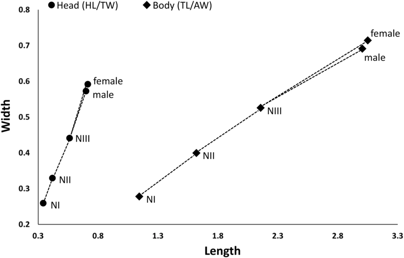
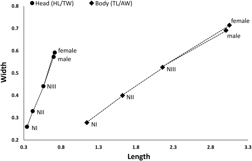
Mulcticola macrocephalus is the first species of the genus for which all preimaginal stages are fully described. First-instar nymphs have no tergal or sternal plates on the abdomen, second-instar nymphs present roughly rounded lateral and medial tergal plates, whereas third nymphs are similar to second instars but with distinct sternal plates in each side of segments III–VIII. Regarding their growth, we had fewer specimens for statistical analyses than employed by Kéler (Reference Kéler1952) and Lonc and Modrzejewska (Reference Lonc and Modrzejewska1986, Reference Lonc and Modrzejewska1989). However, the analysis of the ratio between the length and width of constant structures such as head and whole body show results similar to those achieved by those authors (Kéler Reference Kéler1952; Lonc and Modrzejewska Reference Lonc and Modrzejewska1986, Reference Lonc and Modrzejewska1989). Our plot of these ratios (Fig. 5 ) confirms that the dimensions of the head and body of chewing lice are good diagnostic characters to determine each nymphal stage (Lonc and Modrzejewska Reference Lonc and Modrzejewska1986, Reference Lonc and Modrzejewska1989; Modrzejewska and Złotorzycka Reference Modrzejewska and Złotorzycka1987). In M. macrocephalus, body length increases in greater proportion to corresponding abdominal width during overall growth, whereas the temporal width of the head increases in greater proportion to head length (Fig. 5).
Ratios between head and total lengths compared to greatest head and abdominal widths, respectively, in the postembryonic stages of Mulcticola macrocephalus. Males in blue; females in red after third-instar nymph. All measurements in millimetres. AW, abdominal width; HL, head length; TL, total length; TW, temporal width.
Our results support the ontogenetic classification proposed by Mey (Reference Mey1994) and revised by Smith (Reference Smith2001). The authors classify the nymphal stages in three different groups, based on the morphology of the clypeal region. Species of Mulcticola could be classified as part of the group C (sensu Mey, Reference Mey1994), in which all the nymphal instars possess noncircumfasciated heads, with a medially interrupted marginal carina and a dorsal anterior head plate. This characteristic suggests that Mulcticola species are part of a more derived group within Philopteridae s.l.
In the present paper, we define for the first time the pattern of development for abdominal sclerotisation for a Mulcticola species. The only information known on the abdominal structures of the preimagoes of this genus confirms Mey’s (Reference Mey1994) conclusion. The first nymphal stages lack tergal sclerotisation, and nymphs II and III have two independent plates on each abdominal segment (Valim and Kuabara Reference Valim and Kuabara2015). The ontogenetic path for these abdominal plates corresponds to the group IIIb (sensu Mey, Reference Mey1994).
Descriptions are needed for nymphs from a greater diversity of genera and species from all parts of the world before their morphological characters can become integrated into higher levels of phylogenetic analysis for the Phthiraptera. We intend our description of the nymphs of M. macrocephalus to encourage other researchers and contribute to such analyses.
Acknowledgements
This study was partially supported by the FAPESP – São Paulo Research Foundation (to M.P.V.: 2011/11420-5 and 2012/06951-4; K.D.M.K.: 2013/08685-2 and 2015/07135-4). We thank the Canadian Wildlife Service, Environment Canada, which issued the scientific permits necessary to carry out louse collections from common nighthawks. Dave Holder, Lisa Babey, and many student assistants helped wash nighthawks in the laboratory. Funding was provided in part to T.D.G. by Discovery Grants from the Natural Sciences and Engineering Research Council of Canada. We also thank the Department of Entomology and the Faculty of Agricultural and Food Sciences, University of Manitoba, for their continued support for research on ectoparasites of wildlife.