Introduction
The principal requirement for biomaterials is biocompatibility, and an ideal biocompatible biomaterial should perform its function without causing undue host response or resulting adverse clinical reaction [Reference Williams1, Reference Li, Yang, Qin, Maitz, Zhou and Huang2]. As regards cardiovascular implants that contact blood directly, such as pacemaker components, heart valves, and endovascular stents, blood compatibility is the first consideration. To improve blood compatibility, anticoagulant (heparin, bivalirudin, hirudin, etc.) and zwitterionic (materials that possess moieties with both cationic and anionic groups, such as MPC and glycine betaine) polymers were immobilized on a substrate. Endothelialization of the modified surface needs to be further improved. Recently, nitric oxide (NO) as an important biological signal molecule was used to improve hemocompatibility and endothelial cells (ECs) compatibility [Reference Tousoulis, Kampoli, Tentolouris, Papageorgiou and Stefanadis3]. Systemic use of NO in studies related to thrombosis has led to the consensus that NO prevents platelet adhesion and activation, and similar in response to anticoagulant agents (heparin and hirudin) without the long-term side effects of prolonged therapeutic use [Reference Lei, Vodovotz, Tzeng and Billiar4]. NO is produced and released endogenously by ECs through enzymatic conversion of L-arginine by nitric oxide synthase (NOS) at a rate of approximately (0.5–4.0) × 10−10 mol/(cm2 min) [Reference Naghavi, De, Alavijeh, Cousins and Seifalian5, Reference de Mel, Murad and Seifalian6], Simultaneously, NO has an effect in enhancing EC adhesion and proliferation, and hence, NO-exuding materials have the potential to promote graft endothelialization [Reference Taite, Yang, Jun and West7, Reference Li, Qiu, Gao, Yang, Yang and Huang8]. To immobilize the NO donor (N-diazeniumdiolates and S-nitrosothiols) on the surface of biomaterials, many polymers can be used for mixing the donors and immobilized onto the surface of biomaterials. Hwang [Reference Hwang, Cha and Meyerhoff9] report the synthesis of a modified CuII–cyclen (cyclen = 1,4,7,10-tetraazacyclododecane) complex covalently attached to a cross-linked poly(2-hydroxyethyl methacrylate) (pHEMA) and demonstrate that this new material can catalytically generate NO from naturally occurring S-nitrosothiols (RSNOs) at the physiological pH value. The RSNOs have been proposed as important intermediates in NO (NO•) metabolism, storage, and transport as well as mediators in numerous NO-signaling pathways. Yang [Reference Yang, Yang, Xiong, Li, Qi, Tu, Jing, Weng, Wang and Huang10] immobilized 3,3-diselenodipropionic acid on an amine-bearing plasma polymerized allylamine surface, and the modified surface showed long-term and continuous ability to catalytically decompose endogenous RSNO and generate NO. The polymers had many amino groups for immobilizing NO catalytic materials.
Native ECs are highly organized at the microscale [Reference Zorlutuna, Annabi, Camci-Unal, Nikkhah, Cha, Nichol, Manbachi, Bae, Chen and Khademhosseini11], but cells in vitro on the surface of biomaterials are disorderly. Surface micropatterning technology, known as spatially controlling the cellular micro-organization, has become an effective approach for the surface modification of biomaterials, controlling cell behavior, such as smooth muscle cells spreading, ECs proliferation, apoptosis, and cell function [Reference Zhang, Li, Yang, Qin, Li and Huang12]. Many biomimetic platforms were applied in cardiovascular fields, such as aligned nanofibers [Reference Chen, Ge, Guo, Gao and Ma13, Reference Wang, Wu, Guo and Ma14, Reference Wu, Wang, Guo and Ma15]; the three-dimensional engineered construction can elongate and promote cell differentiation and endothelialization. Biomimetic micropattern was fabricated to regulate cell behaviors [Reference Wu, Wang, Hu, Ma and Guo16], especially ECs behaviors [Reference Nikkhah, Eshak, Zorlutuna, Annabi, Castello, Kim, Dolatshahi-Pirouz, Edalat, Bae and Yang17, Reference Versaevel, Grevesse and Gabriele18].
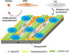
Polydopamine can be deposited on many surfaces of organic and inorganic substrates [Reference Liu, Ai and Lu19, Reference Lee, Dellatore, Miller and Messersmith20]; moreover, the polydopamine that contains amino and catechol groups can form metal complexes with copper ions, where the copper ions can be used as a catalyst for releasing NO [Reference Ren, Wu, Xi, Lehnert, Major, Bartlett and Meyerhoff21, Reference Pant, Goudie, Hopkins, Brisbois and Handa22, Reference Harding23, Reference Wonoputri, Gunawan, Liu, Barraud, Yee, Lim and Amal24]. Copper ions were mixed with dopamine solution (pH: 8.5) and form particles, where the copper ions was wrapped in polydopamine coating [Reference Jia, Li, Zhao, Frazer, Qian, Borguet, Ren and Dikin25, Reference Lee, Rho and Messersmith26]. Moreover, the polydopamine contains quinone groups, which can be used to immobilize many proteins, such as bivalirudin [Reference Yang, Zhong, Yang, Maitz, Li, Tu, Qi, Zhang, Qiu and Wang27, Reference Yang, Li, Qiu, Li, Qi, Maitz, You, Shen, Yang, Tian and Huang28, Reference Park, Brust, Lee, Lee, Watts and Yeo29] and trypsin [Reference Lee, Rho and Messersmith26]. The polymer of PMMPC-HD that has amino groups also can be immobilized onto the surface of the polydopamine coating [Reference Ma, Liu, Chen, Zhao, Yang and Huang30]. PMMPC copolymer contains carboxyl groups that can be cross-linked with hexamethylene diamine by the condensation reaction of amino and carboxyl. Simultaneously, the PMMPC-HD can be immobilized onto polydopamine with hexamethylenediamine by Michael addition reaction and Schiff base reaction [Reference Lee, Rho and Messersmith26]. For better adhesion of ECs, peptides Arg-Glu-Asp-Val (REDV) [Reference Wei, Ji, Xiao, Lin, Xu, Ren and Ji31, Reference Liu, Yang Tan, Yuan and Choong32, Reference Massia and Hubbell33, Reference Hahn, Taite, Moon, Rowland, Ruffino and West34] from fibronectin, which has the ability to adhere EC selectively compared to smooth muscle cell, was widely applied in the field of surface modification of biomaterials. It is because the ligand of REDV can specifically bind with integrin α4β1 in the membrane of ECs, but vascular smooth cells and platelets do not have it [Reference Hubbell, Massia, Desai and Drumheller35]. REDV [Reference Wei, Ji, Xiao, Lin, Xu, Ren and Ji31, Reference Chen, Li, Wang, Zhu, Zhu, Zheng, Yang and Guan36] was used to graft to the side chain of polymer of MPC, and the results of surface modification with the polymer demonstrated that the competitive adhesion and the growth of ECs over SMCs could be enhanced by the synergic action of the nonspecific resistance to PC and the specific recognition of the REDV peptide. In this study, our objective was as follows: Ti plates were chosen as the substrate and polydopamine mixed with copper ions was deposited on their surface. After the polymer of MPC was immobilized, the peptides GREDVY was immobilized on polydopamine by Michael addition reaction and Schiff base reaction. The biomimetic micropattern with PMMPC-HD/GREDVY for regulating cell behaviors, and the copper in polydopamine can generate NO from RSNOs. Therefore, the hypothesis of the study was as follows: surface modification with NO-generating materials and micropattern could be beneficial to the improvement of hemocompatibility and endothelialization.
Results
Characterization of the micropattern
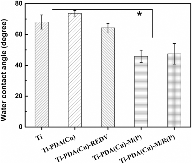
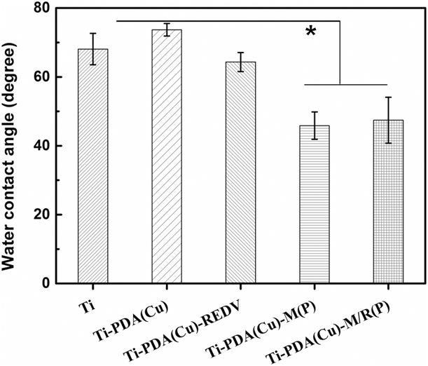
Water contact angle can be used to characterize the hydrophilicity of the surface, which respond to the change of chemical groups on the surface. As shown in Fig. 1, the water contact angle of Ti was 68°, when polydopamine was deposited on Ti, the angle of Ti-PDA(Cu) rise to 73.7°. It can be concluded that the polydopamine coating was more hydrophobic than Ti. After the PMMPC-HD was immobilized on the surface of Ti-PDA(Cu), the water contact angle of Ti-PDA(Cu)-M(P) and Ti-PDA(Cu)-M/R(P) decreases to 45.8° and 47.4°. It was because the polymer PMMPC has many zwitterions that are hydrophilic [Reference Wu, Lin, Wang, Chen and Chang37].
Water contact angle of samples. Data presented as mean ± SD, n = 6. *P < 0.05.
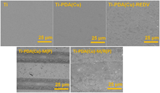
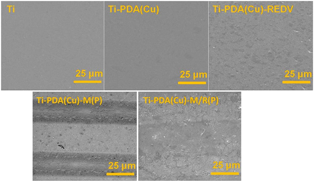
As is shown in Fig. 2, the surface of Ti and Ti-PDA(Cu) was uniform. The micropattern of PMMPC-HD on polydopamine coating was regular stripes. After peptide GREDVY was immobilized on polydopamine, the surface was not uniform, but the micropattern on Ti-PDA(Cu)-M/R(P) was clear. It can be demonstrated that the micropattern was fabricated successfully, the width of them was 25 μm equal. The micropattern can be applied to regulate cell behaviors.
SEM image of the samples.
FTIR
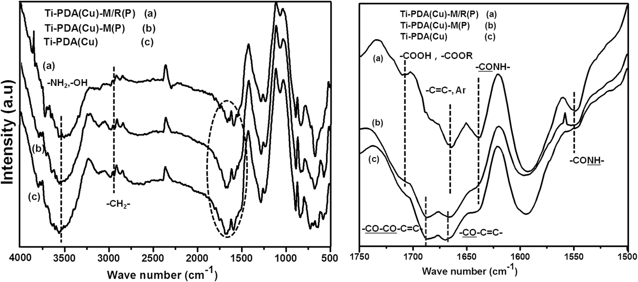
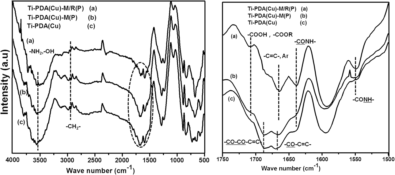
FTIR was an important measurement for characterization of the change of chemical groups on the surface. As is shown in Fig. 3, the stretching vibration of O–H and N–H in amino and hydroxyl group at 3500 cm−1, which demonstrated the sample Ti-PDA(Cu), Ti-PDA(Cu)-M(P), and Ti-PDA(Cu)-M/R(P), had the groups; moreover, the blue-shift of amino and hydroxyl groups showed the copper ions have a chemical reaction with polydopamine, such as complexation reaction, enfolded by polydopamine. The peak at 2940 cm−1 was attributed to –CH2– stretching vibration, which was in the polydopamine, PMMPC-HD, and peptide GREDVY. The peak at 1740 cm−1 was attributed to the C=O stretching vibrations of –COOH and –COOR groups in the PMMPC-HD and GREDVY; moreover, the peak around 1670 cm−1 was ascribed to amide I band  $\left( { – \underline {{\rm{CO}}} {\rm{NH}} – ,\;{\rm{I}}} \right)$, and amide II band
 $\left( { – {\rm{C}}\underline {{\rm{ONH}}} – ,\;{\rm{II}}} \right)$ was at 1551 cm−1, which showed that the PMMPC-HD and GREDVY were immobilized on polydopamine successfully. Additionally, the Ti-PDA(Cu)-M/R(P) had the peak 1630 cm−1, and it can be concluded that the peaks were stretching vibration of C=C of phenyl (with a hydroxyl group) in GREDVY. Also demonstrating the peptide was successfully immobilized on the polydopamine coating.
FTIR spectra of samples: Ti-PDA(Cu) (c), Ti-PDA(Cu)-M(P) (b), and Ti-PDA(Cu)-M/R(P) (a).
NO release
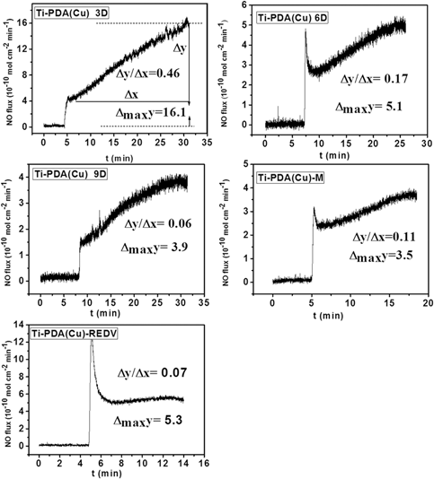
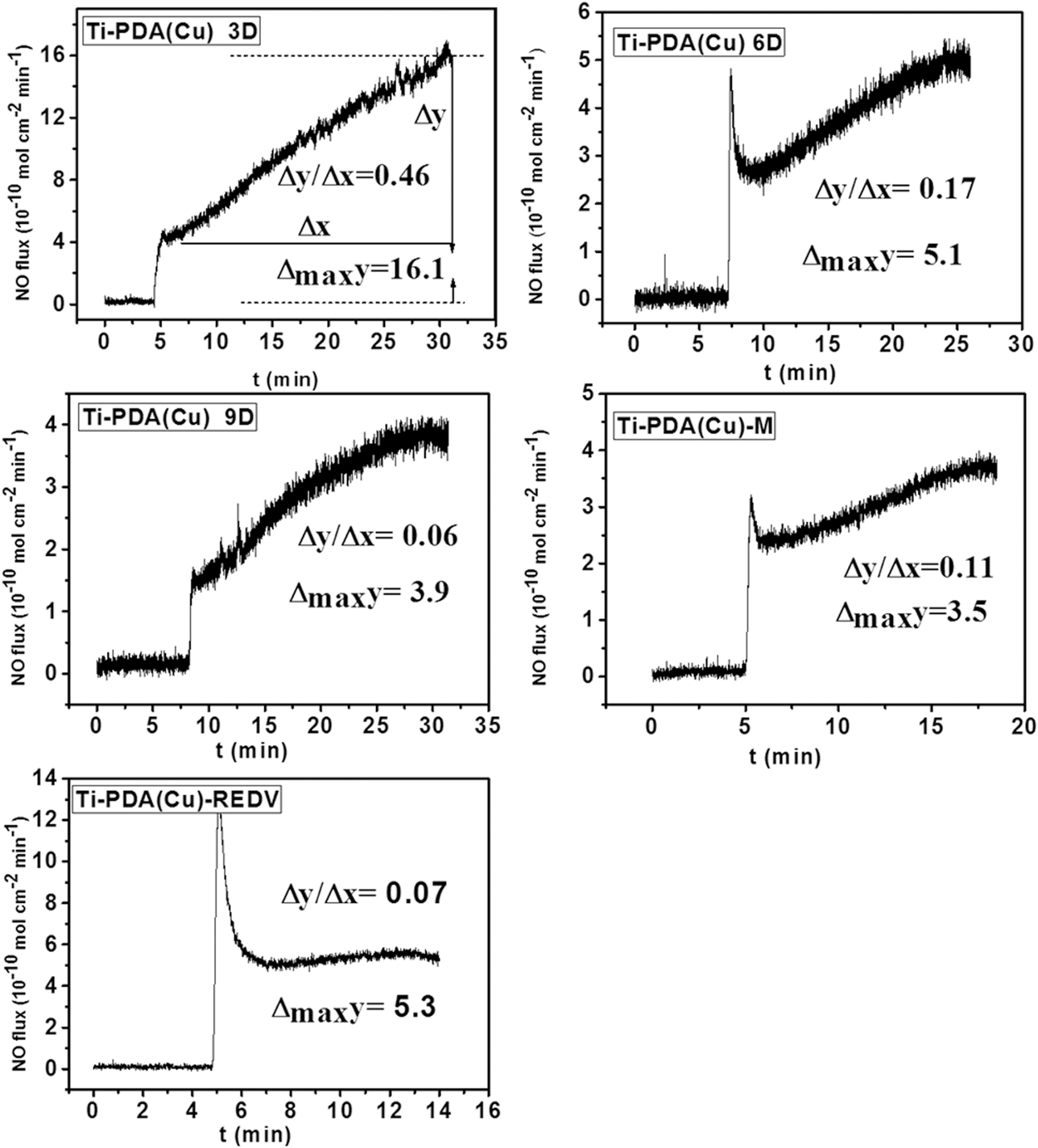
The NO was catalytically generated from GSNO donors by the copper ions in polydopamine. The flat samples were used because it was easy to prepare and can be used to characterize the surface of PMMPC-HD and GREDVY single. To avoid initial burst release, the Ti-PDA(Cu) sample was immersed in PBS solution (pH: 7.4) for 3, 6, and 9 days (3D, 6D, and 9D) to release the copper ions. As shown in Fig. 4, the release rate of NO of Ti-PDA(Cu) 3D was around 4.2 × 10⁻¹⁰ mol/(cm2 min), and after 30 min the rate was about 16.1 × 10⁻¹⁰ mol/(cm2 min). The initial release rate of NO of Ti-PDA(Cu) 6D and Ti-PDA(Cu) 9D decreased to 2.5 × 10⁻¹⁰ mol/(cm2 min) and 1.5 × 10⁻¹⁰ mol/(cm2 min); moreover, the increment was decreasing. The data demonstrated that the burst release of copper ions of Ti-PDA(Cu) 9D was weaker than that of Ti-PDA(Cu) 3D (Fig. S1). When the PMMPC-HD was immobilized on the polydopamine coating, the release rate of NO of Ti-PDA(Cu)-M was around 2.5 × 10⁻¹⁰ mol/(cm2 min); the increment was smaller than that of Ti-PDA(Cu). Similarly, the release rate of NO of Ti-PDA(Cu)-REDV was steadier than that of Ti-PDA(Cu), maintaining around 5.3 × 10⁻¹⁰ mol/(cm2 min). In all, the polydopamine coating mixed copper ions immobilized PMMPC-HD and GREDVY can catalytically generate NO from GSNO maintaining at a relatively stable rate, which was beneficial to improve hemocompatibility and EC compatibility.
NO release from GSNO catalyzed by Ti-PDA(Cu), Ti-PDA(Cu)-M, and Ti-PDA(Cu)-REDV.
Platelets adhesion
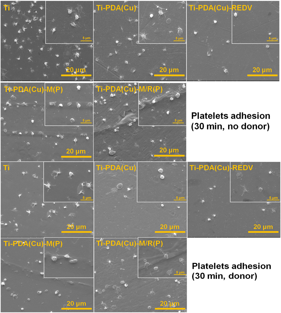
The in vitro platelet adhesion test is used to investigate the blood compatibility of biomaterials [Reference Huang, Yang, Hu, Lin, Sun and Vogler38, Reference Liu, Yuan, Li, Tang, Wang, Chen, Chen and Brash39]. SEM images were used to detect the adhesion and morphology of platelets on various samples surfaces after incubation in platelet-rich plasma (PRP) for 30 min. As shown in Fig. 5, platelets adhered on the Ti and Ti-PDA(Cu) surface without NO donor were marked shape change (spreading) and pseudopod formation. However, platelets adhered on the Ti-PDA(Cu)-REDV, Ti-PDA(Cu)-M(P), and Ti-PDA(Cu)-M/R(P) surface were significantly reduced, and platelets maintained their round shapes with no formation of pseudopods. No obvious shape difference on PMMPC-HD and GREDVY immobilized surfaces was observed, indicating that the PMMPC-HD and GREDVY could not cause platelet activation.
SEM of platelets adhesion on samples without GSNO donor (no donor) and with GSNO donor (donor).
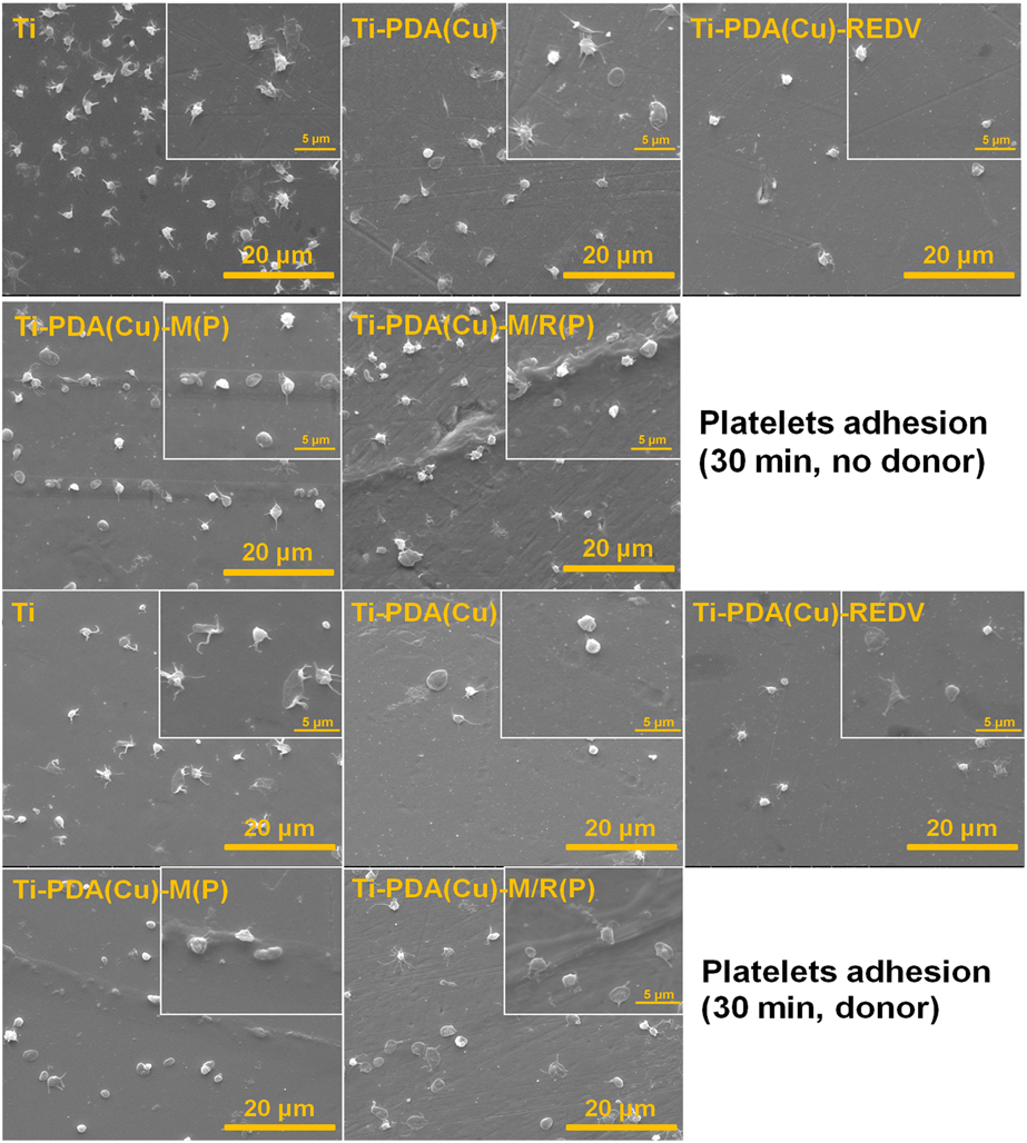
Platelets adhered on the Ti surface with NO donor were marked shape change (spreading) and pseudopod formation, but on the Ti-PDA(Cu) surface with NO donor were round shapes with no formation of pseudopods. Compared to Ti-PDA(Cu) without NO donor and Ti with NO donor, the Ti-PDA(Cu) had better compatibility because the copper ions in polydopamine release could catalytically generate NO from the donor GSNO. Similarly, the Ti-PDA(Cu)-REDV, Ti-PDA(Cu)-M(P), and Ti-PDA(Cu)-M/R(P) could also release copper ions, and the platelets on them (with NO) were round without pseudopod formation. From the SEM image, it can be found that the platelets adhered easily to the edge of micropattern. It suggested that the edge of micropattern had more sites for adhering. It was the reason that the platelets on micropattern were more than the sample of Ti-PDA(Cu)-REDV.
ECs culture
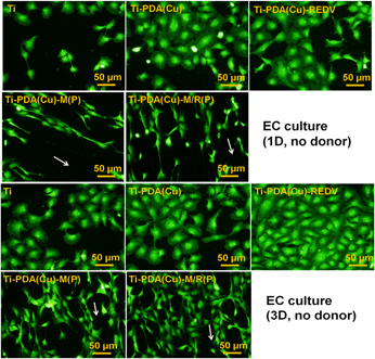
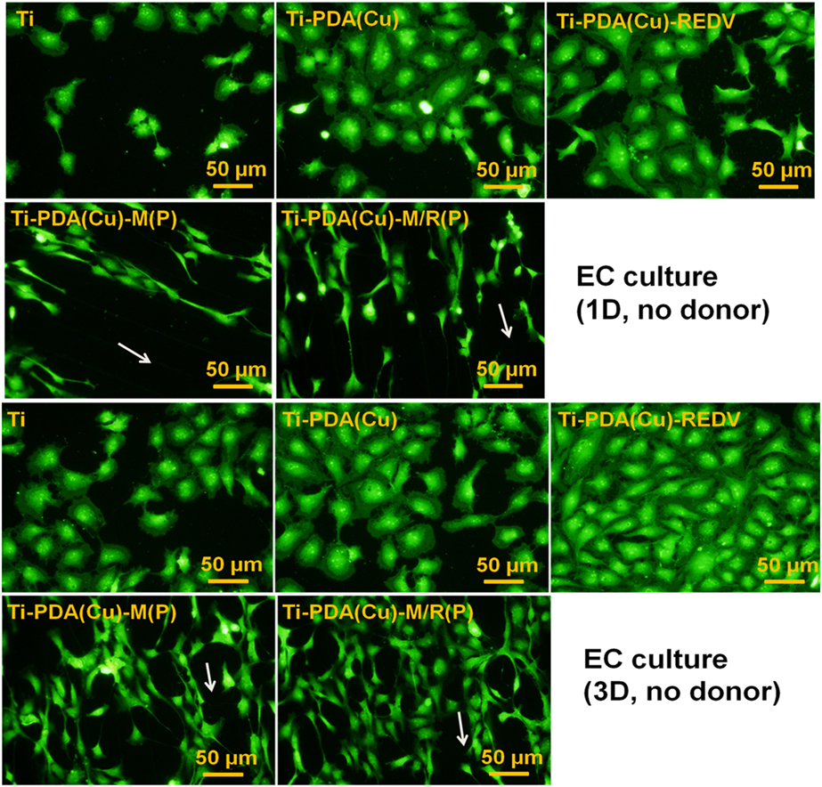
As is shown in Fig. 6, under the condition without NO donor, the ECs on the surface of Ti, Ti-PDA(Cu), and Ti-PDA(Cu)-REDV without micropattern were distributed freely, and the cell morphology was ellipsoidal. On the Ti-PDA(Cu)-M(P) and Ti-PDA-M/R(P), the cells were highly ordered and elongated. The long axis of the cells was in the direction of the stripe of the micropattern. After 3 days of culture, the number of ECs increased significantly.
Fluorescent images of ECs after 1 day (1D) and 3 days’ (3D) culture without GSNO donor (no donor).
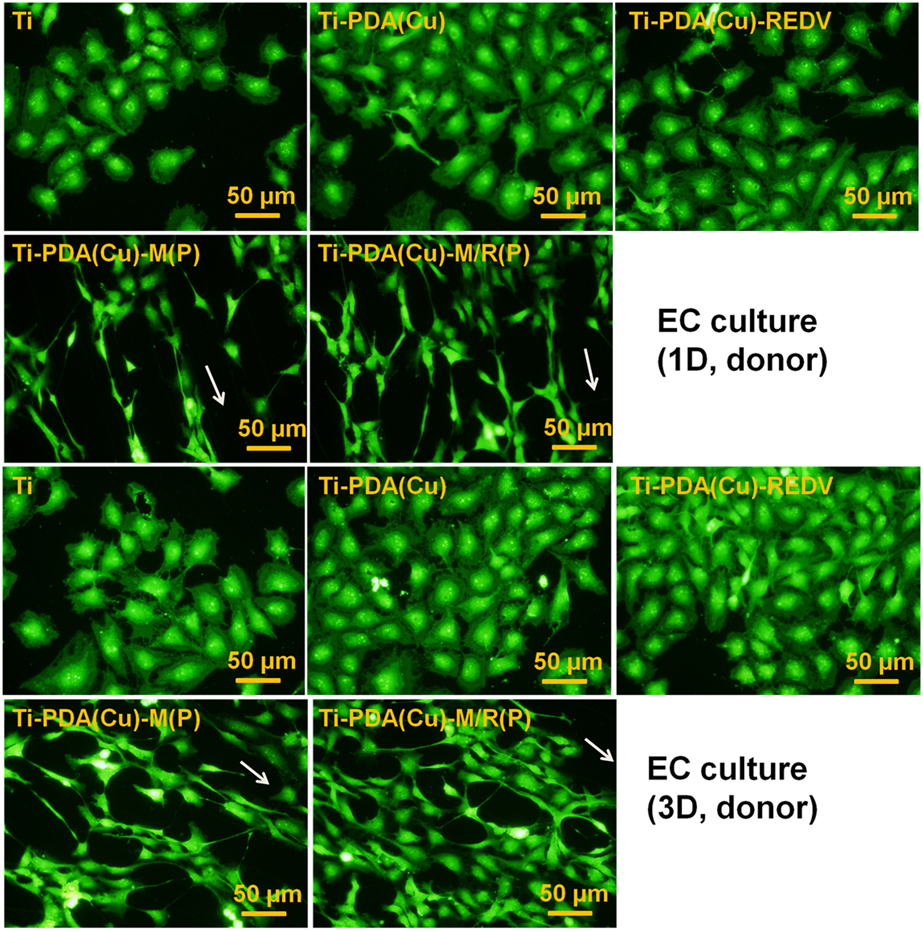
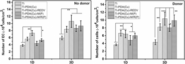
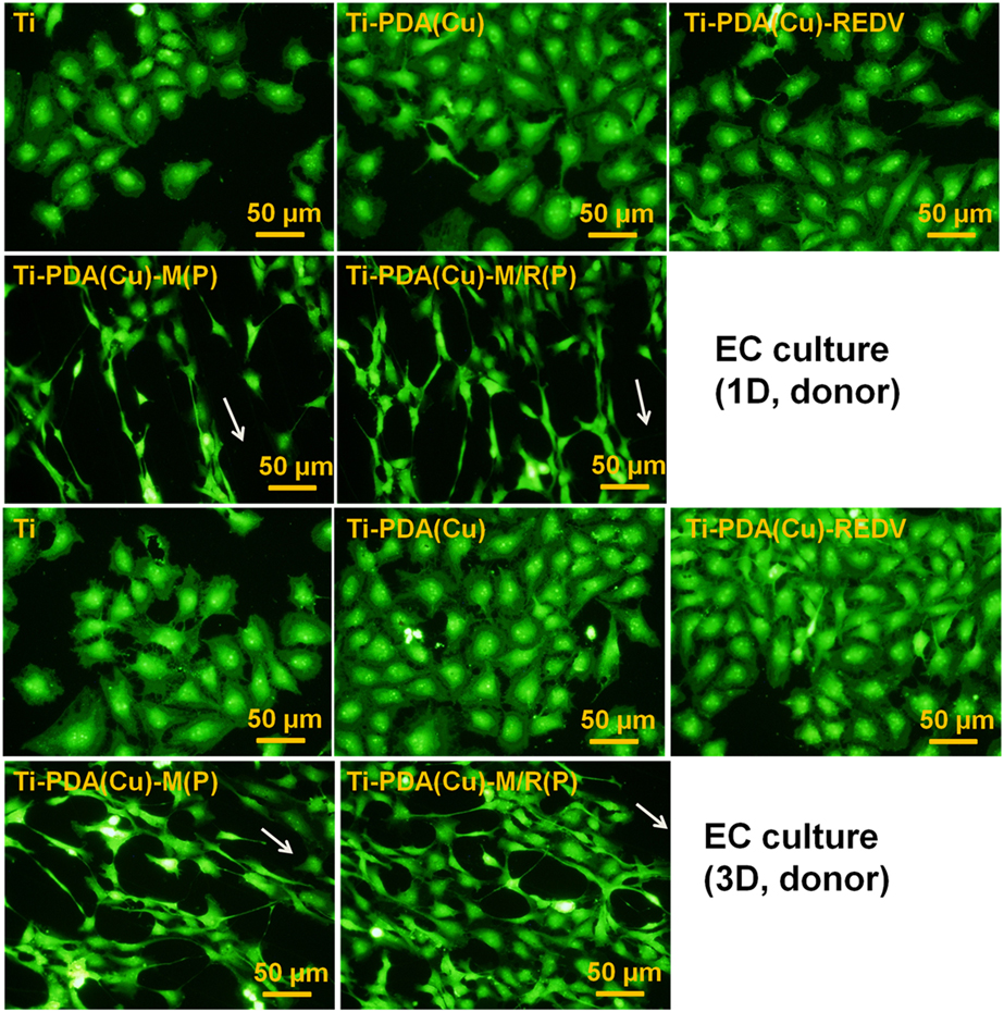
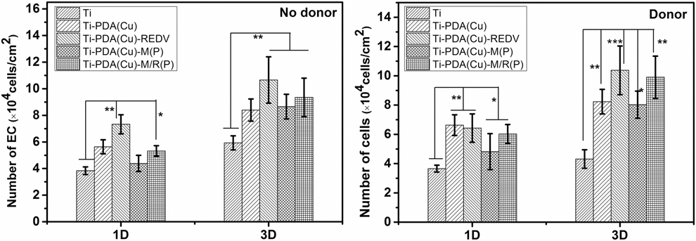
As is shown in Fig. 7, under the condition with NO donor, the number of ECs on Ti-PDA(Cu) was much bigger than those on Ti-PDA(Cu) without NO donor. However, the number of ECs on Ti was not increased. The data showed that the copper ions in polydopamine can catalyze GSNO donor release NO, which was beneficial to the proliferation of ECs. The number of ECs on Ti-PDA(Cu)-REDV was not significantly increased. It was concluded that REDV can capture ECs whether with NO donor or without NO donor. ECs on the Ti-PDA(Cu)-M(P) and Ti-PDA(Cu)-M/R(P) were highly ordered and elongated. The number of ECs on Ti-PDA(Cu)-M/R(P) was bigger than those on Ti-PDA(Cu)-M(P) after 3 days’ culture (as shown in Fig. 8); the data showed that REDV and NO release were beneficial to the proliferation of ECs. In all, ECs on Ti-PDA(Cu)-M/R(P) were elongated and ordered, and NO release catalysis by copper ion was good for the proliferation of ECs. The data on ECs proliferation were consistent with the reported literature [Reference Ren, Wu, Xi, Lehnert, Major, Bartlett and Meyerhoff21].
Fluorescent images of ECs after 1 day (1D) and 3 days’ (3D) culture with GSNO donor (donor).
Number of ECs on samples without GSNO donor (no donor) and with GSNO donor (donor).
Discussion
The data for FTIR, SEM, and water contact angle demonstrated that Ti-PDA(Cu)-M/R(P) micropattern was fabricated successfully. Platelets adhesion test showed that the Ti-PDA(Cu)-M/R(P) micropattern inhibited platelet adhesion. ECs on Ti-PDA(Cu)-M/R(P) micropattern were regulated, and the morphology of ECs was elongated. The long axis of ECs on Ti-PDA(Cu)-M/R(P) was in the direction of micropattern.
MPC polymer has the property of inhibiting adhesion of proteins and cells, because MPC polymer has many zwitterionic groups, and about eight water molecules are tightly bound with one zwitterionic (MPC) unit. The hydrated MPC groups have a weak interaction with proteins (fibrinogen) and the cells, which might illustrate the reason for the resistance of zwitterionic materials to nonspecific protein adsorptions. The PMMPC retained lots of MPC groups, which can be hydrated and can inhibit platelet adhesion and activation. The Ti-PDA(Cu)-M/R(P) micropattern had GREDVY for ECs adhesion. It can be concluded that the width of The Ti-PDA(Cu)-M/R(P) micropattern affects the adhesion, cytoskeletal organization, and cell proliferation.
NO was catalytically generated from GSNO donors by the copper ions in polydopamine, and the release rate of NO was a benefit to the proliferation of ECs. Similarly, the ECs was elongated and orderly. Ti-PDA(Cu)-M/R(P) micropattern will be a promising platform for NO release and surface modification of cardiovascular materials.
Conclusion
Polydopamine incorporated with copper ions was deposited on titanium successfully. Micropatterns of PMMPC-HD polymer and GREDVY were immobilized on polydopamine. Water contact angle, SEM, FTIR, and XPS demonstrated that the Ti-PDA(Cu)-M/R(P) micropattern was fabricated. NO release catalyzed by copper ions was 1.5–5.3 × 10⁻¹⁰ mol/(cm2 min), which can inhibit platelet adhesion and activation and can benefit the proliferation of ECs on Ti-PDA(Cu)-REDV and Ti-PDA(Cu)-M/R(P). The data of ECs culture demonstrated that the morphology of ECs on Ti-PDA(Cu)-M/R(P) micropattern was elongated.
Experimental procedure
Ti deposited with polydopamine loaded copper
Ti was cut into wafers of 10 mm in diameter, then polished, and ultrasonic cleaned with ethanol, acetone, and deionized water. The Ti plates were immersed into dopamine solution, which was prepared by way of dissolving dopamine hydrochloride (2 mg/mL) in tris-base buffer (10 mM, pH = 8.5), and then CuCl2 was added into the solution (0.27 mg/mL). After that, polydopamine-coated titanium was ultrasonically washed with distilled water (10 min × 3 times). The above process was repeated for 4 times, and the surface of the Ti wafer covered with polydopamine was obtained and named Ti-PDA(Cu). The polydopamine coating was prepared according to the method reported by Lee [Reference Lee, Dellatore, Miller and Messersmith20, Reference Ma, Yang, Li, Li, Li, Zhao and Huang40].
Synthesis of PMMPC-HD and fabrication of micropattern
Synthesis of PMMPC (MPC/MA = 7/3) was as follows: first, monomer MPC (5 g, 0.017 mol) and MA (0.625 g, 0.0075 mol) were put into a three-necked flask with 120 mL deoxygenated ethanol, and stirred for 15 min. Second, 0.028 g of azodiisobutyronitrile (AIBN) was added and maintained at 70 °C under Ar gas for 36 h. Finally, the reaction solution was concentrated and precipitated with tetrahydrofuran (THF) three times, and PMMPC was obtained using freeze drying. The process of synthesis of PMMPC was according to the literature [Reference Chen, Li, Zhao, Li, Chen, Yang, Maitz and Huang41].
To cross-link PMMPC with hexamethylene diamine, the PMMPC (100 mg) was put into a flask with 95 mL deionized water and 5 mL ethanol, and then 100 mg of EDC and 25 mg of NHS were added, for activation of carboxyl. After being stirred for 15 min, 10 mg of hexamethylene diamine (maintaining the molar ratio of –NH2 to –COOH was 1:1) was added. After being stirred for 3 h at room temperature under Ar gas, the solution of mixed cross-linked PMMPC-HD was concentrated and transferred into dialysis tubing (molecular weight cut off: 8000) and stirred in deionized water for 48 h to remove the small molecules. The cross-linked polymer called PMMPC-HD was obtained through freeze drying.
A cotton swab was wetted with a solution of the mixed cross-linked polymer of MPC named PMMPC-HD (10 mg/mL), in which the PMMPC [mol%, MPC/MA = 7/3: monomer contain MPC and methacrylic acid (MA), 10 mg/mL] was cross-linked with hexamethylene diamine (HD). And then the swab was dragged across the surface of Ti-PDA(Cu). Polydimethylsiloxane (PDMS) stamps (ridge/groove: 25/25 μm) was prepared following the instruction on the silicon master. The PDMS stamp was then dried with a stream of nitrogen and brought into contact with the samples of Ti-PDA(Cu) for about 30 min, after which the stamp was peeled away from the substrate. The samples were named Ti-PDA(Cu)-M(P). The whole process was fabricated according to the literature [Reference Ma, Liu, Chen, Zhao, Yang and Huang30, Reference Anderson and Hinds42]. The Ti-PDA(Cu)-M(P) were immersed in a solution of GREDVY (300 μg/mL) in PBS (pH: 7.4) for 2 h at 37 °C and named Ti-PDA(Cu)-M/R(P). Identically, the Ti-PDA(Cu) samples immersed in PMMPC-HD and GREDVY were named Ti-PDA(Cu)-M and Ti-PDA(Cu)-REDV, and the samples were flat, not micropatterned.
Characterization of the micropattern
The morphologies of bare Ti, modified Ti, and micropatterned samples were observed by scanning electron microscopy (SEM; JSM-7001F, Japan Electronics Co., Ltd. (JEOL), Tokyo, Japan), and the surface chemical composition was examined using Fourier transform infrared spectrometer (FTIR; NICOLET 5700, Thermo Fisher Scientific, Waltham, Massachusetts) with the mode of infrared grazing. Static water contact angles were measured using a DSA100 Drop Shape Analyzer (Kruss Scientific Instruments, Inc., Hamburg, Germany) at room temperature. At least 6 contact angles in different areas were measured and averaged. The samples were washed with ethanol and dried in an Ar stream immediately before the measurement.
NO release, platelets adhesion, and ECs culture
The NO release catalyzed by copper ions was tested using chemiluminescence NO analyzer (NOA) (Seivers 280i, Boulder, Colorado). For the chemiluminescence method, Ti discs (0.5 × 1 cm2) modified by polydopamine coating was used for NO quantitative analysis. In brief, the NO catalytically produced by samples was purged from the test solution containing s-nitrosoglutathione (GSNO) and reducing agent glutathione (GSH), and transported to the NO analyzer by a stream of N2 (g). The calculation of the amount of NO generated by samples was based on the calibration curves of the NOA, which was in detail shown elsewhere [Reference Cha and Meyerhoff43].
The samples for NO release platelet adhesion test were conducted to evaluate the adhesion, distribution, behavior, and activation of the platelets in vitro. First, fresh whole blood containing anticoagulant with 0.109 M solution of sodium citrate at a dilution ratio of 9:1 (blood/sodium citrate solution) was centrifuged at 1500 rpm for 15 min to separate the blood corpuscles and get PRP. Second, 300 μL PRP was added on the samples and incubated at 37 °C for 30 min with GSNO and without GSNO. Subsequently, the samples were rinsed three times with PBS and fixed with 2.5% glutaraldehyde for 4 h. Finally, the samples were also observed by way of SEM.
HUVECs were derived from a human umbilical vein. The detailed processes of cell culture are described elsewhere [Reference Yang, Lei, Wang, Luo, He, Sun and Huang44]. For the evaluation of HUVEC growth behavior, the use of cell media with and without donor supplement (10 mM GSNO, 10 mM GSH) was necessary to do a comparison. HUVECs were seeded on the specimen surfaces at a density of 5 × 10⁴ cells/cm2 for 1 and 3 days, respectively. For 1 and 3 days of culture, the donor was supplemented in culture medium every 4 h. Here, fluorescence staining was performed to analyze the morphology of the cells. To study the number of HUVECs on samples, we collected at least nine sight fields and counted the HUVECs for statistical analysis.
Statistical analysis
Statistical significance was performed using one-way analysis of variance (ANOVA) with SPSS 17.0 (SPSS Inc., Chicago, Illinois) statistical software. Data are presented as mean ± SD.
Acknowledgments
This work was supported by financially supported by the National Key Research and Development Program of China (2016YFC1100402), National Natural Science Foundation of China (Nos. 31570963 and 81330031), and Key Basic Research Project (No. 2011CB606204).