Introduction
The symbiotic relationship that exists between GI microbes and the host is critical for proper function of nutritional, developmental, immunological, and physiologic processes in animals, and thus contributes to host health (Mackie et al., Reference Mackie, Sghir and Gaskins1999; Hooper et al., Reference Hooper, Wong, Thelin, Hansson, Falk and Gordon2001). The resident microbiota forms an integral part of the intestinal barrier, which protects the host from pathogens. This protective mechanism is known as colonization resistance and includes competition for nutrients and mucosal adhesion sites, which creates a physiologically restrictive environment for invading pathogens (Kanauchi et al., Reference Kanauchi, Matsumoto, Matsumura, Fukuoka and Bamba2005). Furthermore, GI microbes contain enzymes that digest complex carbohydrates from the diet that are not digested by host enzymes and ferment endogenous products such as sloughed epithelial cells and mucus. The primary end-products of the fermentation process, the short-chain fatty acids (SCFA), namely acetate, propionate, and butyrate, provide energy for host epithelial cell growth and metabolism (Sunvold et al., Reference Sunvold, Fahey, Merchen, Titgemeyer, Bourquin, Bauer and Reinhart1995). SCFA also have immuno-modulatory properties and modulate colonic and intracellular pH, both of which indirectly alter the composition of gut microbes (e.g., reduces the growth and activity of potential pathogens) and have application to GI inflammatory diseases (Vernia et al., Reference Vernia, Annese, Bresci, d'Albasio, D'Inca, Giaccari, Ingrosso, Mansi, Riegler, Valpiani and Caprilli2003). Decreased GI pH also results in decreased solubility of bile acids, thus decreased reabsorption and enterohepatic circulation of bile acids, increased absorption of minerals, and reduced absorption of ammonia (Vince et al., Reference Vince, Killingley and Wrong1978; Metzler-Zebeli et al., Reference Metzler-Zebeli, Hooda, Mosenthin, Ganzle and Zijlstra2010), all of which may have implications for the nutritional status of the host and fermentation that occurs in the hindgut.
Although microbial populations are present in many areas of the body, including the oral cavity, urinary and genital tracts, and skin, the GI tract harbors the largest microbial population. The bacterial density ranges from 104 to 105 CFU/g in the stomach to 105 to 107 CFU/g in the small intestine to 109 to 1011 CFU/g in the colon (Benno et al., Reference Benno, Nakao, Uchida and Mitsuoka1992; Johnston, Reference Johnston1999; Mentula et al., Reference Mentula, Harmoinen, Heikkila, Westermarck, Rautio, Huovinen and Kononen2005). Various host factors, such as genetic background, age, sex, initial environmental exposure and diet contribute to the development and maintenance of the core intestinal microbiota (Louis et al., Reference Louis, Scott, Duncan and Flint2007; Khachatryan et al., Reference Khachatryan, Ktsoyan, Manukyan, Kelly, Ghazaryan and Aminov2008; Mariat et al., Reference Mariat, Firmesse, Levenez, Guimaraes, Sokol, Dore, Corthier and Furet2009). Although the upper GI tract harbors many microbes, the colon has the richest microbial population, most of which are anaerobes, and is the main site of the anaerobic digestion process known as fermentation. These microbes metabolize and survive on substrates from the host's diet (e.g., non-digestible carbohydrates and; undigested proteins) and endogenous secretions (Rowland et al., Reference Rowland, Mallett and Wise1985). Despite the dog's evolution in the order Carnivora and a lower consumption of non-digestible carbohydrates than most domestic livestock species, commercial dog foods may contain ingredients that significantly affect the population and/or activity of GI microbes and the fermentation process. Even when dogs are fed high-fiber diets, they do not rely heavily on the GI microbiota to meet their daily metabolizable energy requirement. A balanced microbial population, however, is very important for overall host health.
The emergence of metabolic and intestinal dysbiosis in humans has generated interest in gut microbes in recent years, as gut microbiome changes have been associated not only with colon cancer (Flint et al., Reference Flint, Duncan, Scott and Louis2007) and inflammatory bowel disease (IBD; Friswell et al., Reference Friswell, Campbell and Rhodes2010) but also with complex metabolic diseases such as obesity (Ley et al., Reference Ley, Backhed, Turnbaugh, Lozupone, Knight and Gordon2005; Turnbaugh et al., Reference Turnbaugh, Backhed, Fulton and Gordon2008) and diabetes (Wen et al., Reference Wen, Ley, Volchkov, Stranges, Avanesyan, Stonebraker, Hu, Wong, Szot, Bluestone, Gordon and Chervonsky2008). Similarly in dogs, the intestinal microbiota has been associated with IBD (Suchodolski et al., Reference Suchodolski, Xenoulis, Paddock, Steiner and Jergens2010) and chronic diarrhea (Bell et al., Reference Bell, Kopper, Turnbull, Barbu, Murphy and Mansfield2008; Jia et al., Reference Jia, Frantz, Khoo, Gibson, Rastall and McCartney2010). Recent advances in DNA sequencing technology and application of a variety of ‘omic’ technologies in a systems biology framework, have allowed for a better phylogenetic characterization of the gut microbiome. Furthermore, the metagenomics approach has improved our understanding of microbial function in the GI tract. A deeper understanding of the gut microbiome will provide reference values for healthy populations and assist in diagnosing and treating diseased animals. In addition, dietary interventions to prevent dysbiosis and thus maintain gut health in companion animals may be achieved.
Canine gut microbiome in the pre-sequencing era
Stomach and small intestine
The physiologic changes that occur throughout the GI tract, including the acidic nature of the stomach, and the bile salts and enzymes present in the small intestine, have an effect on what species of microbiota inhabit each region. Traditional studies reported that a mixture of aerobes and anaerobes inhabited the stomach, dominated by Gram-positive bacteria (Greene, Reference Greene1998). Similar to humans, the Gram-negative Helicobacter spp. (e.g., Helicobater felis, Helicobater Bizzozeronii, and Helicobater salomonis) are also present and considered to be potential pathogens in the canine stomach (Fox, Reference Fox and Greene1998), although their pathological role is not clear (Neiger and Simpson, Reference Neiger and Simpson2000). Eubacterium, Bacteroides, Clostridium, Fusobacterium, Bifidobacterium, and Lactobacillus spp. are predominant in the canine duodenum and jejunum (Hermanns et al., Reference Hermanns, Kregel, Breuer and Lechner1995; Johnston, Reference Johnston1999). The published results generated using bacterial cultivation techniques revealed that Bacteroides, Eubacterium, Fusobacterium, Clostridium, Lactobacillus, and Bifidobacterium are the most common anaerobic bacteria in the canine ileum (Davis et al., Reference Davis, Cleven, Balish and Yale1977; Benno et al., Reference Benno, Nakao, Uchida and Mitsuoka1992). Likewise, Streptococcus, Staphylococcus, Pasteurella, Escherichia, and Enterobacter are the most common aerobic and facultative anaerobic bacteria in the canine ileum. Potential pathogenic species present in the small intestine include Clostridium perfringens, Clostridium difficile, Klebsiella pneumoniae, Campylobacter jejuni, Salmonella Typhimurium, enteric Helicobacter spp., Yersinia enterocolitica, and enteropathogenic Escherichia coli (Marks and Kather, Reference Marks and Kather2003).
Large intestine
The large intestine has the highest density and diversity of bacteria (109–1010 CFU/g of content; Greene, Reference Greene1998). In 1977, 84 species within 27 genera were cultivated from ileal, cecal, and colonic contents of dogs (Davis et al., Reference Davis, Cleven, Balish and Yale1977). Although many of the microbes present in the large intestine were also detected in the small intestine, their abundance was much greater in the large intestine. Predominant genera included Bacteroides, Bifidobacterium, Fusobacterium, Peptostreptococcus, Eubacterium, Clostridium, Peptococcus, and Lactobacillus (Davis et al., Reference Davis, Cleven, Balish and Yale1977). Similarly, the prevalence of potential pathogenic bacteria such as C. perfringens, and E. coli were also reported in the large intestine (Davis et al., Reference Davis, Cleven, Balish and Yale1977). Prior to the 21st century, most of the current knowledge about canine GI microbiota was generated using cultivation assays. In addition to being very laborious, time consuming, and requiring immediate processing, cultivation techniques do not allow for identification and differentiation of most microbial strains. As there is a lack of information regarding the growth requirements of most microbes, only a minor fraction of GI microbes have been isolated and studied in culture to date (Eckburg et al., Reference Eckburg, Bik, Bernstein, Purdom, Dethlefsen, Sargent, Gill, Nelson and Relman2005).
Canine gut microbiome in the sequencing era
To overcome the limitations of culture, molecular-based techniques have been used to study GI microbial ecology over the past decade. The primary target for molecular approaches is the 16S ribosomal RNA (rRNA) gene because of its presence in all microbes and because it contains both conserved and variable sequence regions across species (Clarridge, Reference Clarridge2004). Molecular fingerprinting techniques, including denaturing gradient gel electrophoresis (DGGE), temperature gradient gel electrophoresis (TGGE), and terminal restriction fragment length polymorphism (T-RFLP), were initially used to study qualitative differences in GI microbial ecology. For identification, DNA bands of interest may be isolated and sequenced. These techniques are rather insensitive, however, and provide information on only the most predominant bacterial groups. Quantitative PCR (qPCR) and fluorescent in situ hybridization (FISH), which are more sensitive than the gel-based techniques, have been widely used over the last decade to quantify select bacterial groups. The primary issue with qPCR and FISH is that they are limited in scope, only providing data on a few specific microbial groups.
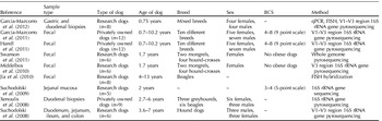
Recently, DNA-sequencing techniques that use automated high-throughput sequencing platforms have increased our knowledge regarding GI microbial identification and classification. These techniques are being used in the human microbiome projects to characterize the entire microbial community present in the GI tract, mouth, vagina, nasal cavity, skin and other body regions (Peterson et al., Reference Peterson, Garges, Giovanni, McInnes, Wang, Schloss, Bonazzi, McEwen, Wetterstrand, Deal, Baker, Di Francesco, Howcroft, Karp, Lunsford, Wellington, Belachew, Wright, Giblin, David, Mills, Salomon, Mullins, Akolkar, Begg, Davis, Grandison, Humble, Khalsa, Little, Peavy, Pontzer, Portnoy, Sayre, Starke-Reed, Zakhari, Read, Watson and Guyer2009). These high-throughput DNA-sequencing techniques have recently been applied to describe the phylogenetic structure and functional capacity of the canine GI microbiome. Details pertaining to study design, dog number and characteristics (e.g., age, breed, sex and body condition score), and method of analyses used in these experiments are summarized in Table 1. Although the samples studied have included some mucosa and digesta samples from various segments of the GI tract, most of the studies have focused on bacteria in fecal samples as a proxy for gut bacteria. As bacterial populations are known to differ between intestinal biopsy and fecal samples (Momozawa et al., Reference Momozawa, Deffontaine, Louis and Medrano2011); however, data from fecal samples may not be completely representative of data in the more proximal parts of the intestine.
Animals, experimental design, and analytical methods used in selected studies aimed at characterizing the canine GI microbiome

Stomach of healthy dogs
Garcio-Mazcorro et al. (Reference Garcia-Mazcorro, Suchodolski, Jones, Clark-Price, Dowd, Minamoto, Markel, Steiner, Dossin and Marchesi2012) used FISH, qPCR, 16S rRNA gene libraries, and 454 pyrosequencing of 16S rRNA gene amplicons (V1–V3 region) to study the microbiome of the canine stomach. Gastric biopsies were collected from eight clinically healthy mixed-breed research dogs. qPCR results indicated 105 16S rRNA gene copies of total bacteria present, with a predominance of Helicobacter and Lactobacillus spp. The total sequence number obtained by pyrosequencing from the stomach was 142,026 and ranged from 1199 to 7734 per sample. A median of 36 operational taxonomic units (OTUs) (range of 18–119) were detected per dog. The majority of the sequences belonged to Proteobacteria (99.6%), with only a few Firmicutes (0.3%). The Proteobacteria included the genus Helicobacter (98.6%) in gastric biopsies (Garcia-Mazcorro et al., Reference Garcia-Mazcorro, Suchodolski, Jones, Clark-Price, Dowd, Minamoto, Markel, Steiner, Dossin and Marchesi2012).
Duodenum of healthy dogs
The microbial communities of canine duodenal mucosa and contents were recently studied using 16S rRNA gene clone libraries and 454 pyrosequencing (Suchodolski et al., Reference Suchodolski, Camacho and Steiner2008; Xenoulis et al., Reference Xenoulis, Palculict, Allenspach, Steiner, Van House and Suchodolski2008; Garcia-Mazcorro et al., Reference Garcia-Mazcorro, Suchodolski, Jones, Clark-Price, Dowd, Minamoto, Markel, Steiner, Dossin and Marchesi2012). The microbial community of duodenal biopsy samples of healthy client-owned dogs was represented by six primary phyla: Firmicutes (46.4%), Proteobacteria (26.6%), Bacteroidetes (11.2%), Spirochaetes (10.3%), Fusobacteria (3.6%), and Actinobacteria (1.0%) (Xenoulis et al., Reference Xenoulis, Palculict, Allenspach, Steiner, Van House and Suchodolski2008). Similarly, Garcia-Mazcorro et al. (Reference Garcia-Mazcorro, Suchodolski, Jones, Clark-Price, Dowd, Minamoto, Markel, Steiner, Dossin and Marchesi2012) reported the predominance of Firmicutes (46.9%), Proteobacteria (21.2%), and Bacteroidetes (6.9%), which comprised more than 80% of all sequences in duodenal biopsies of healthy research dogs. Within Firmicutes, the orders Clostridiales (19.6%), Lactobacillales (14.1%), and Bacillales (12%) and families Clostridiaceae (13.9%), Carnobacteriaceae (7.4%), and Turicibacteriaceae (8.9%), were most abundant in duodenal mucosa samples (Xenoulis et al., Reference Xenoulis, Palculict, Allenspach, Steiner, Van House and Suchodolski2008). The family Helicobacteriaceae was the most abundant family within Proteobacteria (13.2%), followed by Pasteurellaceae (6.5%) and Moraxellaceae (1.9%). Bacteroidetes (11.2%) and Spirochaetes (10.2%) were two other phyla present in the duodenal mucosa, represented by the families Porphyromonadaceae (7.2%) and Spirochaetaceae (10.3%), respectively (Xenoulis et al., Reference Xenoulis, Palculict, Allenspach, Steiner, Van House and Suchodolski2008).
Suchodolski et al. (Reference Suchodolski, Camacho and Steiner2008) examined the bacterial microbial communities of duodenal, jejunal, ileal, and colonic digesta of healthy research dogs. The bacterial diversity indices increased gradually along the GI tract from the duodenum to the colon. Similar to mucosal samples, duodenal digesta samples were dominated by phyla Firmicutes and orders Clostridiales (~40%) and Lactobacillales (~22%) (Suchodolski et al., Reference Suchodolski, Camacho and Steiner2008). The distribution of two predominant Clostridium clusters (XI and XIVa) was represented by 25% and 31% of the duodenal digesta samples, respectively. Proteobacteria (32%), including E. coli-like organisms, was also abundant in duodenal digesta of these dogs. Anaerobic Fusobacteriales and Bacteroidales were only present sporadically in duodenal digesta (Suchodolski et al., Reference Suchodolski, Camacho and Steiner2008).
Jejunum of healthy dogs
A detailed analysis of the microbiome inhabiting the jejunal mucosa of healthy research dogs was conducted by Suchodolski et al. (Reference Suchodolski, Dowd, Westermarck, Steiner, Wolcott, Spillmann and Harmoinen2009) using 16S rRNA gene pyrosequencing. The predominant bacterial phyla measured were Proteobacteria (46.7%), Firmicutes (15.0%), Actinobacteria (11.2%), Spirochaetes (14.2%), Bacteroidetes (6.2%), and Fusobacteria (5.4%). That study also identified four additional phyla that were not reported in dogs previously: Tenericutes, Cyanobacteria, Verrucomicrobia, and Chloroflexi, which were all present in low abundance (<0.1%). The most abundant bacterial classes in jejunal mucosa samples were Gamma-proteobacteria (~40%), followed by Spirochaetes (~13%), Actinobacteria (~12%), Clostridia (~10%), Bacteroidetes (~6%), Fusobacteria (~4%), and Alpha-proteobacteria (~3%). Predominant bacterial families included Moraxellaceae (33.7%), Spirochaetaceae (14.1%), Corynebacteriaceae (7.0%), Clostridiaceae (5.5%), Enterobacteriaceae (4.6%), and Fusobacteriaceae (5.4%).
Suchodolski et al. (Reference Suchodolski, Camacho and Steiner2008) used 16S rRNA gene clone sequencing to examine the microbial communities of canine jejunal digesta contents in healthy research dogs. Firmicutes was the most abundant phylum, with Clostridiales (~40%), Lactobacillales (10%), and Enterobaceriales (~30%) being the most abundant bacterial orders. Fusobacteriales was also present at ~10% and at greater concentrations than in duodenal digesta samples (~3%) of these dogs. Clostridium cluster XI was present in greater concentrations (51%) than cluster XIVa (11%), with Clostridium hiranonis, Clostridium glycolicum, and Clostridium propionicum being the predominant species.
Ileum of healthy dogs
The ileal digesta contents collected from healthy research dogs are dominated by Fusobacteria, Firmicutes, and Bacteroidetes (Suchodolski et al., Reference Suchodolski, Camacho and Steiner2008), with the orders Fusobacteriales (~30%), Clostridiales (25%), Bacteroidales (22%), and Enterobacteriales (~20%) predominating (Suchodolski et al., Reference Suchodolski, Camacho and Steiner2008). Both Clostridium clusters XI and XIVa were present in ileal digesta, but at low abundance (8–18%). In contrast to digesta sampled from the duodenum and jejunum, ileal contents had much lower abundance of Lactobacillus spp. (1.4%).
Colon of healthy dogs
Colon contents of healthy research dogs were assessed using 16S rRNA gene clone sequencing and were co-dominated by Fusobacteria, Bacteroidetes, and Firmicutes (Suchodolski et al., Reference Suchodolski, Camacho and Steiner2008). Fusobacteriales (~30%) and Bacteroidales (~30%) were the most abundant bacterial orders in the colonic contents of healthy dogs. The presence of Fusobacteria (108 CFU per ml of intestinal content) also has been demonstrated using culture-based techniques (Davis et al., Reference Davis, Cleven, Balish and Yale1977). The next abundant order present was Clostridiales (~18%), with Clostridium cluster XIVa being the predominant member (50%). The presence of Clostridium cluster XIVa was likely due to functional and metabolic capacity of the members of this group. This cluster includes Eubacterium, Roseburia, and Ruminococcus groups that are dietary fiber fermenters. Proteobacteria, including E. coli-like organisms, appeared to be present at low concentrations (1.4%) in the digesta of the canine colon. Lactobacillales in colonic digesta (~10%) were higher than in ileal digesta (~1.4%) (Suchodolski et al., Reference Suchodolski, Camacho and Steiner2008). Studies using FISH indicated the presence of mucosa-associated bacteria in the healthy colon, mainly on the outer mucous layer and not adherent to epithelial cells (Simpson et al., Reference Simpson, Dogan, Rishniw, Goldstein, Klaessig, McDonough, German, Yates, Russell, Johnson, Berg, Harel, Bruant, McDonough and Schukken2006).
Feces of healthy dogs
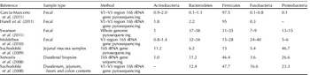
FISH was performed on fecal samples of eight healthy beagle dogs between 4 and 13 years of age (Jia et al., Reference Jia, Frantz, Khoo, Gibson, Rastall and McCartney2010). The most abundant phylum present in healthy dog feces was Firmicutes (31%) and included Clostridium, Eubacterium, and the Lactobacillus–Enterococcus group. Actinobacteria (12%) was another predominant phylum in the fecal samples of healthy dogs, with the presence of the Atobium cluster and bifidobacteria (108 cells/g of feces). In the same study, Bacteroidetes was present at about 108 cells/g of feces and consisted of <1% of total bacteria in the feces of dogs. Middelbos et al. (Reference Middelbos, Vester Boler, Qu, White, Swanson and Fahey2010) was the first to use 16S rRNA gene pyrosequencing to study fecal microbial communities of healthy adult research dogs. Sequencing of the V3 hypervariable region resulted in 17,828 high-quality sequences from five individual fecal samples of dogs fed a control diet and 21,660 sequences from five individual fecal samples of dogs fed a beet pulp-containing diet (7.5% of diet). The results indicated the predominance of the phyla Firmicutes (15–28%), Bacteroidetes (32–34%), Fusobacteria (24–40%), Actinobacteria (0.8–1.4%), and Proteobacteria (5–6%) (Table 2). Although Firmicutes and Bacteroidetes have been reported to co-dominate the human fecal microbiome that study suggested that canine feces were co-dominated by Firmicutes, Bacteroidetes, and Fusobacteria. Fecal samples from this study were also analyzed using shotgun sequencing to assess the functional and metabolic capacity of the canine fecal microbiome in addition to its phylogenetic composition (Swanson et al., Reference Swanson, Dowd, Suchodolski, Middelbos, Vester, Barry, Nelson, Torralba, Henrissat, Coutinho, Cann, White and Fahey2011). Shotgun sequencing identified the same phyla; however, the abundance of Bacteroidetes and Firmicutes (~35%) was much higher than that of Fusobacteria (7–8%), suggesting a bias involved with the generation of amplicons for the 16S rRNA gene-based method. Within Firmicutes, the class Clostridia was predominant (83–90%).
Bacterial phyla (expressed as percentage of sequences) in fecal and mucosal samples of select canine GI microbiome studies

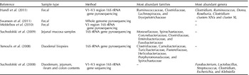
Two recent studies (Handl et al., Reference Handl, Dowd, Garcia-Mazcorro, Steiner and Suchodolski2011; Garcia-Mazcorro et al., Reference Garcia-Mazcorro, Lanerie, Dowd, Paddock, Grutzner, Steiner, Ivanek and Suchodolski2011) examined fresh fecal samples collected from 12 client-owned dogs of a wide age range (0.7–10.2 years) and of 10 breeds. These studies used V1–V3-specific primers to generate 16S rRNA amplicons for pyrosequencing (Table 1). Both studies reported the predominance of Firmicutes (95–97.5%) (Table 2), comprising, the classes Clostridia, Bacilli, and Erysipelotrichi (Table 2). Ruminococcaceae, Clostridiaceae, Lachnospiraceae, and Erysipelotrichaceae were the most abundant families present in the fecal samples of these dogs (Table 3). The clostridia class was dominated by the Clostridium (22.7%) and Ruminococcus (17.4%) genera. Dorea (7%) and Roseburia (5%) were the other prevalent genera reported. Within Clostridium, clusters XIVa (~60% of all Clostridiales sequences) and cluster XI (~34%) were most abundant. Both clusters have been linked to GI health outcomes, as members of both clusters actively produce SCFA, especially butyrate. Butyrate is the preferred energy source of colonocytes and is important for GI health via stimulation of cell proliferation, promotion of apoptosis, and prevention of colon cancer (Wong et al., Reference Wong, de Souza, Kendall, Emam and Jenkins2006). In contrast to the results of Middelbos et al. (Reference Middelbos, Vester Boler, Qu, White, Swanson and Fahey2010) Middelbos et al. (Reference Middelbos, Vester Boler, Qu, White, Swanson and Fahey2010) these two studies reported a very low abundance of Bacteroidetes (2.2%), and its predominant genera Prevotella (0.04%) and Bacteroides (0.022%). The abundance of Actinobacteria was also quite low (1.8%). Coriobacteriaceae, composed of the genera Collinsella and Slackia, was the main family present within Actinobacteria. In contrast to Middelbos et al. (Reference Middelbos, Vester Boler, Qu, White, Swanson and Fahey2010), Fusobacteria were reported to be present at very low abundance (0.3%) in that study. Although a more in-depth evaluation of the canine GI microbiota is needed, these initial studies highlight the spatial differences that exist among the various sections of intestine and fecal samples.
Most abundant bacterial families and genera in fecal and mucosal samples of select canine GI microbiome studies

–, Data not provided.
Although 16S rRNA gene pyrosequencing has many advantages over traditional culture, its limitations cannot be ignored. Differences in DNA extraction methods and PCR primers may result in the over- or under-representation of some microbial groups. The focus on the 16S rRNA gene also only provides phylogenetic information. Thus, other techniques are needed to understand microbial metabolism and host–microbe interactions. A metagenomics approach that uses shotgun sequencing provides phylogenetic and functional information on the microbiome. Using 1,008,341 DNA sequences from two pooled samples from healthy adult research dog feces, Swanson et al. (Reference Swanson, Dowd, Suchodolski, Middelbos, Vester, Barry, Nelson, Torralba, Henrissat, Coutinho, Cann, White and Fahey2011) reported the gene content of the canine microbiome. Primary functional categories were not significantly affected by diet and were associated with carbohydrates (12.5–13.0% of sequences); protein metabolism (8.1–9.1%); DNA metabolism (7.1–7.4%); cell wall and capsule (7.0–7.6%); amino acids and derivatives (6.8–6.9%); virulence (6.2–7.2%); and cofactors, vitamins, prosthetic groups and pigments (5.7–6.0%).
Effects of dietary intervention on canine GI microbiota
Many commercial dog foods contain ingredients that significantly affect the population and/or activity of GI microbes and the fermentation process. Similar to what is done with human foods, dietary fiber, prebiotics, and probiotics are often added to canine diets as a means to enhance GI health. The notion that commensal GI microbes are beneficial is not new. In fact, the modulation of GI microbes by probiotics was established early in the 20th century. Manipulation of canine GI microbiota to improve health via dietary intervention, however, did not begin until the early 1990s. Many studies have indicated the beneficial effects of feeding dietary non-digestible carbohydrates, including resistant starch, non-starch polysaccharides, and non-digestible oligosaccharides, to dogs. At the proper concentrations, non-digestible carbohydrates can improve laxation and stool quality, and alter microbial populations and fermentative end-product concentrations. Dietary fiber and prebiotics have been the major nutritional strategies studied to modulate the canine GI microbiota. Fructans, including chicory root extract, inulin, oligofructose, and short-chain fructooligosaccharides (scFOS) have been studied in greater detail in dogs than other ingredients with prebiotic potential. Other ingredients used in this regard include yeast cell wall (YCW) extracts, galactooligosaccharides, lactosucrose, lactulose, maltodextrin-like oligosaccharides, transgalactooligosaccharides and xylooligosaccharides, pullulan, and γ-cyclodextrins.
Very few clinical experiments have been performed to test the ability of dietary fiber and prebiotics to promote GI health in dogs. More commonly, an array of GI health indicators, including fecal scores, pH, microbiota, and fermentation end-products, have been used to evaluate prebiotic efficacy in healthy dog populations. Due to their relationship with GI health, increased fecal bifidobacteria, lactobacilli, and butyrate are traditionally considered beneficial, while increased C. difficile or C. perfringens, E. coli, and protein catabolites (e.g., ammonia, phenols, indoles, biogenic amines, and volatile sulfur-containing compounds) are traditionally considered detrimental. As only a brief overview of this area is provided below, the authors refer to Swanson and Fahey (Reference Swanson, Fahey, Gibson and Rastall2006) and Vester and Fahey (Reference Vester, Fahey, Cho and Finocchiaro2010) for an in-depth description of these prebiotic-based studies in dogs.
Fecal bifidobacteria and lactobacilli populations have long been used in human and animal studies as a gauge of prebiotic potential and GI health. Several forms of fructans, the most studied prebiotic class in dogs, have been reported to have prebiotic activity. Russell et al. (Reference Russell1998) conducted three experiments to study the effects of scFOS or chicory on fecal microbiota populations. Compared to the control, dogs fed scFOS (1%) or chicory (3%) had greater fecal bifidobacteria (1×10⁹ CFU/g versus 8.2×10¹⁰ CFU/g versus 4.5×10¹⁰ CFU/g, respectively) and lower fecal clostridia (1.6×10¹¹ CFU/g versus 1.3×10¹⁰ CFU/g versus 4.2×10¹⁰ CFU/g, respectively) concentrations in the first experiment. A second and a third experiment that evaluated increasing chicory (1.5, 3.0, and 5.0%) and chicory (0, 0.5, 2.0, and 4.0%) and soy (0, 6.0, and 12.0%) concentrations demonstrated similar effects. Beynen et al. (Reference Beynen, Baas, Hoekemeijer, Kappert, Bakker, Koopman and Lemmens2002) reported greater fecal bifidobacteria in dogs fed 1% oligofructose compare to control (8.8×10¹⁰ versus 7.8×10¹⁰ CFU/g), while Swanson et al. (Reference Swanson, Grieshop, Flickinger, Healy, Dawson, Merchen and Fahey2002a) demonstrated similar effects in healthy adult dogs fed 4 g scFOS (~1.3% of food intake)+2 g YCW extract (~0.67% of food intake) per day. In that study, not only were fecal bifidobacteria greater (9.4×10¹⁰ versus 10.0×10¹⁰ CFU/g), but ileal lactobacilli (7.6×10¹⁰ versus 8.7×10¹⁰ CFU/g) and fecal lactobacilli (8.2×10¹⁰ versus 9.8×10¹⁰ CFU/g) were greater in dogs fed scFOS+YCW extract compared to controls. Swanson et al. (Reference Swanson, Grieshop, Flickinger, Bauer, Healy, Dawson, Merchen and Fahey2002b,Reference Swanson, Grieshop, Flickinger, Bauer, Chow, Wolf, Garleb and Fahey2002c) demonstrated similar effects in healthy adult dogs fed 2 g (~0.5% of food intake) scFOS+2 g (~0.5% of food intake) YCW extract and 4 g (~0.8% of food intake) scFOS+2×10⁹ CFU Lactobacillus acidophilus probiotic per day, respectively. In healthy geriatric dogs, consumption of 1% chicory or 1% YCW extracts resulted in greater fecal bifidobacteria compared to control dogs (10.1×10¹⁰ versus 10.5×10¹⁰ versus 10.6×10¹⁰ CFU/g) (Grieshop et al., Reference Grieshop, Flickinger, Bruce, Patil, Czarnecki-Maulden and Fahey2004). Blends of fermentable carbohydrates (beet pulp, FOS and YCW extract) have also been demonstrated to increase fecal bifidobacteria compared to a non-fermentable fiber (cellulose) control (Middelbos et al., Reference Middelbos, Fastinger and Fahey2007).
The prebiotic potential of other carbohydrate-based ingredients has been tested in healthy dogs. For example, lactosucrose, a trisaccharide produced from lactose and sucrose, has been shown to increase bifidobacteria populations (8.9 CFU log 10/g at baseline versus 9.4 CFU log 10/g 14 days of supplementation; Terada et al., Reference Terada, Hara, Oishi, Matsui, Mitsuoka, Nakajyo, Fujimori and Hara1992). In that study, lecithinase-positive clostridia populations, which include C. perfringens, were decreased after 14 days (3.1 versus 6.0 CFU log 10/g) of lactosucrose feeding. Inclusion of pullulan (4 g/day), a naturally occurring fermentation product produced by Aureobasidium pullulans, increased ileal bifidobacteria (~0.7 log unit increase) and lactobacilli (~0.9 log unit increase) populations in healthy adult research dogs (Spears et al., Reference Spears, Karr-Lilienthal, Grieshop, Flickinger, Wolf and Fahey2005). Finally, Faber et al. (Reference Faber, Hopkins, Middelbos, Price and Fahey2011) reported a quadratic increase in bifidobacteria after supplementation of increasing dietary galactoglucomannanoligosaccharide concentrations (0.0, 0.5, 1, 2, 4, and 8%). An approximate 2 log unit increase (those fed 8.0 versus 9.8 CFU log 10/g) was noted between dogs fed the control and 8% galactoglucomannanoligosaccharides.
Although the majority of prebiotic studies have focused on fecal samples of healthy adult dogs, Apanavicius et al. (Reference Apanavicius, Powell, Vester, Karr-Lilienthal, Pope, Fastinger, Wallig, Tappenden and Swanson2007) performed an experiment aimed at evaluating the protective effects of 1% scFOS or 1% inulin during a pathogen (Salmonella Typhimurium) challenge in weanling (12-week-old) puppies. Results indicated that infected puppies consuming fructans had a lower severity of enterocyte sloughing than those fed the control diet. Puppies fed inulin also had higher fecal acetate, total SCFA concentrations and lactobacilli. Thus, this study indicated that prebiotics such as inulin and FOS appeared to attenuate some of the negative effects of Salmonella challenge and may provide protection against infection in weanling puppies.
Although the above studies have provided evidence that prebiotics and fibers have the ability to manipulate the GI microbiota of dogs, the field as a whole has many limitations. First, most prebiotic studies have used traditional plating techniques or qPCR to quantify a limited number of bacteria (e.g., Lactobacillus, Bifidobacteria, C. perfringens, and E. coli) to form conclusions regarding prebiotic potential. Such studies do not provide a widespread phylogenic and taxonomic characterization of gut microbial communities. Second, most studies were performed in healthy adult research populations rather than the ill or susceptible (e.g., weanling and geriatrics) animals most likely to benefit from such dietary intervention. Third, most studies have analyzed fecal samples rather than mucosal or digesta samples within the GI tract, so application to disease is sometimes questioned. Lastly, a wide range of prebiotic dosages have been tested across the experiments, often making it difficult to compare one study to another.
Recent canine fiber and prebiotic experiments incorporated the use of 454 pyrosequencing to provide a more in-depth view of the GI microbial communities and how they may be impacted by diet. The first nutritional intervention study in dogs that used pyrosequencing was performed by Middelbos et al. (Reference Middelbos, Vester Boler, Qu, White, Swanson and Fahey2010), with a goal to assess the effects of beet pulp fiber on fecal microbial composition. Dogs fed a control diet were compared to dogs fed a diet containing 7.5% beet pulp in a crossover design with 14 days periods. Beet pulp is a very common dietary fiber source in commercial dog foods and consists of a complex mixture of fermentable and non-fermentable carbohydrates. In that study, the abundance of Actinobacteria and Fusobacteria was lower when dogs were fed the beet pulp diet compared to control. Within Firmicutes, Clostridia abundance was greater in beet pulp-fed dogs compared to control dogs. Eubacterium halii, a butyrate producer, was only present in beet pulp-fed dogs. Furthermore, an overrepresentation of Faecalibacterium prausnitzii and an under-representation of Fusobacteria was observed when dogs were fed the beet pulp-containing diet. Similar to E. halii, F. prausnitzii is another butyrate producer and has been shown to possess anti-inflammatory properties.
The effects of a synbiotic formulation that included a probiotic (5×10⁹ CFU of a mixture of seven bacterial species) and prebiotic (fructooligosaccharides, arabinogalactans) in dogs were reported recently (Garcia-Mazcorro et al., Reference Garcia-Mazcorro, Lanerie, Dowd, Paddock, Grutzner, Steiner, Ivanek and Suchodolski2011). Changes in fecal microbes were evaluated using 454 pyrosequencing. The synbiotic capsules were fed to privately owned dogs for 21 days. The most abundant phylum in feces of all dogs was Firmicutes, followed by Actinobacteria and Bacteroidetes, regardless of synbiotic treatment. Synbiotic treatment increased the abundance of family Eubacteriaceae. The proportion of the Eubacterium genus was greater after synbiotic treatment as compared to baseline values. Similarly, the proportion of phylum Fusobacteria and all phylogenetic levels down to the genus Fusobacterium were significantly higher during synbiotic administration. Massive parallel 454 pyrosequencing is a great tool that allows the characterization of bacterial groups at various taxonomic levels. More research using this technology is needed to expand our knowledge of GI tract microbial communities in healthy versus diseased dog populations and to study the effects of dietary intervention.
Effects of disease on canine GI microbiota
The invasion and/or colonization of the GI tract with specific pathogens may profoundly disturb the integrity of the intestinal epithelial barrier (Viswanathan et al., Reference Viswanathan, Hodges and Hecht2009). Several potential GI pathogens are recognized in dogs, including C. difficile, C. perfringens, C. jejuni, Salmonella spp., and E. coli (Marks et al., Reference Marks, Rankin, Byrne and Weese2011). However, most of those are recognized commensal in the canine GI tract and have been isolated at similar frequencies from dogs with signs of GI disease and clinically healthy dogs (Marks and Kather, Reference Marks and Kather2003). Therefore, the cause–effect relationships between those organisms and GI disease need to be interpreted with caution.
Non-specific alterations of the GI microbiota have also been regarded as a pivotal factor for the development of GI disease. As mentioned above, the balanced GI microbiota is responsible for crucial effects on the host. Therefore, a disturbed GI microbiota may result in intestinal disorders (e.g., bacterial deconjugation of bile acids may lead to malabsorption of fat and lipid-soluble vitamins; Hall, Reference Hall2011). Small intestinal bacterial overgrowth (SIBO) and IBD are associated with a disturbed GI microbiota in humans. In dogs, the use of the term SIBO (typically >105 CFU/ml small intestinal content) is under debate, since a study has shown that much higher bacterial counts are commonly found in clinically healthy dogs (German et al., Reference German, Day, Ruaux, Steiner, Williams and Hall2003). Therefore, the alternative terms antibiotic-responsive diarrhea or small intestinal dysbiosis are currently used for dogs with this syndrome.
Recent studies have attempted to characterize the fecal microbiota of diarrheic dogs. Bell et al. (Reference Bell, Kopper, Turnbull, Barbu, Murphy and Mansfield2008) assessed changes in the GI microbiota associated with acute episodes of diarrhea by using T-RFLP and qPCR, and observed large-scale changes of the GI microbiota during episodes of diarrhea. These included increased abundance of Clostridiales (especially C. perfringens) and Enterococcus spp. Jia et al. (Reference Jia, Frantz, Khoo, Gibson, Rastall and McCartney2010) utilized FISH and DGGE to investigate the composition of fecal microbiota in dogs with chronic diarrhea. FISH analysis revealed a significantly higher Bacteroides count in dogs with chronic diarrhea compared to control dogs. The proportion of the Lactobacillus–Enterococcus group was 8% higher, albeit this was not significant, in control dogs than in dogs with chronic diarrhea. Bifidobacteria were not significantly different between the two groups (Jia et al., Reference Jia, Frantz, Khoo, Gibson, Rastall and McCartney2010).
IBD is a common chronic GI disorder in dogs. Similarly to humans, interplay between the intestinal microbiota together with an underlying genetic susceptibility of the host and dietary and environmental factors, are implicated in the development of IBD (Virgin and Todd, Reference Virgin and Todd2011). Technological advances have allowed a more detailed understanding of the composition of the intestinal microbiota in IBD. In human IBD, lower abundance of Firmicutes and Bacteroides, and higher abundance of Proteobacteria compared to healthy subjects have been observed (Seksik, Reference Seksik2010). Similar changes in the small intestinal microbiota have been observed in dogs with IBD. Xenoulis et al. (Reference Xenoulis, Palculict, Allenspach, Steiner, Van House and Suchodolski2008) constructed 16S rRNA gene clone libraries from duodenal brush samples from healthy dogs and dogs with IBD. In this study, a significant lower species richness and significantly higher proportion of Enterobacteriaceae were observed in dogs with IBD (Xenoulis et al., Reference Xenoulis, Palculict, Allenspach, Steiner, Van House and Suchodolski2008). Suchodolski et al. (Reference Suchodolski, Xenoulis, Paddock, Steiner and Jergens2010) applied the same technique to duodenal biopsies from dogs with IBD. These dogs had a significantly higher abundance of Proteobacteria and a significantly lower abundance of Clostridia (Suchodolski et al., Reference Suchodolski, Xenoulis, Paddock, Steiner and Jergens2010).
Future directions
Recent advances in sequencing technology and bioinformatics have enabled the molecular characterization of GI microbial populations in humans and animals. Although the human microbiome projects have the support of the National Institutes of Health and will lay a strong foundation of science relating to microbial ecology and host–microbe interactions, more work is desperately needed in the dog population. Despite the recent progress made through the use of high-throughput sequencing techniques, the phylogenetic structure and functional/metabolic capacity of the healthy canine GI microbiome is far from being fully characterized. Moreover, the impact that dog breed, age, diet, and other environmental factors has on microbial groups, and how and which microbes contribute to GI and host metabolic diseases, is far from understood.
In the future, a deeper characterization of the phylogenetic and functional/metabolic capacity of canine GI microbiome, including the mucosal and luminal populations that exist throughout the GI tract, is needed. Furthermore, to obtain a better understanding of how the microbial populations interact with the host in times of health and disease, the application of transcriptomic-, proteomic-, and metabolomic-based techniques to host and microbial samples, will be required. Although these techniques are only now being applied to human GI microbiology, the same is needed in canine GI health. As the use of any single ‘omics’ approach will not be sufficient to characterize and understand the complexity of the host–microbe relationships that exist in the gut, an integrated approach is the key to understanding the role of the microbiome in canine health and disease.