




No CrossRef data available.
The prenatal diagnosis of, and short-term outcome for, patients with congenitally corrected transposition
Published online by Cambridge University Press: 21 January 2005
Abstract
Congenitally corrected transposition is a rare congenital anomaly, with only a few cases diagnosed and reported prenatally even in the largest fetal series. To determine the morphologic features and outcome for the lesion as recognized during fetal life, we reviewed the fetal and postnatal echocardiograms and medical records of 11 consecutive cases of congenitally corrected transposition. These were identified among 230 (4.7%) consecutive cases of structural cardiac disease referred to our fetal cardiology unit over a period of 4 years. The mean gestational age at diagnosis was 24.7 weeks. Reasons for referral were suspected complete transposition, abnormal position of the heart, and bradyarrhythmias. Associated cardiac lesions included an abnormal cardiac position in 6 cases, ventricular septal defect in 8, obstruction of the subpulmonary outflow tract in 6, tricuspid valvar displacement in 5, and complete atrioventricular block in 2. Only 3 of the cases had mild tricuspid regurgitation prior to birth. Termination was chosen in 4 cases with severe obstruction to pulmonary flow. Of the remaining cases, 2 patients died at 3 and 12 months after birth, respectively. Both developed significant tricuspid regurgitation associated with unexpected major arrhythmias. The remaining 5 patients are alive and relatively well at a mean follow-up of 25.4 months. An epicardial pacemaker was inserted in 1 because of complete atrioventricular block. We conclude that prenatal counseling must be guarded following the diagnosis of congenitally corrected transposition, even in fetuses with an apparently favorable state at initial examination. Some of these cases may undergo major and unexpected changes, particularly with regard to cardiac rhythm and tricuspid valvar function, with concomitant significant changes in prognosis.
Information
- Type
- Original Article
- Information
- Copyright
- © 2004 Cambridge University Press
In postnatal series, congenitally corrected transposition accounts for 0.6–1.4% of all forms of congenital cardiac disease.1, 2 Although the segmental combination can occur in isolated form, almost all cases seen at autopsy have been associated with other intracardiac abnormalities.3 These include a ventricular septal defect, obstruction to the subpulmonary outflow tract, tricuspid valvar abnormalities, and atrioventricular block. Less frequently, the associated cardiac malformations are straddling of either atrioventricular valve, hypoplasia of one ventricle, aortic stenosis, and lesions of the aortic arch.4–6 Cases with absence of the left atrioventricular connection, and/or double-outlet right ventricle, have been included in some series, but these lesions cannot be associated with congenitally corrected transposition, since both would remove the double discordance that is the essence of the malformation. Although patients without associated defects may remain asymptomatic, and undetected until late in life,7 congenitally corrected transposition is not a benign condition, and survival is substantially reduced in most adults.8–15 Mortality can be the consequence not only of the associated cardiac malformations, but also to the anatomic condition itself, particularly to tricuspid valvar insufficiency, right ventricular dysfunction, and atrioventricular block.11, 13, 14 Up to one-third of these deaths can be sudden.10 It is reported that complications develop at an older age in patients without associated lesions, and less frequently in cases undergoing palliative interventions or no surgery compared with those who had intracardiac operations.12, 14 Congenitally corrected transposition can be detected prenatally, but even the largest series thus far reported contain a small number of cases.16–19 Such series show a distorted spectrum of cardiac malformations, in which the prognosis is poor. This spectrum includes lesions that are more easily detected in a first level obstetrical scan, particularly severe Ebstein’s malformation of the tricuspid valve, or a diminutive ventricle.17 Because of the rarity of congenitally corrected transposition, and the paucity of data from fetal groups, there is still insufficient knowledge about the clinical course and survival in these patients for adequate parental counseling subsequent to prenatal diagnosis.
Materials and methods
Among 230 consecutive cases of structural congenital heart disease referred to our Fetal Cardiology Unit from January 1999 to July 2003, we retrospectively identified 11 (4.7%) cases of congenitally corrected transposition that have not previously been published. All fetal echocardiograms were performed with Acuson Sequoia 512 (Acuson Inc., Mountain View, California) ultrasound system, interfaced with 7 or 5 MHz transducers. Fetal examination was carried out following the segmental approach and using standard anatomical planes. The technique of Cordes et al.20 was used to assess the fetal right/left axis, and consequently the atrial arrangement and cardiac position. Cross-sectional echocardiography was complemented with color Doppler flow imaging and spectral Doppler study of the fetal heart in all cases. Criterions for inclusion in the study were:
- usual or mirror-imaged atrial arrangement;
- discordant atrioventricular and ventriculo-arterial connections;
- complete follow-up data. These includ postnatal echocardiograms, surgical records and autopsy findings in case of interruption or death.
Cases with absence of one atrioventricular connection, or double-outlet right ventricle, have been excluded.
All fetal echocardiographic studies were reviewed with particular attention to tricuspid valvar insertion and function, ventricular septal defects, obstruction to the subpulmonary outflow tract, and cardiac rhythm. In order to define the degree of displacement of the tricuspid valve, we expressed the distance between the annular insertion of the mitral and the tricuspid valve as a percentage of the total length of the ventricular septum as seen in a four-chamber view at end-diastole. Displacement was defined as mild when less than 25%, moderate when between 25% and 50%, and severe when more than 50%. The severity of tricuspid regurgitation was judged by color Doppler on the basis of spatial extension of the regurgitant jet and its relation to the atrial size. Cross-sectional anatomy of the ventricular septum was investigated for location, number, and size of defects. The ventricular septal defect was judged as large when its size was greater than one-third of the diameter of the aorta. The direction of interventricular shunting was studied with both pulsed and color Doppler ultrasound. The ratio of the diameter of the aortic root to that of the pulmonary trunk was calculated by a single observer (A.S.) unaware of the follow-up data for each patient. Internal diameters of the great arteries were taken following the method described by Sharland and Allan.21 Doppler studies of these vessels and of the arterial duct were performed in all cases.
In uninterrupted pregnancies, we reviewed the medical records, postnatal echocardiograms, electrocardiograms, operative, and autopsy reports in all patients. Serial echocardiograms were performed in every patient. The dimensions of ventricular septal defect at birth are reported. The severity of tricuspid regurgitation was judged by color Doppler imaging. The severity of obstruction to the pulmonary flow was assessed by the highest Doppler peak gradient obtained after the 1st month of age. Postnatal echocardiograms were reviewed in a non-blinded fashion.
Guidelines to prenatal diagnosis
Prenatal diagnosis of congenitally corrected transposition has to be based on precise identification of morphologic characteristics of the cardiac chambers and the great arteries, independent of their position in the chest. This is particularly important in view of common anomalies of the location of the heart in this setting. If the heart lies mostly in the right or left side of the chest, the finding most readily detected in the four-chamber view is the posterior position of the morphologically right ventricle (Fig. 1a). The mirror-imaged position of the ventricles in these cases will be appreciated also in the short-axis view of the heart at the level of the two atrioventricular valves (Fig. 2b). This view will demonstrate the posterior position of the morphologically right ventricle, with the tricuspid valve attached to the ventricular septum. Although useful, this method does not apply when the heart lies in the midline. Indeed, the ventricles are often nearly side-by-side, so it will be very difficult to appreciate which one is the most posterior ventricle (Fig. 1b). Whatever may be the cardiac position, the most reliable finding in the four-chamber view is noticing how the morphologically left atrium, which receives the pulmonary venous drainage, is connected with the morphologically right ventricle (Fig. 1a, b), so demonstrating the discordant atrioventricular connections.

Figure 1. Transverse sections of the fetal thorax in caudal-to-cranial perspective showing the four-chamber view in two cases of congenitally corrected transposition, (a) corresponds to the 11th, and (b) to eighth case in this series. In (a), with usual atrial arrangement, the heart is mostly located in the left side of the chest while in (b), which shows mirror-imaged atrial arrangement, the heart is in the midchest. Both pictures show how the morphologically left atrium, which receives the pulmonary venous drainage, is connected with the morphologically right ventricle. The flap valve of the oval foramen is oriented into this atrium. LA: left atrium; RA: right atrium; mLV: morphologically left ventricle; mRV: morphologically right ventricle; AO: aorta; A: anterior; P: posterior; L: left; R: right.

Figure 2. Parasagittal sections of the fetal thorax imaging the short axis of the heart at the level of the two atrioventricular valves in a normal fetus (a) and in the sixth case of congenitally corrected transposition (b), in our series. The heart is located mostly in the left side of the chest, but unlike the normal situation, (b) shows how the posterior ventricle is the morphologically right ventricle, with a tricuspid valve attached to the ventricular septum. The morphologically left ventricle is in anterior position, and possesses a bileaflet atrioventricular valve unattached to the septum. mLV: morphologically left ventricle; mRV: morphologically right ventricle; ST: stomach; A: anterior; P: posterior; I: inferior; S: superior.
In both regular and congenitally corrected transposition, the great arteries exit from the heart in parallel. In both cases, the aorta arises from the morphologically right ventricle, almost always in anterior position relative to the pulmonary trunk. Due to the different position of the ventricles, the aorta is usually anterior and to the right in regular transposition, whereas it is typically anterior and to the left in congenitally corrected transposition. Parasagittal views of the fetus can easily demonstrate the parallel arrangement of the great arteries (Fig. 3a, b), but not their relative spatial relationship. If the sonographer relies only on the parallel arrangement, therefore, it is easy to mistake congenitally corrected transposition for regular transposition.

Figure 3. This shows the parallel course of the great arteries arising from the heart in a case of regular transposition (a), and in a case of congenitally corrected transposition (b). It is very difficult to differentiate between these two cardiac malformations utilizing only on this echocardiographic sign. PT: pulmonary trunk; AO: aorta.
The three-vessel view, however, can easily distinguish between these two cardiac malformations. In regular transposition the three vessels appear “piled up”, with the ascending aorta anterior and to the right of the pulmonary trunk (Fig. 4b). In congenitally corrected transposition, the pulmonary trunk will always be seen between the superior caval vein and the ascending aorta. The aorta will usually be anterior to the left in usual atrial arrangement and anterior to the right in mirror-imaged atrial arrangement (Fig. 4c,d).

Figure 4. Transverse sections of the upper fetal thorax in caudal-to-cranial perspective showing the three-vessel view in: a normal fetus (a), a case with regular transposition (b), and two cases with congenitally corrected transposition (c, d). In the normal situation, the three vessels are arranged in a straight line with a decreasing order of diameters. The pulmonary trunk is the most leftward and anterior vessel. In regular transposition, the three vessels appear “piled up” and the ascending aorta is displaced anteriorly and to the right of the pulmonary trunk. In congenitally corrected transposition, the pulmonary trunk lies characteristically in between the aortic root and the superior caval vein. In usual atrial arrangement, the ascending aorta is usually anterior and to the left of the pulmonary trunk (c), whereas in mirror-imaged atrial arrangement, the aorta is usually anterior and to the right (d). SCV: superior caval vein; PT: pulmonary trunk; AAO: ascending aorta; DAO: descending aorta; A: anterior; P: posterior; L: left; R: right.
Statistical analysis
Descriptive statistics, Shapiro–Wilk test for normal distribution and Pearson correlation have been used for determining the significance of the ratio of the diameters of the fetal aorta and pulmonary trunk, and for the Doppler peak gradient across the pulmonary outflow after birth. Statistical analysis has been performed by Statistica 5.5, StatSoft Inc., Tulsa, OK, USA.
Results
Prenatal diagnosis
The mean gestational age at first diagnosis was 24.7 weeks, with a range from 20 to 34 weeks. Both sexes were equally distributed. All patients had normal karyotype, and no extracardiac malformation was noted. The findings are summarized in Table 1.
Table 1. Prenatal diagnostic features in fetuses with congenitally corrected transposition.

Reason for referral
Regular transposition had been suspected at the obstetric scan in five patients. Cardiac malposition had been noted in four cases, bradyarrhythmia in two cases, and hypoplasia of the left ventricle was suspected in one case.
Atrial arrangement and cardiac location
In 10 cases, there was usual atrial arrangement, with mirror-imaged arrangement in the other case. The heart was abnormally positioned in six cases. Of these, it was in the midline in 5, with the apex pointing to the opposite side expected from the atrial arrangement (Fig. 1b). In one case, with usual atrial arrangement, the heart was mostly in the right hemithorax (Fig. 5a, b).

Figure 5. (a) Shows the autopsied specimen from our fifth case, prepared to simulate the four-chamber view. The heart was located in the right chest with the apex to the right. The morphological illustration shows the displacement of the posterior part of the tricuspid valve (white arrowhead). This is not appreciable in (b), which represents a four-chamber view at 20 weeks gestational age in the same case, imaging a more anterior portion of this valve. RA: right atrium; LA: left atrium; mLV: morphologically left ventricle; mRV: morphologically right ventricle.
Fetal cardiac rhythm
At first examination, there was sinus rhythm in 10 cases, albeit that one of these (case 1) developed 2:1 atrioventricular block (Fig. 6) at 34 weeks, and had complete atrioventricular block at birth. In cases 2 and 4, recurrent episodes of sinus bradycardia were noted, apparently unrelated to the compression of the maternal abdomen by the probe. Our sixth fetus had complete atrioventricular block when first seen at 34 weeks, and this persisted until term.

Figure 6. Color Doppler M-mode tracing from our first case at 34 weeks gestational age, showing 2:1 atrioventricular block. There are two atrial contractions to each ventricular contraction. The two lines mark the onset of ventricular outflow into the aorta. The ventricular rate is 64 beats/min. a: atrial contraction; V: ventricular contraction.
Ventricular septal defect
A ventricular septal defect was detected in 8 cases. The defect was in subpulmonary position and peri-membranous in six fetuses, and located in the outlet and trabecular parts of the muscular septum in one case each. A large defect was present in three cases, all associated with severe obstruction to pulmonary flow (Fig. 7b). All other cases had small defects. Color flow mapping showed a unidirectional shunt, from the morphologically left to the morphologically right ventricle through the defect, whenever significant obstruction to the pulmonary outflow was present (Fig. 8b). The shunt was bidirectional in the other cases.

Figure 7. (a) The external view of the autopsied specimen from our fourth case shows a midline heart with the apex pointing to the right and a large anterior aorta. (b) A simulated four-chamber view of the same specimen showing a large muscular-outlet ventricular septal defect (white arrow) and the small size of the posterior pulmonary trunk, which had an atretic valve and was retrogradely perfused through the arterial duct. RAA: right atrial appendage; LAA: left atrial appendage; mLV: morphologically left ventricle; mRV: morphologically right ventricle; AO: aorta; PT: pulmonary trunk; A. Duct: arterial duct.

Figure 8. Cross-sectional four-chamber view in our seventh case, which had moderate displacement of the tricuspid valve (a). Color flow mapping in the same projection (b) shows the small trabecular ventricular septal defect with shunt from the morphologically left to the morphologically right ventricle. This unidirectional shunt was due to significant obstruction to pulmonary flow. The three-vessel view of this patient (c) illustrates the small size of the pulmonary trunk compared with that of the ascending aorta. LA: left atrium; RA: right atrium; mLV: morphologically left ventricle; mRV: morphologically right ventricle; VSD: ventricular septal defect; SCV: superior caval vein; PT: pulmonary trunk; AAO: ascending aorta; DAO: descending aorta.
Tricuspid valve
Mild displacement of the tricuspid valve was detected in four cases, with two of these having mild regurgitation. Moderate displacement was noted in another fetus, which had no evidence of regurgitation (Fig. 8a). In all the other fetuses, we noted normal insertion and function of the tricuspid valve.
Pulmonary outflow obstruction
Obstruction to pulmonary flow was suspected in six fetuses with an aorta-to-pulmonary trunk diameter ratio greater than 1.1 (Fig. 8c). In the fourth case, with a ratio of 2.6, the pulmonary valve was atretic, with retrograde flow noted through the arterial duct at fetal examination. All other cases had antegrade pulmonary flow. Of the cases, 3 also had a large ventricular septal defect.
Results
Interrupted pregnancies
Interruption was chosen in 4 out of the 8 cases (50%) diagnosed within 24 weeks of gestation. These fetuses, which had the highest aorta-to-pulmonary trunk ratio in this series, had severe obstruction to pulmonary flow and no echocardiographic evidence of tricuspid valvar displacement and regurgitation. Autopsy confirmed prenatal diagnosis in all cases but the fifth, where an Ebstein-like displacement of the tricuspid valve was found, undetected on fetal echocardiography (Fig. 5a, b).
Uninterrupted pregnancies
The data are summarized in Table 2. No intrauterine death occurred. The mean gestational age, and the mean weight at birth, was 38.5 weeks and 3071 grams, respectively. The two patients with complete atrioventricular block at term were delivered through a cesarean section. Of the patients, our first and seventh cases died within the first year of life. The first patient had received a pacemaker a few days after birth because of complete atrioventricular block associated with rapidly progressive tricuspid insufficiency (Fig. 9). At 6 months, the pulmonary trunk was banded to retrain the morphologically left ventricle for a double switch operation. Sudden death occurred at 12 months due to a lead fracture of the pacemaker. The seventh patient had trivial tricuspid regurgitation at birth, with mild subvalvar and valvar pulmonary obstruction, with a peak gradient of 25 mmHg. Shortly after birth, there were recurrent crises of sustained supraventricular tachycardia. The surface electrocardiogram showed no signs of pre-excitation. At 1 month of age, the patient had at least moderate tricuspid valve regurgitation, with a peak gradient across the pulmonary outflow tract of 75 mmHg. The supraventricular tachycardia was refractory to several single-drug antiarrhythmic therapies, and sudden death occurred at 3 months while the patient was receiving amiodarone at 6 mg/kg/day. The remaining five patients are alive and relatively well, with no medication at a mean follow-up of 25.4 months, with a range from 2 to 45 months. Our sixth patient, who had complete atrioventricular block since the 34th week of gestation, had a pacemaker implanted at 5 months because of failure to thrive.
Table 2. Postnatal characteristics and outcome of fetuses with congenitally corrected transposition in continuing pregnancies.


Figure 9. This shows a four-chamber view with color flow Doppler of tricuspid regurgitation in our first case 34 weeks gestational age, and at 71 days postnatally. In this fetus, the tricuspid valve was mildly displaced.
The ratio of the diameter of the aorta to the pulmonary trunk measured at fetal echocardiography showed a good correlation to the presence of significant obstruction to pulmonary flow. Severe obstruction was confirmed at autopsy in all cases that underwent termination of pregnancy. Among continuing pregnancies, two patients with ratio measured at greater than 1.1 during fetal life developed a significant gradient across the pulmonary outflow tract after birth (Fig. 10). Figure 11 shows the correlation between the measured ratio and the peak gradient across the pulmonary outflow tract as estimated by Doppler at 1 month of age (r = 0.86, p = 0.012).

Figure 10. This shows the rapid increase in the peak gradient across the pulmonary outflow at 3 days of life in our nineth case, compared with the normal velocity in the pulmonary trunk at 32 weeks of gestation.

Figure 11. This shows the correlation between the measured ratio and the peak gradient across the pulmonary outflow tract as estimated by Doppler at 1 month of age. AO: aorta; PT: pulmonary trunk; POG: peak pulmonary outflow gradient.
Discussion
Prevalence
The prevalence (4.7%) of congenitally corrected transposition observed in our study is relatively high. Congenitally corrected transposition may have been overrepresented in our series because fetuses with lethal chromosomal anomalies, or severe extracardiac malformations, are not systematically referred to our unit of fetal cardiology. Moreover, the period of our study reflects the substantial progress made in ultrasonic systems, and the technical skill of operators, that has occurred in recent years. The series of Allan et al.,16 including cases from the early 80s to 1992, had a very low incidence of congenitally corrected transposition (0.2%). The Italian Multicentric Study,18 whose cases were mostly collected from the mid 80s to 1996, reported an incidence of 2.1%. In our study, the reason for referral was suspected regular transposition in 4 cases diagnosed at an early stage. This is probably the result of the advanced training program in the Piedmont region, aimed at including the assessment of ventriculo-arterial connections in the routine ultrasonic screening at midtrimester. Some cases of congenitally corrected transposition may also have been classified into different categories in other series, such as double-outlet right ventricle, pulmonary atresia with ventricular septal defect, or hypoplastic left heart syndrome, because of diagnostic difficulties due to the position of the heart and complex associated cardiac anomalies. This is more probable in case of interruption, and when postmortem examinations are not performed by an experienced cardiac pathologist.
Atrial arrangement and position of the heart
In agreement with postnatal series of congenitally corrected transposition,3 usual arrangement was prevalent in our study, with mirror-imaged arrangement observed in one case (case 8). We think that precise evaluation of cardiac connections in the latter setting represents one of the most challenging tasks in prenatal diagnosis, with an increased risk of misinterpretation and inappropriate counseling.
Cardiac malposition was present in just over half of our cases, with one having the heart mostly in the right hemithorax, and five with midline hearts with the apex pointing to the opposite site expected from the atrial arrangement. Congenitally corrected transposition, therefore, should be strongly suspected whenever such a cardiac malposition is found during fetal ultrasound.
Arrhythmias
Bradyarrhythmias were the prevailing arrhythmia, with complete atrioventricular block in almost one-fifth of our cases. We found that complete atrioventricular block could be present at the first fetal examination, as in our sixth case, or could develop progressively before birth, as in the first case. Such a progression, a well-known complication in postnatal series, occurring at about 2% per year, was described in a fetus with discordant atrioventricular connections by Schmidt et al.22 We did not observe the intermittent appearance of atrioventricular block,2, 23, 24 although we do not know what the evolution might have been in the second and fourth fetuses, who had episodes of sinus bradycardia and underwent interruption.
Supraventricular tachyarrhythmias, although rare, have been described in congenitally corrected transposition. Atrial fibrillation or flutter usually come late in life, with dilation of the left atrium secondary to tricuspid valvar regurgitation usually considered the anatomic substrate.9, 10 Supraventricular tachycardia can also appear earlier, due to dual atrioventricular nodal pathways or accessory muscular atrioventricular connections, the latter usually associated with Ebstein’s malformation of the tricuspid valve and ventricular septal defect.25, 26 In our seventh case, sustained supraventricular tachycardia presented a major problem. We do not know if prophylactic implantation of a pacemaker could have avoided death. On the other hand, our first patient is the unlucky example of sudden death related to a malfunctioning pacemaker.
Tricuspid valvar displacement and dysfunction
The prevalence of tricuspid valvar displacement in our fetal series was 45%, lower than the 90% reported in anatomical studies.4 In continuing pregnancies, postnatal echocardiographic assessment of such displacement left the prenatal assessment substantially unchanged. The lower sensitivity of fetal echocardiography may derive from the limited ability to highlight minor forms of displacements, particularly when located in the posterior portion of the valve. On the other hand, postnatal echocardiographic study of the tricuspid valve is often limited. Due to the common malposition of the heart, the sonographer is dealing with difficult transthoracic windows, and the apical four-chamber view is often foreshortened. Our fifth patient, who had a mild Ebstein-like malformation of the tricuspid valve, was detected only at autopsy (Fig. 5), supporting this hypothesis. Mild tricuspid regurgitation was present at fetal echocardiography only in two cases, both with mildly displaced valves. Rapid and unexpected progression of tricuspid regurgitation after birth developed in our first and seventh patients (Fig. 9a, b). This seems to support the hypothesis that postnatal tricuspid regurgitation in congenitally corrected can be related to malformations of the valve in themselves,11 and to the increased load on the morphologically right ventricle.8, 27 Moreover, tricuspid insufficiency in our first patient, possibly related to complete atrioventricular block through ventricular dilation and higher stroke volume, was not reversed by dual-chamber pacing. Our seventh patient had long episodes of sustained supraventricular tachycardia refractory to different drug therapies. In a series of patients with congenitally corrected transposition older than 18 years of age, Connelly et al.10 documented atrial arrhythmias, including supraventricular tachycardia, in 5 out of 9 patients who died. We suspect that this arrhythmia, and antiarrhythmic therapy, could have contributed to right ventricular dysfunction, tricuspid regurgitation, and sudden death in our patient.
Obstruction to pulmonary flow
Pulmonary outflow obstruction was present in just over half of our fetal cases. The measured ratio of the arterial trunks showed a good correlation with the presence of significant obstruction after birth. The pulmonary outflow gradient increased progressively within the first few weeks after birth.
Ventricular septal defect
Our fetal series confirms the high incidence of ventricular septal defect seen in association with congenitally corrected transposition. Large defects are usually associated with significant obstruction to pulmonary flow, and are easy to be identified during fetal life. Small defects can be detected at fetal echocardiography when there is unidirectional shunting because of obstruction to pulmonary flow. The only false negative case at fetal examination, the eighth case in our series, had multiple small trabecular defects and no significant obstruction to pulmonary flow.
In the combined series of Allan and colleagues17 of 2136 fetal cardiac malformations, there were 19 cases of congenitally corrected transposition prospectively diagnosed, with three having absence of the left atrioventricular connection. As far as we know, this is the largest series of prenatal diagnosis of congenitally corrected transposition reported by a single institution not including cases of absent atrioventricular connection or double-outlet right ventricle.
We conclude that congenitally corrected transposition can be more common in prenatal series than previously reported. Cardiac malposition, ventricular septal defect, displacement of the tricuspid valve, and pulmonary outflow obstruction are common associated lesions. Congenitally corrected transposition can be commonly mistaken for regular transposition at the first level examination. Increased detection rates are expected when assessment of ventriculo-arterial connections of the fetal heart is included in obstetrical ultrasound screening programs. An increased ratio of the diameter of the aorta relative to the pulmonary trunk is an easy and reliable predictor of obstruction to pulmonary flow. Complete atrioventricular block may be present at the first fetal examination, or be the result of progressive block even before birth. Cases undergoing termination of pregnancy in our series had severe obstruction to pulmonary flow. In most cases of continuing pregnancies, a relatively good quality of life is expected over the short-term period of follow-up. Nevertheless counseling must be guarded, even when an apparently favorable morphological and functional state is found at the first fetal examination. Some cases may undergo major and unexpected changes, particularly as regards cardiac rhythm and tricuspid valvar function, with concomitant major changes in prognosis.
References
Fontana RS, Edwards JE. Frequency of occurrence of various anatomic types of malformation among patients with congenital cardiac disease. In: Congenital Cardiac Disease: A Review of 357 Cases Studied Pathologically. WB Saunders, Philadelphia, 1962, pp 28–65.
Losekoot TG, Anderson RH, Becker AE, Danielson GK, Soto B. Congenitally Corrected Transposition. Churchill Livingstone, New York, 1983, pp 15–16.
Allwork SP, Bentall HH, Becker AE, et al. Congenitally corrected transposition of the great arteries. Morphologic study of 32 cases. Am J Cardiol 1976; 38: 910–923.Google Scholar
Freedom RM, Dyck JD. Congenitally corrected transposition of the great arteries. In: Moss AJ, Adam FJ (eds). Heart Disease in Infants, Children, Adolescents. Lippincott Williams & Wilkins, Philadelphia, 2001, pp 1085–1101.
Zahn EM, Smallhorn JF, Freedom RM. Congenitally corrected transposition of the great arteries with hypoplasia of the morphologically left ventricle in the setting of situs inversus. Int J Cardiol 1992; 36: 9–12.Google Scholar
Celermajer DS, Cullen S, Deanfield JE, Sullivan ID. Congenitally corrected transposition and Ebstein’s anomaly of the systemic atrioventricular valve: association with aortic arch obstruction. J Am Coll Cardiol 1991; 18: 1056–1058.Google Scholar
Presbitero P, Somerville J, Rabaioli F, Stone S, Conte MR. Corrected transposition of the great arteries without associated defects in adult patients: clinical profile and follow up. Br Heart J 1995; 74: 57–59.Google Scholar
Hutha JC, Danielson GK, Ritter DG, Ilstrup DM. Survival in atrioventricular discordance. Pediatr Cardiol 1985; 6: 57.Google Scholar
Lundstrom U, Bull C, Wise RKH, Somerville J. The natural and “unnatural” history of congenitally corrected transposition. Am J Cardiol 1990; 65: 1222–1229.Google Scholar
Connelly MS, Liu PP, Williams WG, Webb GD, Robertson P, McLaughlin PR. Congenitally corrected transposition of the great arteries in the adult: functional status and complication. J Am Coll Cardiol 1996; 27: 1238–1243.Google Scholar
Prieto LR, Allan JH, Secic M, Rosenbaun MS, Gersony WM. Progressive tricuspid valve disease in patients with congenitally corrected transposition of the great arteries. Circulation 1998; 98: 997–1005.Google Scholar
Voskuil M, Hazekamp MG, Kroft LJM, et al. Postsurgical course of patients with congenitally corrected transposition of the great arteries. Am J Cardiol 1999; 83: 558–562.Google Scholar
Graham TP Jr, Bernard YD, Mellen BG, et al. Long term outcome in congenitally corrected transposition of the great arteries. A multi-institutional study. J Am Coll Cardiol 2000; 36: 255–261.Google Scholar
Rutledge JM, Nihill MR, Fraser CD, O’Brian Smith E, McMahon CJ, Bezold LI. Outcome of 121 patients with congenitally corrected transposition of the great arteries. Pediatr Cardiol 2002; 23: 137–145.Google Scholar
Beauchesne LM, Warnes CA, Connoly HM, Ammash NM, Tajik AJ, Danielson GK. Outcome of the unoperated adult who presents with congenitally corrected transposition of the great arteries. J Am Coll Cardiol 2002; 40: 285–290.Google Scholar
Allan LD, Sharland GK, Milburn A, et al. Prospective diagnosis of 1006 consecutive cases of congenital heart disease in the fetus. J Am Coll Cardiol 1994; 23: 1452–1458.Google Scholar
Allan LD. Atrioventricular discordance. In: Allan LD, Hornberger LK, Sharland GK (eds). Textbook of Fetal Cardiology. Greenwich Medical Media, London, 2000, pp 183–189.
Fesslová V, Nava S, Villa L, and The Fetal Cardiology Study Group of the Italian Society of Pediatric Cardiology. Evolution and long term outcome in cases with fetal diagnosis of congenital heart disease: Italian Multicenter Study. Heart 1999; 82: 594–599.Google Scholar
Paladini D, Rustico M, Todros T, et al. Conotruncal anomalies in prenatal life. Ultrasound Obstet Gynecol 1996; 8: 241–246.Google Scholar
Cordes TM, O’Leary PW, Seward JB, Hagler DJ. Distinguishing right from left: a standardized technique for fetal echocardiography. J Am Soc Echo 1994; 7: 47.Google Scholar
Sharland GK, Allan LD. Normal fetal cardiac measurements derived by cross-sectional echocardiography. Ultrasound Obstet Gynecol 1992; 2: 175–181.Google Scholar
Schmidt KG, Ulmer HE, Silverman NH, Kleinman CS, Copel JA. Perinatal outcome of fetal complete atrioventricular block: a multicenter experience. J Am Coll Cardiol 1991; 17: 1360–1366.Google Scholar
Honey M. The diagnosis of corrected transposition of the great vessels. Br Heart J 1963; 25: 313.Google Scholar
Friedberg DZ, Nadas AS. Clinical profile of patients with congenital corrected transposition of the great arteries. New Engl J Med 1970; 282: 1053.Google Scholar
Daliento L, Corrado D, Buja G, John N, Nava A, Thiene G. Rhythm and conduction disturbances in isolated, congenitally corrected transposition of the great arteries. Am J Cardiol 1986; 58: 314–318.Google Scholar
Bahrati S, Rosen K, Steinfeld L, Miller RA, Lev M. The anatomic substrate for preexcitation in corrected transposition. Circulation 1980; 62: 831–842.Google Scholar
Acar P, Sidi D, Bonnet D, Aggoun Y, Bonhoffer P, Kachaner J. Maintaining tricuspid valve competence in double discordance: a challenge for the paediatric cardiologist. Heart 1998; 80: 479–483.Google Scholar

Transverse sections of the fetal thorax in caudal-to-cranial perspective showing the four-chamber view in two cases of congenitally corrected transposition, (a) corresponds to the 11th, and (b) to eighth case in this series. In (a), with usual atrial arrangement, the heart is mostly located in the left side of the chest while in (b), which shows mirror-imaged atrial arrangement, the heart is in the midchest. Both pictures show how the morphologically left atrium, which receives the pulmonary venous drainage, is connected with the morphologically right ventricle. The flap valve of the oval foramen is oriented into this atrium. LA: left atrium; RA: right atrium; mLV: morphologically left ventricle; mRV: morphologically right ventricle; AO: aorta; A: anterior; P: posterior; L: left; R: right.

Parasagittal sections of the fetal thorax imaging the short axis of the heart at the level of the two atrioventricular valves in a normal fetus (a) and in the sixth case of congenitally corrected transposition (b), in our series. The heart is located mostly in the left side of the chest, but unlike the normal situation, (b) shows how the posterior ventricle is the morphologically right ventricle, with a tricuspid valve attached to the ventricular septum. The morphologically left ventricle is in anterior position, and possesses a bileaflet atrioventricular valve unattached to the septum. mLV: morphologically left ventricle; mRV: morphologically right ventricle; ST: stomach; A: anterior; P: posterior; I: inferior; S: superior.

This shows the parallel course of the great arteries arising from the heart in a case of regular transposition (a), and in a case of congenitally corrected transposition (b). It is very difficult to differentiate between these two cardiac malformations utilizing only on this echocardiographic sign. PT: pulmonary trunk; AO: aorta.

Transverse sections of the upper fetal thorax in caudal-to-cranial perspective showing the three-vessel view in: a normal fetus (a), a case with regular transposition (b), and two cases with congenitally corrected transposition (c, d). In the normal situation, the three vessels are arranged in a straight line with a decreasing order of diameters. The pulmonary trunk is the most leftward and anterior vessel. In regular transposition, the three vessels appear “piled up” and the ascending aorta is displaced anteriorly and to the right of the pulmonary trunk. In congenitally corrected transposition, the pulmonary trunk lies characteristically in between the aortic root and the superior caval vein. In usual atrial arrangement, the ascending aorta is usually anterior and to the left of the pulmonary trunk (c), whereas in mirror-imaged atrial arrangement, the aorta is usually anterior and to the right (d). SCV: superior caval vein; PT: pulmonary trunk; AAO: ascending aorta; DAO: descending aorta; A: anterior; P: posterior; L: left; R: right.

Table 1.

(a) Shows the autopsied specimen from our fifth case, prepared to simulate the four-chamber view. The heart was located in the right chest with the apex to the right. The morphological illustration shows the displacement of the posterior part of the tricuspid valve (white arrowhead). This is not appreciable in (b), which represents a four-chamber view at 20 weeks gestational age in the same case, imaging a more anterior portion of this valve. RA: right atrium; LA: left atrium; mLV: morphologically left ventricle; mRV: morphologically right ventricle.

Color Doppler M-mode tracing from our first case at 34 weeks gestational age, showing 2:1 atrioventricular block. There are two atrial contractions to each ventricular contraction. The two lines mark the onset of ventricular outflow into the aorta. The ventricular rate is 64 beats/min. a: atrial contraction; V: ventricular contraction.

(a) The external view of the autopsied specimen from our fourth case shows a midline heart with the apex pointing to the right and a large anterior aorta. (b) A simulated four-chamber view of the same specimen showing a large muscular-outlet ventricular septal defect (white arrow) and the small size of the posterior pulmonary trunk, which had an atretic valve and was retrogradely perfused through the arterial duct. RAA: right atrial appendage; LAA: left atrial appendage; mLV: morphologically left ventricle; mRV: morphologically right ventricle; AO: aorta; PT: pulmonary trunk; A. Duct: arterial duct.

Cross-sectional four-chamber view in our seventh case, which had moderate displacement of the tricuspid valve (a). Color flow mapping in the same projection (b) shows the small trabecular ventricular septal defect with shunt from the morphologically left to the morphologically right ventricle. This unidirectional shunt was due to significant obstruction to pulmonary flow. The three-vessel view of this patient (c) illustrates the small size of the pulmonary trunk compared with that of the ascending aorta. LA: left atrium; RA: right atrium; mLV: morphologically left ventricle; mRV: morphologically right ventricle; VSD: ventricular septal defect; SCV: superior caval vein; PT: pulmonary trunk; AAO: ascending aorta; DAO: descending aorta.
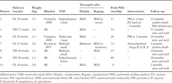
Table 2.

This shows a four-chamber view with color flow Doppler of tricuspid regurgitation in our first case 34 weeks gestational age, and at 71 days postnatally. In this fetus, the tricuspid valve was mildly displaced.

This shows the rapid increase in the peak gradient across the pulmonary outflow at 3 days of life in our nineth case, compared with the normal velocity in the pulmonary trunk at 32 weeks of gestation.

This shows the correlation between the measured ratio and the peak gradient across the pulmonary outflow tract as estimated by Doppler at 1 month of age. AO: aorta; PT: pulmonary trunk; POG: peak pulmonary outflow gradient.
 You have
Access
- 15
- Cited by