Introduction
Lymphatic filariasis (LF) is caused by mosquito-borne filarial nematodes such as Wuchereria bancrofti, Brugia malayi and Brugia timori (WHO, 2013). WHO launched a global programme to eliminate LF in 2000, implementing mass drug administration (MDA) of the anthelmintics diethylcarbamazine, albendazole and ivermectin (WHO, 2013). The tremendous efforts made to date reduced the estimated number of infected individuals from 199 million in 2000 to 51 million in 2018 (Cromwell et al., Reference Cromwell, Schmidt and Kwong2020). However, LF remains a public health problem in focal areas of Africa and Southeast Asia. Furthermore, since the current LF elimination programme relies heavily on MDA, the risk of the development and elevation of drug resistance in filarial parasites is of increasing concern (Cobo, Reference Cobo2016), and the development of complementary and alternative tools to eliminate LF is still required.
Infection with filarial parasites occurs when a host is bitten by a mosquito carrying the filarial infective third-stage larvae (L3) (WHO, 2013). During blood feeding, filarial L3 are deposited on the skin surface by a mosquito (Ewert, Reference Ewert1967; Ewert & Ho, Reference Ewert and Ho1967). Immediately after the mosquito withdraws its mouthparts from the puncture wound made by the bite, the L3 presumably migrate and penetrate the skin via the wound and enter the host body. Bargmann & Mori (Reference Bargmann, Mori, Riddle, Blumenthal and Meyer1997) suggest that water-soluble chemicals tend to diffuse slowly in the soil and may provide short-range chemosensory cues for Caenorhabditis elegans, a soil-dwelling nematode, whereas volatile chemicals diffuse more rapidly and, thus, can be used for long-range chemotaxis. Since filarial L3 are carried to a host by an intermediate host mosquito, the L3 may not need to use volatile chemicals for long-range detection of the host (host-finding) or for staying on or near the host's feces, as do L3 of skin-penetrating human-parasitic threadworm Strongyloides stercoralis and hookworm Ancylostoma ceylanicum (Gang et al., Reference Gang, Castelletto, Yang, Ruiz, Brown, Bryant, Grant and Hallem2020). Moreover, the distance travelled by filarial L3 deposited on the skin by a mosquito during the blood feeding may be short (site-finding). This suggests that filarial L3 use water-soluble chemicals as chemosensory cues to reach the wound. In fact, L3 of Brugia pahangi are attracted to water-soluble mammalian-serum, sodium ions and tris(hydroxymethyl)aminomethane (Tris), but are not attracted to volatile mammalian skin and sweat odorants such as urocanic acid and 3-methyl-1-butanol (Gunawardena et al., Reference Gunawardena, Fujimaki and Aoki2003; Kusaba et al., Reference Kusaba, Fujimaki, Vincent and Aoki2008; Mitsui et al., Reference Mitsui, Miura, Bome and Aoki2012, Reference Mitsui, Ureña-Tatis and Aoki2018; Wheeler et al., Reference Wheeler, Heimark, Airs, Mann, Bartholomay and Zamanian2020), even though the two odorants are attractants for S. stercoralis L3 (Safer et al., Reference Safer, Brenes, Dunipace and Schad2007; Castelletto et al., Reference Castelletto, Gang, Okubo, Tselikova, Nolan, Platzer, Lok and Hallem2014; Gang & Hallem, Reference Gang and Hallem2016; Gang et al., Reference Gang, Castelletto, Yang, Ruiz, Brown, Bryant, Grant and Hallem2020). On the other hand, L3 of Ancylostoma caninum are attracted to a host serum and dialysed host serum (Wauters et al., Reference Wauters, Klaver-Wesseling and Vetter1982; Vetter et al., Reference Vetter, Vingerhoed, Schoeman and Wauters1985); Strongyloides ratti L3 are attracted to dialysed rat serum, mammalian serum and egg albumins, and sodium chloride (Koga &Tada, Reference Koga and Tada2000; Tobata-Kudo et al., Reference Tobata-Kudo, Higo, Koga and Tada2000); and S. stercoralis L3 are attracted to sodium chloride (Forbes et al., Reference Forbes, Ashton, Boston and Schad2003). These facts suggest that L3 of mammalian-parasitic threadworms and hookworms, like filarial L3, may also use water-soluble chemicals as attractants when they penetrate the skin to enter the host body.
The chemical attractants for filarial L3 provide an indispensable foothold for the elucidation of the mechanism of filarial skin-penetrating behaviours. To date, only sodium ions and Tris have been identified as attractants for L3 of B. pahangi (Mitsui et al., Reference Mitsui, Miura, Bome and Aoki2012, Reference Mitsui, Ureña-Tatis and Aoki2018). Sodium ions, a major component of mammalian blood, are easily acceptable as an attractant for filarial L3. However, since Tris is not an endogenous substance, it can be assumed that, in natural infection to a host, filarial L3 recognizes an endogenous Tris-related compound. Furthermore, a few amino acids have been identified as water-soluble attractants for second-stage juveniles (J2) of Meloidogyne incognita, a plant-parasitic nematode (Fleming et al., Reference Fleming, Maule and Fleming2017), suggesting that the amino acids may also attract filarial L3.
The objective of the present study was to assess the chemotactic responses of B. pahangi L3 for Tris-related compounds and amino acids and to identify endogenous attractants for L3 using the modified agar-plate assay of Mitsui et al. (Reference Mitsui, Ureña-Tatis and Aoki2018).
Materials and methods
Chemicals
Hank's balanced salt (HBS) was obtained from Nissui Pharmaceutical Co., Ltd. (Tokyo, Japan); 2-amino-1,3-propanediol (APD, known as serinol), 2-amino-2-methyl-1,3-propanediol (AMPD), 2,2-dimethyl-1,3-propanediol (DPD), 2-hydroxymethyl-1,3-propanediol (HMP), 1,1,1-tris(hydroxymethyl)ethane (THE), 1,1,1-tris(hydroxymethyl)propane (THP), arginine (Arg), asparagine, aspartic acid and histidine (His) were purchased from Sigma-Aldrich (St. Louis, Missouri, USA). Tris, (R)-(-)-2-amino-1-propanol (R-AP), (S)-(+)-2-amino-1-propanol (S-AP), 2-amino-2-methyl-1-propanol (AMP), 2-aminoethanol (2-AE), cysteine and lysine (Lys) were purchased from FUJIFILM Wako Pure Chemical Corporation (Osaka, Japan). Alanine, glutamic acid, glutamine, glycine, isoleucine, leucine, methionine, phenylalanine, proline, serine (Ser), threonine, tryptophan, tyrosine (Tyr) and valine were purchased from Peptide Institute, Inc. (Osaka, Japan). All the other chemicals were of pure chemical grade.
Test solutions
Test solutions for the assays of chemotactic responses of B. pahangi L3 were prepared according to the test solutions used in previous studies (Mitsui et al., Reference Mitsui, Miura, Bome and Aoki2012, Reference Mitsui, Ureña-Tatis and Aoki2018). For the assay for chemotactic response of filarial L3 to Tris-related compounds and amino acids, all Tris-related compounds were dissolved in deionized water (DW) at a concentration of 200 mm, while amino acids were dissolved in DW at concentrations of 50, 150 and 200 mm in view of the differences in solubility. Only Tyr was excluded from the assay because of the poor solubility at a concentration of 5 mm. In the assay to determine the effect of hydrogen chloride (HCl) concentrations on the chemoattractant activity of 200 mm Arg, APD and AMPD for filarial L3, 0.5 ml of 0 (DW alone), 100, 200, 300, 400 and 500 mm HCl was added to 0.5 ml of a 400 mm solution of 348 mg Arg, 182 mg APD and 212 mg AMPD dissolved in 5 ml of DW. The concentration of the resulting solutions was 200 mm Arg, APD and AMPD plus 0, 50, 100, 150, 200 or 250 mm HCl, respectively. In the assay to determine the effect of concentrations of Arg, APD and AMPD on the chemoattractant activity for filarial L3, a 200 mm solution of 34.8 mg Arg, 18.2 mg APD and 21.2 mg AMPD dissolved in 1 ml of DW was serially diluted with DW to a concentration of 100, 50, 25, 12.5, 6.25 and 3.125 mm. The potential of hydrogen (pH) in all the test solutions was measured with a pH meter.
Parasite strain
The strain of B. pahangi used in the present study had been maintained for many years with Mongolian jirds (Meriones unguiculatus) and Aedes aegypti (Liverpool strain) mosquitoes at the Animal Research Center for Tropical Infections in the Institute of Tropical Medicine, Nagasaki University (Chuang et al., Reference Chuang, Nakajima and Aoki1979). Brugia pahangi L3 were obtained from mosquitoes fed two weeks earlier on microfilaremic jirds, using the modified technique of Ash & Riley (Reference Ash and Riley1970). Briefly, the infected mosquitoes were collected using a mouth aspirator, stunned by immersion in an ice bucket and put into a bag (around 50 × 50 mm) made from a piece of Kimwipe® (S-200, Nippon Paper Crecia Co., Ltd., Tokyo, Japan) paper. The bag was placed on top of the lid of a 90 mm plastic petri dish. All the mosquitoes in the bag were then lightly crushed by rolling a 15 ml plastic centrifuge tube over them, and immersed in 15 ml of a HBS solution (pH: 7.4) in a 90 mm petri dish at a temperature of 25°C. Ten minutes later, the filarial L3 that migrated out of the bag to the HBS solution were collected using a micropipette under a dissecting microscope, washed twice with HBS solution and used for the chemotactic assay.
Chemotactic assay
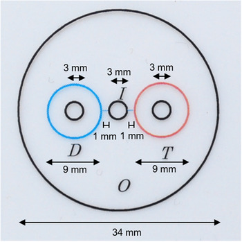
The assays of chemotactic responses of filarial L3 to test solutions were conducted by the modified method described previously by Mitsui et al. (Reference Mitsui, Ureña-Tatis and Aoki2018). Briefly, a 35 mm petri dish filled with 2 ml of 0.6% (w/v) agar (referred to as ‘agar plate’) was placed over a template transparent sheet (fig. 1). One microliter of DW was spotted in the I-area of the agar plate. Immediately, approximately ten L3 were placed into the I-area using a fine needle. Two microliters of DW and 2 μl of DW (control) or a test solution were spotted on the D- and T-areas, respectively. Then, the agar plate was placed on a hot plate adjusted to 30–35°C. After 25–30 min, the numbers of L3 accumulating in the I-area, D-, T- and O-zones were counted under a dissecting microscope. The assay was repeated five times for each test solution.
A template transparent sheet showing areas and zones to assess the chemotactic responses of Brugia pahangi infective larvae for test solutions. The small circles of I, D and T indicate the areas on which 1 μl of DW and filarial larvae, 2 μl of DW and 2 μl of a test solution are placed, respectively. The large concentric circles of D and T are the respective zones. O indicates the zone outside of the I-area, the two D- and T-zones.

Statistical analysis
The proportion of filarial L3 accumulating in the T-zone of DW (control) or test solutions was calculated according to the procedure of Mitsui et al. (Reference Mitsui, Ureña-Tatis and Aoki2018) as 100× (number of L3 accumulating in the T-zone) / (total number of L3 applied to the five agar plates using approximately ten L3 in each plate). A 95% confidence interval of the proportion was calculated using the method of Wilson (Reference Wilson1927). Furthermore, comparisons of the proportions of L3 accumulating in the T-zone of a test solution with that of DW (control group) were conducted using the Fisher's exact test in Epi-Info software (version 7, Centers for Disease Control and Prevention, Atlanta, Georgia, USA).
Results
Chemotactic response of B. pahangi infective L3 to compounds structurally related to Tris
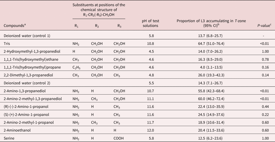
Table 1 shows the chemotactic responses of filarial L3 to test solutions of Tris and its structurally related compounds dissolved in DW at a concentration of 200 mm. A test solution of Tris had a pH of 10.8 and significantly attracted 65% of L3 compared with DW (14%, P < 0.01), while test solutions of HMP, THE, THP and DPD showed an acidic pH ranging from 4.5 to 4.8, and did not significantly attract L3 compared with DW. Test solutions of APD and AMPD had an alkaline pH of 10.7 and 11.1, and significantly attracted 56 and 60% of L3, respectively, compared with DW (14%, P < 0.01 and P < 0.01, respectively). In contrast, test solutions of R-AP, S-AP, AMP and 2-AE had an alkaline pH ranging from 11.6 to 12.0, and neither attracted nor repelled L3 significantly compared with DW. In addition, a test solution of Ser had an acidic pH of 5.8 and did not significantly attract L3.
Chemotactic responses of Brugia pahangi infective L3 to Tris and its structurally related compounds.

a All test compounds were diluted in deionized water (DW) at a concentration of 200 mm.
b The proportion of L3 accumulating in the T-zone of a test solution was calculated as 100× (total number of L3 accumulating in the T-zone) / (total number of L3 applied to five agar plates using approximately ten L3 in each plate, ranging from 49 to 56) and a 95% confidence interval of the proportion is shown as 95% CI.
c The proportion of L3 accumulating in the T-zone of each test solution was compared with that of DW as control 1 or 2 according to the Fisher's exact test.
Chemotactic responses of B. pahangi infective L3 to water-soluble amino acids
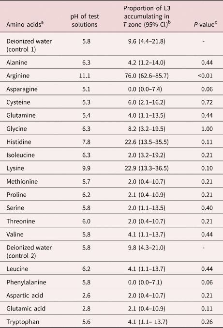
Table 2 shows the chemotactic responses of filarial L3 to test solutions of 19 water-soluble amino acids. Among the test solutions, a solution of 200 mm Arg had a pH of 11.1 and significantly attracted 76% of L3 compared with DW (10%, P < 0.01). A test solution of 200 mm His or Lys had an alkaline pH of 7.8 or 9.9, and slightly attracted 23 or 23% of L3, respectively, compared with DW (10%, P = 0.11, and P = 0.10). In contrast, the other 16 amino acids dissolved in DW showed an acidic pH ranging from 2.6 to 6.3, and neither attracted nor repelled L3 in a significant manner.
Chemotactic responses of Brugia pahangi infective L3 to 19 water-soluble amino acids.

a All test amino acids were diluted in deionized water (DW) at a concentration of 200 mm, except for leucine and phenylalanine at 150 mm, and aspartic acid, glutamic acid and tryptophan at 50 mm.
b The proportion of L3 accumulating in the T-zone of a test solution was calculated as 100× (total number of L3 accumulating in the T-zone) / (total number of L3 applied to five agar plates using approximately ten L3 in each plate, ranging from 48 to 53) and a 95% confidence interval of the proportion is shown as 95% CI.
c The proportion of L3 accumulating in the T-zone of each test solution was compared with that of DW as the corresponding control 1 or 2 according to the Fisher's exact test.
Effect of HCl concentration on the chemoattractant activity of Arg, APD and AMPD for B. pahangi infective L3
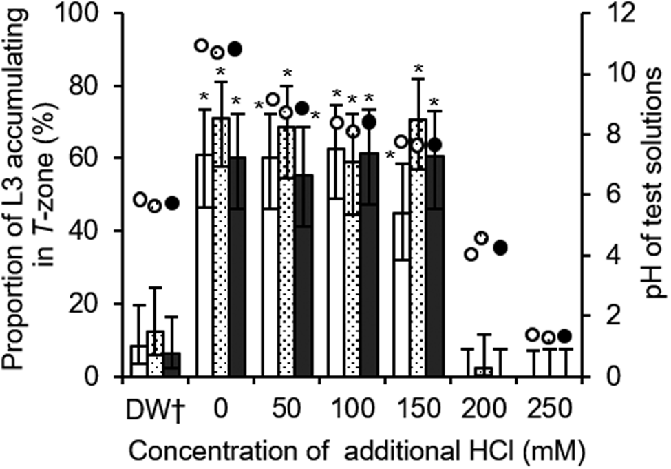
Solutions of 200 mm Arg, APD and AMPD dissolved in DW had a pH of 11.1, 10.7 and 11.0, respectively, and significantly attracted 61, 71 and 60% of filarial L3, respectively, compared with DW (corresponding controls: 8%, P < 0.01; 12%, P < 0.01; and 6%, P < 0.01) (fig. 2). As the amount of HCl was gradually increased to 50, 100 and 150 mm in the respective solutions, the pH of the solutions decreased to 9.2, 8.8 and 8.0, 8.7, 8.1 and 7.6, and 8.8, 8.3 and 7.5, respectively. The proportion of the L3 accumulating in the T-zone of 200 mm Arg, APD and AMPD plus additional 50, 100 and 150 mm HCl was similar to that of 200 mm Arg, APD and AMPD alone. When 200 mm HCl was added to a solution of 200 mm Arg, APD and AMPD (pH of test solutions: 3.5, 4.5 and 4.0, respectively), the L3 apparently avoided the test solutions.
Effect of additional amounts of HCl on the chemotactic responses of Brugia pahangi infective L3 to 200 mm Arg, APD and AMPD. The proportions of L3 accumulating in the T-zone of Arg, APD and AMPD plus additional amounts of HCl are expressed as white, dotted and black bars, respectively. Each error bar shows the 95% confidence interval for the proportions of L3. White, dotted and black circle symbols show the pH of control and test solutions of Arg, APD and AMPD, respectively. *P < 0.01, the proportion of L3 accumulating in the T-zone of 200 mm Arg, APD and AMPD plus additional amounts of HCl, was compared with that of DW alone as the corresponding control, Fisher's exact test. †Control.

Effect of the concentrations of Arg, APD and AMPD on the chemoattractant activity for B. pahangi infective L3
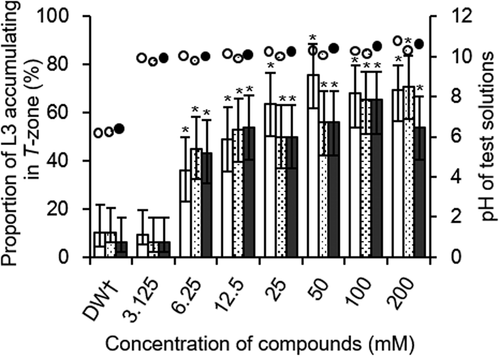
The chemotactic responses of filarial L3 to Arg, APD and AMPD were assessed in concentrations ranging from 0 to 200 mm (fig. 3). The pH of solutions of Arg, APD and AMPD at concentrations between 3.125 and 200 mm ranged between 9.8 and 10.9, 9.7 and 10.1, and 9.9 and 10.3, respectively. The three compounds significantly attracted L3 at concentrations between 6.25 and 200 mm compared with DW (corresponding control). The significant attractant activity of the three compounds was not observed at a concentration of 3.125 mm.
Effect of concentrations of Arg, APD and AMPD on the chemotactic responses of Brugia pahangi infective L3. The proportions of filarial L3 accumulating in the T-zone of Arg, APD and AMPD are expressed by white, dotted and black bars, respectively. Each error bar shows the 95% confidence interval for the proportions of L3. White, dotted and black circle symbols show the pH of control and test solutions of Arg, ADP and AMPD, respectively. *P < 0.01, the proportion of L3 accumulating in the T-zone of Arg, APD and AMPD at different concentrations, was compared with that of DW alone as the corresponding control, Fisher's exact test. †Control.

Discussion
Exogenous Tris was reported to be an attractant for B. pahangi L3, suggesting the existence of an endogenous and Tris-related attractant for the L3 (Mitsui et al., Reference Mitsui, Ureña-Tatis and Aoki2018). Since then, there had been little progress in investigations regarding the attractant of filarial L3. Thus, the present study assessed the chemotactic responses of B. pahangi L3 to Tris-related compounds in order to identify an endogenous attractant. At first, to determine the effect of functional groups of Tris on the attractant activity of Tris for filarial L3, the chemotactic responses of L3 to various Tris-related compounds lacking one primary amino, one or two hydroxymethyl groups of Tris, respectively, were assessed (table 1). Among these compounds, APD and AMPD, possessing one primary amino and two hydroxymethyl groups, were confirmed as potent attractants for the L3. Furthermore, the chemotactic response of filarial L3 to amino acids was also assessed (table 2). Among all tested amino acids, Arg was confirmed to be a potent attractant for filarial L3, while Lys and His only slightly attracted the L3. Since Arg and Lys were identified as attractants for M. incognita J2 (Fleming et al., Reference Fleming, Maule and Fleming2017), the preference of filarial L3 for water-soluble amino acids was similar to that of M. incognita J2.
The present study also shows the preference of filarial L3 for acidic or alkaline solutions of Tris-related compounds and amino acids dissolved in DW (tables 1 and 2). Among Tri-related compounds, filarial L3 were attracted to mild alkaline Tris, APD and AMPD (pH of test solutions: 10.7–11.1) rather than strong alkaline R-AP, S-AP, AMP and 2-AE (pH of test solutions: 11.6–12.0) and acidic HMP, THE, THP and DPD (pH of test solutions: 4.5–4.8). Furthermore, filarial L3 were attracted to mild alkaline amino acids such as Arg, His and Lys (pH of test solutions: 7.8–11.1) potentially or slightly, whereas the L3 were not attracted to the other 16 acidic amino acids (pH of test solutions: 2.6–6.3). These results suggest that filarial L3 may prefer mild alkaline compounds to strong alkaline or acidic compounds. Furthermore, the chemoattractant activity of Arg, APD and AMPD for B. pahangi L3 was affected by the pH of a test solution (fig. 2). Solutions of 200 mm Arg, APD and AMPD dissolved in DW had a pH of 11.1, 10.7 and 11.0, respectively, and attracted 61, 71 and 60% of filarial L3, respectively. Even after 150 mm HCl was added to the solutions of 200 mm Arg, APD and AMPD (pH of test solutions: 8.0, 7.6 and 7.5, respectively), the L3 were attracted to the test solution each at a level similar to that of 200 mm Arg, APD and AMPD alone. After 200 mm HCl was added to solutions of 200 mm Arg, APD and AMPD (pH of test solutions: 3.5, 4.5 and 4.0, respectively), the L3 apparently avoided the test solutions. These results show that the chemoattractant activity of Arg, APD and AMPD for filarial L3 is exhibited in neutral to mild alkaline but not acidic conditions, a result similar to those of Tris (Mitsui et al., Reference Mitsui, Ureña-Tatis and Aoki2018), suggesting that the three compounds can serve as attractants for filarial L3 at around the pH 7.4 of host blood. In addition, solutions of 6.25 mm Arg, APD and AMPD (pH of test solutions: 10.0, 9.7 and 9.9, respectively) significantly attracted filarial L3, while solutions of 3.125 mm Arg, APD and AMPD (pH of test solutions: 9.8, 9.7 and 9.8, respectively) did not significantly attract filarial L3 (fig. 3). These results strongly suggest that the chemotactic activity of the three compounds for filarial L3 is due to their concentrations and that the L3 are not attracted to only mild alkaline solutions.
The minimal concentration of Arg, APD and AMPD attracting filarial L3 was 6.3, 6.3 and 6.3 mm, respectively (fig. 3), but that of Tris and sodium ions attracting the L3 was reported to be 25 and 50 mm using an agar-plate assay similar to the present assay (Mitsui et al., Reference Mitsui, Miura, Bome and Aoki2012, Reference Mitsui, Ureña-Tatis and Aoki2018). Hence, the attractant activity of Arg, APD and AMPD for the L3 may be equal to or slightly higher than that of Tris and sodium ions. Arg and APD are especially endogenous compounds. Arg is a conditionally essential amino acid in humans, and the mean concentrations of free Arg in plasma and peripheral blood mononuclear cells of healthy subjects are reported to be 97.8 ± 18.6 (±standard deviation) and 717 ± 261 μM, respectively (Davids & Teerlink, Reference Davids and Teerlink2013). Furthermore, although the blood concentration of free APD has not been reported, Ito et al. (Reference Ito, Anada, Tani, Ikeda, Sano, Kihara and Igarashi2007) reported that APD derivatives such as sphingosine-1-phosphate (S1P) and sphingosine (SPH) are present in whole blood at levels of 1155 and 79 nM, respectively. These results suggest that the concentration of Arg or APD that would be encountered by filarial L3 in the infection to a host is much lower than the minimal concentration of Arg or APD attracting the L3 (6.1 and 6.1 mm, respectively). Therefore, it is surmised that, in natural infection to a host, filarial L3 may recognize and move toward, not only sodium ions in host blood (normal blood sodium level: 135–145 mm) leaking from the puncture wound made by a mosquito bite, but also other endogenous compounds such as Arg and APD as complementary attractants.
There is no report that APD is an attractant for organisms such as bacteria, fungi and nematodes. However, sphingolipids (SLs) containing a APD moiety play an important role as structural components in cellular membranes as well as some SL metabolites, including ceramide, SPH and S1P, functioning as signalling molecules in the regulation of cell growth, differentiation, senescence and apoptosis (Bartke & Hannun, Reference Bartke and Hannun2009). Thus, APD may function as one of the key signalling molecules in the chemotactic behaviour of filarial L3. On the other hand, since the biological activity of AMPD is rarely reported, its biological role in the chemotactic behaviour of filarial L3 remains unclear. In contrast, Arg has been reported to attract bacteria such as Pseudomonas aeruginosa (Craven & Montie, Reference Craven and Montie1985), Azorhizobium caulinodans (Liu et al., Reference Liu, Xie, Wang, Sun, Dang and Sun2019) and Halobacterium salinarum (Storch et al., Reference Storch, Rudolph and Oesterhelt1999). Moreover, Shivakumara et al. (Reference Shivakumara, Dutta, Chaudhary, von Reuss, Williamson and Rao2019) demonstrated that the knockdown of the M. incognita tax-2 and tax-4 genes via RNA interference results in defective chemotaxis of M. incognita J2 to Arg, suggesting that the genes are related to the chemotactic response of filarial L3 to Arg. In addition, both Brugia osm-9 and tax-4 genes are reported to be involved in the chemotactic behaviour of B. pahangi L3 forward serum (Wheeler et al., Reference Wheeler, Heimark, Airs, Mann, Bartholomay and Zamanian2020). Thus, it can be surmised that osm-9, tax-2 and tax-4 genes may be involved in the chemotactic behaviour of filarial L3 to Arg.
This is the first report demonstrating that Arg, APD and AMPD are potent attractants for B. pahangi L3. Importantly, Arg and APD are endogenous compounds and may be involved in the chemotactic behaviour of filarial L3 in the infection to a host. Therefore, a better understanding of the two attractants will undoubtedly help to elucidate the mechanism of filarial skin-penetrating invasion of a host and to develop new strategies to prevent the invasion of filarial infective larvae to the skin.
Acknowledgements
We would like to thank Prof Brian Burke-Gaffney, Nagasaki Institute of Applied Science, for his helpful advice in the revision of the manuscript.
Financial support
This research received no specific grant from any funding agency, commercial or not-for-profit sectors. The present study was conducted on the basis of current budget allocations from Nagasaki University.
Conflicts of interest
None.
Ethical standards
The authors assert that all procedures contributing to this work comply with the ethical standards of relevant national and institutional guides on the care and use of laboratory animals. The present investigation was reviewed and approved by the Institutional Animal Care and Use Committee of Nagasaki University, and all animal experiments were performed in accordance with the Guidelines for Animal Experimentation of Nagasaki University in the Animal Research Centre for Tropical Infections at the Institute of Tropical Medicine, Nagasaki University.