Introduction
In recent years, the urgent need for rapid and low-cost medical diagnostics—both in developed and developing countries—has become clear. One platform that addresses this need is paper-based microfluidics, which eliminates the need for external equipment to move fluids. Porous materials such as paper are ideal substrates for low-cost assays because they are inexpensive and disposable. One of these materials—porous nitrocellulose—is now one of the most commonly used materials in point-of-care (POC) devices, such as the OraQuick Advance HIV-1/2 test, the BinaxNow tests for a variety of infectious pathogens, and, most commonly, the home pregnancy test. This review focuses on the history and evolution of nitrocellulose-based assays, as well as critical aspects of assay performance: flow in porous media, protein adsorption, dry reagent storage, and analyte detection.
Background
History of nitrocellulose
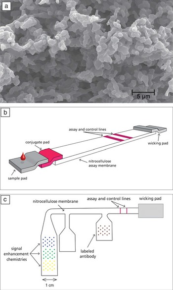
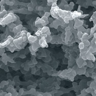
Nitrocellulose is a versatile polymer that has been broadly utilized since the 1800s.Reference Martin and Colombi 1 – Reference Falkowitz and Piech 3 Also known as cellulose nitrate, nitrocellulose is created commercially by the reaction of cellulose (purified from plants, commonly wood pulp and cotton) with nitric acid, replacing the cellulose hydroxyl groups with nitrate groups.Reference Tonkinson and Stillman 4 Today, nitrocellulose membranes are created by phase inversion,Reference Ferry 5 – Reference Zsigmondy and Bachmann 7 in which nitrocellulose is dissolved in an organic solvent that is evaporated in the presence of a nonsolvent, leaving a precipitated nitrocellulose membrane with high porosityReference Tonkinson and Stillman 4 , Reference Wong and Tse 8 ( Figure 1 a). The porosity and pore size of the membrane can be controlled by the solvents used, evaporation speed, temperature, and humidity.Reference Wong and Tse 8 The result is a material with the unique combination of tunable pore size, high surface-to-volume ratio, and very low cost.
Nitrocellulose and nitrocellulose-based assays. (a) SEM image of a blade-cut edge of a porous nitrocellulose assay membrane. (b) Schematic of a traditional lateral flow assay. (c) Schematic of a multistep two-dimensional paper network assay.

Porous nitrocellulose membranes were first used to immobilize biomolecules in the 1960s.Reference Tonkinson and Stillman 4 Nygaard and Hall demonstrated in 1963 that RNA-DNA complexes adsorb onto nitrocellulose membranes, while free nucleic acid strands pass through.Reference Nygaard and Hall 9 Others then began immobilizing nucleic acids on nitrocellulose membranes to probe for interactions between a nucleic acid of interest and other biomolecules.Reference Gillespie and Spiegelman 10 , Reference Flavell, Birfelder, Sanders and Borst 11 In 1975, Southern demonstrated the transfer of DNA from polyacrylamide gels to nitrocellulose.Reference Southern 12 This groundbreaking technique, known as the “Southern blot,” allowed specific nucleic acid fragments to be captured for subsequent analysis. The Southern blot inspired the “Northern blot” for RNA transferReference Alwine, Kemp and Stark 13 and the “Western blot” for protein transfer to nitrocellulose.Reference Towbin, Staehelin and Gordon 14 , Reference Burnette 15 These blotting techniques have been widely employed in biological research and highlight the unique ability of nitrocellulose to interact with three of the most important classes of biomolecules (proteins, DNA, and RNA).Reference Tonkinson and Stillman 4
Stemming from the blotting assays, other assays were developed utilizing porous nitrocellulose as a solid support for diagnostic assays incorporating flow. Both flow-through assays and lateral flow (LF) tests feature capture molecules (e.g., target-specific antibodies) immobilized on nitrocellulose through which a sequence of sample and reagents is passed to detect the presence of a target molecule. The flow-through assay uses flow perpendicular to the membrane, driven by a large absorbent padReference Valkirs and Barton 16 or vacuum manifold.Reference Ijsselmuiden, Herbrink, Meddens, Tank, Stolz and Vaneijk 17 While the flow-through assay has the advantage of being multiplexable, the highly manual nature of the test typically relegates it to the laboratory. However, two examples of POC flow-through assays are the Multispot HIV testReference O’Connell, Agan, Anderson, Malia and Michael 18 and the DxBox.Reference Lafleur, Stevens, McKenzie, Ramachandran, Spicar-Mihalic, Singhal, Arjyal, Osborn, Kauffman, Yager and Lutz 19 For most POC applications, the LF test has become the assay of choice.
Lateral flow technology
Over the last 25 years, nitrocellulose has become a prominent material in POC diagnostics through its role in LF technology. Since the first LF test came to market in 1988,Reference Chard 20 this popular technology has developed into a $2.1 billion market involving over 200 companies.Reference Wong and Tse 8 Because LF tests are inexpensive, rapid, portable, equipment-free, and easy to use, they are now used in many fields, including clinical, environmental, agricultural, veterinary, drug testing, and bio-defense.Reference Wong and Tse 8
The most common LF devices consist of a “strip” with four contacting porous components arrayed linearly: (1) sample pad, (2) conjugate pad containing dried, gold-conjugated detection antibodies specific to the target analyte, (3) nitrocellulose membrane with capture agents immobilized at control and test lines, and (4) wicking pad to draw the sample through the device (Figure 1b).Reference Wong and Tse 8 The nitrocellulose membrane is critical to the functioning of LF devices because analyte binding and detection occur within it. To begin the test, the user adds a microliter-volume of sample (e.g., blood, urine, saliva) to the sample pad. Results are typically available in less than 15 minutes and can be visually read based on the development of color at the test line, due to the accumulation of the gold-conjugated detection antibodies. The device’s simplicity permits easy manufacturing, and the dried reagents eliminate the need for refrigeration and extend shelf life.Reference Posthuma-Trumpie, Korf and van Amerongen 21
While LF devices have revolutionized low-cost testing, they have several drawbacks: they are usually qualitative, have lower sensitivity than gold standards (e.g., nucleic acid amplification tests, enzyme-linked immunoassays (ELISA), and bacterial/viral culture), require additional sample preparation, are rarely multiplexable, and cannot perform multiple automated steps.Reference Posthuma-Trumpie, Korf and van Amerongen 21 Currently, numerous innovations, such as two-dimensional paper networks (2DPNs),Reference Fu, Kauffman, Lutz and Yager 22 – Reference Lutz, Trinh, Ball, Fu and Yager 27 are being developed to overcome these disadvantages. Such innovations require an understanding of the scientific and engineering principles involved in assay design.
Considerations for assay development
Physics of flow in porous media
Understanding flow in porous materials is essential when designing nitrocellulose-based assays. The principle of capillary flow describes the passive wicking of fluids that occurs in porous media. Capillary flow is affected by several characteristics of the porous medium: pore size, pore distribution, and porosity; by characteristics of the fluid (e.g., viscosity, surface tension); and by the interaction between the two: the contact angle of the fluid with the porous material.Reference Washburn 28 – Reference Bird, Stewart and Lightfoot 30
Though most porous media have some tortuosity (i.e., the “twistiness” that is defined by the ratio of the actual path length of a particle flowing through the material to the straight-line distance), they are most easily approximated as a bundle of straight pores. This approximation allows us to use conventional fluidic principles, such as the Reynolds number, to understand the behavior of fluid in these pores. In nitrocellulose membranes (where the most applicable Reynolds number is ~10–3), flow is laminar. Thus, flow streamlines are parallel to the channel walls, and the primary mode of mixing between adjacent streams is diffusion.Reference Bird, Stewart and Lightfoot 30 , Reference Purcell 31
The “wet-out” process of a fluid wicking into a dry porous membrane is governed by the Washburn equation:Reference Washburn 28

where L, the length of the fluid column in the porous material, depends on pore diameter (dp ), fluid surface tension (γ), fluid viscosity (μ), and the cosine of the contact angle (θ). Taking the derivative of L with respect to t, we see that the wet-out linear velocity is inversely proportional to the fluid column length behind the wetting edge:

This slowing is due to the linear increase in viscous resistance as the fluid column lengthens.
From Equation 2, we see that the wet-out rate is inversely proportional to fluid viscosity (μ) and proportional to the membrane pore diameter (dp ). Though the viscosity of a liquid is generally known, the pore diameter within a particular membrane is not. In nitrocellulose membranes, pore size distribution varies widely depending on the material and casting process used. The membrane wet-out rate is also proportional to the cosine of the contact angle (θ) and the surface tension (γ), which account for the liquid-air and liquid-solid interfacial energies. Surfactants are often added to nitrocellulose during manufacturing to decrease the contact angle, thereby improving wettability. 32 The surface tensions of common liquids are well known, though changes in temperature affect wet-out rates, and fluids used in real assays are never pure solvents.
Once a porous membrane has fully wetted out, flow is governed by Darcy’s equation for flow in porous media:Reference Darcy 29

where the volumetric flow rate (Q) depends on the paper’s permeability (κ), cross-sectional paper area (wh), fluid viscosity (μ), and the pressure drop (∆P) that occurs over the length (l) of the paper network. This equation assumes that flow is fed by an infinite source, and, as the wicking pad becomes wetted, the capillary pressure it generates does not decrease. These assumptions are approximately correct as long as the wicking pad does not approach saturation.
The utility of both Washburn’s equation and Darcy’s law for modeling flow in paper devices has been repeatedly demonstrated.Reference Osborn, Lutz, Fu, Kauffman, Stevens and Yager 33 Furthermore, methods such as pH-based electrochemical markingReference Kauffman, Fu, Lutz and Yager 34 have been developed to quantify flow rates in porous devices to enable a systematic validation process for mathematical modeling.
Protein adsorption
The adsorption of biomolecules—typically proteins in commercial lateral flow tests—to the porous nitrocellulose membrane is another key assay consideration. While protein adsorption to nitrocellulose has been observed for years,Reference Brash and Horbett 35 , Reference Horbett and Brash 36 the process is not well understood.Reference Tonkinson and Stillman 4 However, known principles should be considered when trying to achieve protein immobilization at a test line—or to prevent nonspecific adsorption elsewhere.
First, proteins are thermodynamically driven to adsorb to nitrocellulose.Reference Vanoss, Good and Chaudhury 37 This adsorption is governed by noncovalent interactions between the two.Reference Horbett and Brash 36 , Reference Vanoss, Good and Chaudhury 37 Some argue that this attraction is a hydrophobic interaction between the carbon-containing nitrocellulose and the hydrophobic portion of the protein,Reference Wong and Tse 8 while others describe it as a primarily electrostatic interaction between the dipoles of the nitrocellulose nitrate groups and dipoles within the protein. 32 Either way, it is well-established that proteins will adsorb to porous nitrocellulose membranes unless something is done to prevent this process.Reference Wong and Tse 8 In particular, nitrocellulose-based LF assays are often pre-treated with a protein-containing blocking solution to fill unused binding sites.Reference Wong and Tse 8 This blocking step prevents the sample analyte from nonspecifically adsorbing to the membrane before it can reach the capture line.
Second, the kinetics of protein adsorption and desorption to the membrane must be considered, especially for capture molecules immobilized at a test line. Contrary to classic adsorption studies in which protein adsorption—tested in simplified systems at equilibrium—is considered irreversible,Reference Horbett and Brash 36 the complex systems of LF assays render protein adsorption a kinetic, two-way process. Specifically, the fluid flow velocity,Reference Washburn 28 presence of potential stripping agents such as salts and detergents,Reference Hoffman and Jump 38 and reagent drying timeReference Horbett and Brash 36 , Reference Vanoss, Good and Chaudhury 37 are important considerations for assays that depend on protein immobilization.
The most commonly used tools for the deposition of proteins include piezoelectric spotters that can deposit small volumes into precise locations on a strip,Reference Fu, Liang, Spicar-Mihalic, Houghtaling, Ramachandran and Yager 26 , Reference Gonzalez-Macia, Morrin, Smyth and Killard 39 high-volume stripers that “paint” a line of reagent across a large batch of strips,Reference Wong and Tse 8 and inkjet printers.Reference Lutz 40 , Reference Yu, Shang, Cheng, Paik, Kaplan, Andrade and Ratner 41 Each method has advantages and disadvantages; the choice depends on the required throughput, the need for patterning flexibility, and budget. These deposition tools are also important for patterning assay reagents so they may be stored directly on the device.
Dry reagent storage, rehydration, and patterning
When designing POC diagnostics, storing reagents in dry form directly on the device is most desirable. Using dry reagents reduces user steps, removes the need for continuous refrigeration (i.e., cold chain), provides a longer assay shelf life, and facilitates device automation.
Generally, proteins lose function at high temperatures or when dried in an uncontrolled manner.Reference Crowe, Carpenter and Crowe 42 However, a wide body of research has demonstrated that the presence of the non-reducing disaccharides trehaloseReference Crowe, Carpenter and Crowe 42 – Reference Mazzobre, Buera and Chirife 46 and (less frequently) sucroseReference Crowe, Carpenter and Crowe 42 , Reference Stevens, Petri, Osborn, Spicar-Mihalic, McKenzie and Yager 47 can stabilize dried proteins for extended periods at elevated temperatures. It is believed that the high glass transition temperature of trehalose (106°CReference Elias and Elias 48 ) ensures that molecular mobility of embedded proteins is kept low at elevated temperatures, thus protecting the protein against degradation.Reference Crowe, Carpenter and Crowe 42 , Reference Garcia, Kirkham, Hatch, Hawkins and Yager 43 , Reference Mazzobre, Buera and Chirife 46 Furthermore, the sugar hydroxyl groups substitute for the waters of hydration of the protein, which are lost upon drying, helping to preserve the protein’s native conformation.Reference Crowe, Carpenter and Crowe 42
Because dried reagents are important for POC diagnostics, several techniques have been developed for dry reagent storage and subsequent rehydration. Reagents are often stored dry within a conjugate pad placed in line with the fluidic pathways for rehydration at the time of use. Conjugate pads have been used in both LF immunoassaysReference Wong and Tse 8 and, more recently, 2DPNs.Reference Fu, Liang, Spicar-Mihalic, Houghtaling, Ramachandran and Yager 26 Though these pads are effective for reagent storage, they offer minimal control over the spatial and temporal release of rehydrated reagents and require additional materials and manufacturing steps, which add cost. Recent developments have sought to address the latter of these concerns. Abe et al. showed the ability to rehydrate gold-anti-IgG antibody conjugate dried on blocked cellulose membranes and to use it downstream to detect IgG in buffer.Reference Abe, Kotera, Suzuki and Citterio 49 In our group, Fridley et al. developed methods for controlled release of reagents printed directly onto nitrocellulose membranes. By patterning reagents and sugars into specific layouts, we controlled both the spatial and temporal distribution of reagents as they rehydrated on the membrane.Reference Fridley, Le, Fu and Yager 50
Detection
In an ideal assay, the user would be able to detect the lowest clinically relevant concentration of an analyte in a patient sample. In reality, however, the limit of detection (LOD) of rapid tests often does not reach that ideal. Most commonly, LF tests use labels that can be interpreted by eye to eliminate the need for additional machinery. Three promising detection labels are colloidal gold nanoparticles, colored latex beads, and carbon black.Reference Wong and Tse 8 Signal amplification chemistries, such as metallic enhancement of colloidal gold and enzyme-based systems, can also greatly improve the assay LOD and dynamic range.
Numerous labels and chemistries have been developed and are being used in commercial assays.Reference Wong and Tse 8 See Chapter 5 of the Lateral Flow Immunoassay book for a thorough review of detection labels and amplification chemistries.Reference Wong and Tse 8
The Future
As discussed previously, existing LF tests have inadequate sensitivity and specificity for many medical applications.Reference Posthuma-Trumpie, Korf and van Amerongen 21 , Reference Poehling, Griffin, Dittus, Tang, Holland, Li and Edwards 51 , Reference Cazacu, Greer, Taherivand and Demmler 52 Sophisticated paper-based technologies, which are still driven by capillary flow, have evolved from simple LF tests.Reference Fu, Kauffman, Lutz and Yager 22 – Reference Fu, Ramsey, Kauffman, Lutz and Yager 25 , Reference Osborn, Lutz, Fu, Kauffman, Stevens and Yager 33 , Reference Carrilho, Phillips, Vella, Martinez and Whitesides 53 – Reference Martinez, Phillips, Nie, Cheng, Carrilho, Wiley and Whitesides 61 These new paper-based assays demonstrate a breadth of functionalities lacking in LF tests.
Since 2008, our group has pursued the development of next-generation nitrocellulose-based assays. In particular, our work has focused on the development of 2DPNs (Figure 1c), which feature automated multistep processing. We began with the creation of a fluidic toolbox to understand and control fluid flow and reagent delivery in an automated fashion. These tools include methods for fluidic mixing,Reference Osborn, Lutz, Fu, Kauffman, Stevens and Yager 33 volume metering,Reference Fu, Kauffman, Lutz and Yager 22 , Reference Lutz, Trinh, Ball, Fu and Yager 27 fluid transport control,Reference Fu, Lutz, Kauffman and Yager 23 , Reference Fu, Ramsey, Kauffman, Lutz and Yager 25 and staged reagent delivery.Reference Fu, Kauffman, Lutz and Yager 22 , Reference Fu, Lutz, Kauffman and Yager 23 We then utilized many of these tools in a 2DPN device to demonstrate a six-fold signal enhancement using staged delivery of gold nanoparticles and gold enhancement solution.Reference Fu, Kauffman, Lutz and Yager 22 Next, we adapted an existing LF pregnancy test into a 2DPN with gold enhancement, demonstrating a four-fold LOD improvement compared to the conventional strip alone.Reference Fu, Liang, Houghtaling, Ramachandran, Ramsey, Lutz and Yager 24
Finally, we used this body of knowledge to develop two self-contained assays. First, we developed a 2DPN assay for malaria using reagents dried on conjugate pads. This POC assay has the same sensitivity as a laboratory-based malaria ELISA.Reference Fu, Liang, Spicar-Mihalic, Houghtaling, Ramachandran and Yager 26 Based on that successful assay, we developed a gold-enhanced malaria assay with comparable sensitivity that uses only reagents patterned and dried directly on the nitrocellulose membrane. By patterning reagents, both the volume of reagents and the total number of components were decreased.Reference Fridley, Le, Fu and Yager 62 This work exemplifies the promise of 2DPNs to deliver simplified manufacturing and reduced costs for automated multistep assays in the future.
Conclusions
In this article, we have reviewed the past and present of nitrocellulose in assays and provided insights into the technological considerations for development of sophisticated novel diagnostics. Overall, the current state of the art is rich with tools and information that can be exploited to develop improved point-of-care assays. These new capabilities permit features such as multiplexing, on-device sample processing, and signal enhancement, which have the promise of enabling higher-sensitivity assays with rich diagnostic information, while still retaining the low cost and ease of use of traditional lateral flow tests. These assay improvements, especially when combined with recent developments in automated, cell phone-based readers,Reference Martinez, Phillips, Carrilho, Thomas, Sindi and Whitesides 58 , Reference Wang, Zhao, Khimji, Akbas, Qiu, Edwards, Cramer, Ye and Demirci 63 – Reference Coskun, Wong, Khodadadi, Nagi, Tey and Ozcan 65 are poised to spawn the next generation of tools for medical diagnosis at the point of care, whether that be a doctor’s office in Lima, a home in the suburbs of Seattle, or a rural clinic in the mountains of Nepal. Given the strides made in this field already, it is exciting to look forward to future advancements in next-generation nitrocellulose-based devices.
Acknowledgments
The authors would like to thank the University of Washington NanoTech User Facility and Paul Wallace for the use of and assistance with the SEM. Funding was provided by NIH (1 R01 AI 096184–01) and DARPA DSO (HR0011–11–2-0007). This material is based upon work supported by the National Science Foundation under Grant No. DGE-0718124 (GEF and CAH). Any opinions, findings, and conclusions or recommendations expressed in this material are those of the authors and do not necessarily reflect the views of the National Science Foundation.