Extra-nodal marginal zone lymphoma of mucosa-associated lymphoid tissue (MALT lymphoma) is an extra-nodal mature B-cell non-Hodgkin lymphoma that is common in the adult population but is exceedingly rare in children. Reference Claviez, Meyer and Dominick1 Lymphatic vessel dysplasia has been associated with extra-nodal non-Hodgkin lymphoma including MALT lymphoma both as a potential stimulus Reference Grulich, Vajdic and Cozen2,Reference Sokol, Beaugerie and Maynadié3 and a complication Reference Laharie, Degenne and Laharie4,Reference Pratz, Dingli, Smyrk and Lust5 of the lymphoproliferative disorder. Protein-losing enteropathy is a well-described complication of surgical palliation with a cavopulmonary anastomosis (e.g. Fontan) characterised by a loss of lymph often through secondary dysplastic lymphatics into the gastrointestinal tract resulting in significant morbidity and mortality. Reference Umar and DiBaise6,Reference Itkin, Piccoli and Nadolski7 Here, we present the first case of MALT lymphoma in a patient with Fontan-associated protein-losing enteropathy with discussion of the overlap of lymphatic dysplasia and lymphoproliferative disorder.
Case report
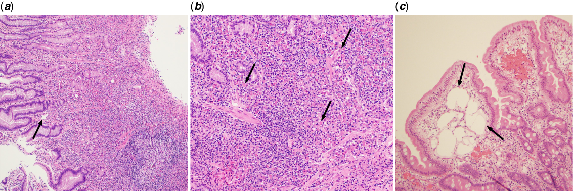
A 7-year-old boy followed by cardiology for hypoplastic left heart syndrome (mitral and aortic atresia) presented to the emergency department for hematemesis. He underwent bilateral pulmonary artery bands without ductal stent placement at 12 days due to 33-week prematurity followed by Norwood procedure with 3.5 mm modified Blalock–Taussig shunt and take down of the pulmonary artery bands at 2 months of age. He underwent hemi-Fontan palliation at the age of 8 months and then fenestrated lateral tunnel Fontan palliation at the age of 3.6 years. His course was complicated by left pulmonary artery stenosis requiring surgical and catheter-based intervention. On the day of presentation, he developed an acute upper gastrointestinal bleed with large volume hematemesis. His bleeding was stabilised with acid suppression and he underwent esophagogastroduodenoscopy that identified friable gastric tissue with a healing gastric ulcer. His biopsy demonstrated lymphoplasmacytic infiltrate consistent with MALT lymphoma of the gastric antrum and fundus (Fig 1a and b). Biopsy was negative for MALT1 rearrangement and Helicobacter pylori. Surveillance positron emission tomography/computed tomography imaging was consistent with retroperitoneal lymph node enlargement, fluorodeoxyglucose uptake in the stomach, mildly avid submandibular and cervical lymph nodes (stage II bulky disease), and note of mild-to-moderate bilateral pleural effusions. He was discharged home and then returned for port-a-cath placement for a treatment course of chemotherapy.
Histopathology of biopsy specimens from the initial esophagogastroduodenoscopy establishing the MALT lymphoma diagnosis. (a) Gastric mucosa demonstrating diffuse lymphoplasmocytic infiltration with numerous CD20 B-cells (not shown) and fluorescent in situ hybridisation negative for MALT1 rearrangement with dilated lymphatic channels (arrow) (H&E 100x). (b) High magnification of gastric mucosa demonstrating many plasma cells (CD138 staining not shown) with Russell bodies seen (arrows) (H&E 400x). There was clonal expansion of kappa light chain and rare lambda light chain cells present. (c) Cluster of dilated lymphatic channels within the duodenum consistent with lymphangiectasia (H&E 200x).

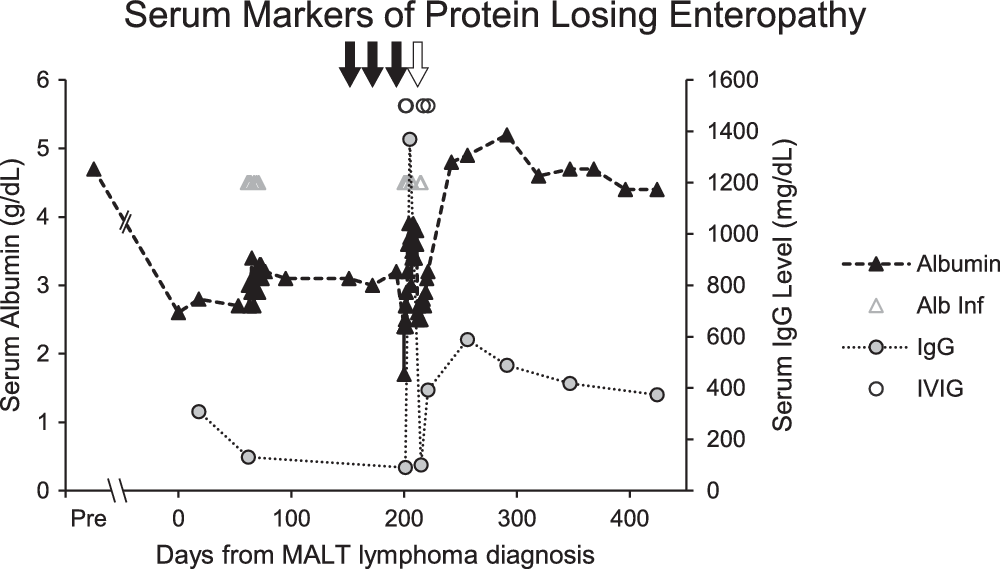
Upon representation for surgery, he was noted to have abdominal distension, periorbital edema, and shortness of breath. After port-a-cath placement, he was noted to have hypoxemia and dyspnea with interval increase in his bilateral pleural effusions and persistent hypoalbuminemia (Fig 2). He was therefore admitted for fluid management and focussed evaluation for protein-losing enteropathy based on initial laboratory findings and clinical history. He was subsequently diagnosed with protein-losing enteropathy based on clinical features with persistent hypoalbuminemia, hypogammaglobulinemia (IgG 130, IgA 36, IgM 59 mg/dl), lymphopenia (600 cell/μl) with low CD3 (208 cell/μl), CD4 (90 cell/μl), and CD8 (74 cell/μl) counts, and stool alpha-1-antitrypsin >2389 mg/dl. Further review of his pathology demonstrated dilated lymphatic vessels (Fig 1c) consistent with lymphangiectasia with the abnormal vessels more pronounced in the duodenum. Cardiac catheterisation was performed and demonstrated Fontan pressure of 13 mmHg, transpulmonary gradient of 5 mmHg, right ventricular end-diastolic pressure of 7–8 mmHg, indexed pulmonary vascular resistance of 1.2 WU × m2, cardiac index of 4.3 L/minute/m2 with qualitatively normal right ventricular function on echocardiogram, two trivial venovenous collaterals, no baffle leak, systemic arterial oxygen saturation of 91% with pulmonary artery oxygen saturation of 61%, and no evidence of left pulmonary artery obstruction or superior caval vein stenosis. He was started on enteric corticosteroids, diuretics, and serial albumin infusion with incomplete clinical improvement. A trial of unfractionated heparin yielded minimal improvement and was discontinued. Sildenafil was added to his enteric corticosteroids and diuretics with stabilisation of albumin levels (Fig 2).
Time course of markers of protein-losing enteropathy (albumin, closed triangle, and IgG, closed circle). Day 0 represents his initial gastric biopsy and diagnosis of MALT lymphoma. Admitted after port-a-cath placement on day 59 with diagnosis of protein-losing enteropathy. Open triangles (albumin) and circles (intravenous immunoglobulins, IVIG) demonstrate need for frequent albumin and IgG supplementation. After treatment course of rituximab (closed arrows) and first biopsy without evidence of MALT lymphoma (open arrow), there is sustained improvement in albumin and IgG levels.

His case was discussed with a multidisciplinary tumor board and the decision was made to manage his protein-losing enteropathy for 3 months then repeat his esophagogastroduodenoscopy and biopsy, which demonstrated persistence of the MALT lymphoma. The decision was made for treatment with four cycles of rituximab given risks of abdominal radiation and anthracycline toxicity with Fontan physiology. On repeat biopsy after three cycles of rituximab, he had decreased but persistent burden of MALT lymphoma. Four days after biopsy (day 199, Fig 2), he was readmitted with toxic shock syndrome secondary to group A strep cellulitis. During admission, he developed massive gastrointestinal bleeding with emergent esophagogastroduodenoscopy performed for haemostasis (day 211, Fig 2). Esophagogastroduodenoscopy identified a large friable mass with ulceration without evidence of lymphoma in the gastrum on biopsy. He was discharged home on unchanged protein-losing enteropathy therapies. Serial biopsies demonstrated no further evidence of MALT lymphoma. The course of chemotherapy was considered effective and complete (three total cycles). Over the following 6 months, his protein-losing enteropathy symptoms improved with resolution of hypoalbuminemia without need of additional albumin on unchanged protein-losing enteropathy therapies of enteral budesonide, sildenafil, furosemide, and enalapril. Repeat stool alpha-1-antitrypsin had decreased to 187 mg/dl after the third dose of rituximab (day 202, Fig 2).
Discussion
In the adult population, MALT lymphoma represents an indolent disease that is associated with Helicobacter pylori and MALT1 translocation Reference Farinha and Gascoyne8 that responds well to therapy. Given the extreme rarity of the disease in children without typical triggers in an additionally rare disease (Fontan-associated protein-losing enteropathy), the presence of protein-losing enteropathy presents a unique question of co-incidence, trigger, or enhanced symptomology in our case. Our best evidence suggests Fontan-associated protein-losing enteropathy is a disease of the lymphatic circulation that can arise in part through the unique haemodynamics of a Fontan circulation and the formation of secondary intestinal lymphangiectasais. Reference Umar and DiBaise6,Reference Itkin, Piccoli and Nadolski7
In terms of enhanced symptomology of protein-losing enteropathy and MALT lymphoma, it is important to consider the interplay of pathology in these diseases. There is evidence that an inflammatory milieu contributes to protein-losing enteropathy pathology. Reference Umar and DiBaise6 This is best demonstrated by response to corticosteroids as a mainstay of treatment for Fontan-associated protein-losing enteropathy Reference Schumacher, Cools and Goldstein9 and that inflammatory bowel disease or autoimmune disease can present with a phenotypically similar protein-losing enteropathy. Reference Umar and DiBaise6 As such the inflammatory microenvironment of the MALT lymphoma may have exacerbated his protein-losing enteropathy symptoms.
As a potential link between the development of MALT lymphoma and Fontan-associated protein-losing enteropathy, there is evidence that the pathophysiology in Fontan-associated protein-losing enteropathy could contribute to lymphoma formation. First, it has been demonstrated that immunodeficiency, particularly dysregulation of humoral immunity, is one of the strongest risk factors of non-Hodgkin lymphoma which increases with the degree of immunodeficiency, including MALT lymphoma. Reference Grulich, Vajdic and Cozen2,Reference Riaz, Faridi, Patnaik and Abraham10 Lymph loss in protein-losing enteropathy yields a pattern of quantitative immune abnormalities with characteristic hypogammaglobulinemia and lymphopenia (specifically T-cell lymphopenia). Reference Magdo, Stillwell and Greenhawt11,Reference Strober, Wochner, Carbone and Waldmann12 While the associated immune abnormalities seen in protein-losing enteropathy is atypical without apparent increase in opportunistic pathogens Reference Magdo, Stillwell and Greenhawt11 and preserved synthetic function, Reference Strober, Wochner, Carbone and Waldmann12 it can lead to skin anergy and long-term acceptance of skin grafts. Reference Strober, Wochner, Carbone and Waldmann12 This suggests protein-losing enteropathy creates at least a disordered immune regulatory state that we hypothesise could be a mechanism to promote the formation of MALT lymphoma. Second, evidence of chronic intestinal inflammation that appears to be present in protein-losing enteropathy is also associated with increased risk of primary intestinal lymphomas as seen in inflammatory bowel disease. Reference Sokol, Beaugerie and Maynadié3 This combination is born out in the association of B-cell lymphoma formation in protein-losing enteropathy secondary to primary intestinal lymphangiectasia, Reference Laharie, Degenne and Laharie4 which has close pathophysiology to Fontan-associated protein-losing enteropathy.
There is also evidence that protein-losing enteropathy can be secondary to the effects of lymphoma. The B-cell lymphoma of Waldenstrom macroglobulinemia can result in a secondary intestinal lymphangiectasia. Reference Pratz, Dingli, Smyrk and Lust5 In the rare cases of overlap of lymphoma with protein-losing enteropathy, protein-losing enteropathy symptoms tend to improve with treatment of the associated lymphoma. Reference Laharie, Degenne and Laharie4,Reference Pratz, Dingli, Smyrk and Lust5 Our case is similar in demonstrating improvement in hypoalbuminemia after biopsy resolution of his MALT lymphoma (Fig 2). Taken together, there is evidence to postulate that the gastric and intestinal lymphatic dysplasia seen in our case contributed to the formation of his gastric lymphoma, and, at the same time, his MALT lymphoma may have contributed to his protein-losing enteropathy symptoms.
Of the limited study of patients with protein-losing enteropathy and endoscopy, the mucosal tissue is often friable and edematous and is associated with ulceration and erosion with gastrointestinal bleeding. Reference Itkin, Piccoli and Nadolski7 However, biopsies are rarely performed, which may miss any potential changes in lymphoid proliferation. Therefore, MALT lymphoma or – more broadly – abnormal lymphoid hyperplasia may represent an under-recognised diagnosis, and it should raise a clinical question on management if clonal lymphoid proliferation is found on biopsy, as occurred in the presented case. This raises the question about the clinical utility of esophagogastroduodenoscopy and biopsy for Fontan-associated protein-losing enteropathy that appears refractory to treatment.
Financial support
This research received no specific grant from any funding agency, commercial or not-for-profit sectors.
Conflicts of interest
None.
Ethical Standard
This article is exempt and does not contain any studies with human participants performed by any of the authors. Informed consent was obtained from the patient/guardian for submission of the case report.