




No CrossRef data available.
Article contents
Surgical treatment of an unusual atrial septal defect: the vestibular defect
Published online by Cambridge University Press: 20 January 2005
Abstract
A 14-year-old female patient underwent surgical treatment of multiple atrial septal defects associated with unroofed coronary sinus and pulmonary valvar stenosis. One of the defects was that of the superior oval fossa and the other a large ellipsoidal defect positioned inferior to the inferior rim of the oval fossa. The patient underwent primary closure of the defects with a favorable result. To the best of our knowledge, this is the first surgical experience of an unusual atrial septal defect or the vestibular defect.
Information
- Type
- Brief Report
- Information
- Copyright
- © 2004 Cambridge University Press
Interatrial communications are currently divided into those within, and those beyond, the confines of the oval fossa. Recently, Sharratt and associates1 reported an unusual form of the defect, together with a novel concept of atrial septation. In this report, we describe surgical experience with a comparable defect of the atrial septum.
Case report
A 14-year-old female patient was diagnosed as having atrial septal defect and pulmonary valvar stenosis and admitted for further evaluation. She had been asymptomatic from early ages and tolerated physical exercise without difficulty. Chest roentgenography showed mild cardiomegaly and pulmonary plethora. Electrocardiography showed regular sinus rhythm, prolonged PQ interval (0.28 s), and right axis deviation of the QRS complex. The apical four-chamber view of cross-sectional echocardiography showed a large defect at the lower level of the atrial septum and side-by-side relationship of the atrioventricular valves with less than usual offsetting of the hinge points (Fig. 1a). The parasternal short axis view showed that the mitral valve opened exclusively to the left ventricle and had normal bifoliate leaflet (Fig. 1b). Catheter examination revealed bidirectional shunt at the atrial level with a ratio of pulmonary to systemic flows of 2.4 and a pressure gradient across the pulmonary valve of 44 mmHg. Angiocardiography demonstrated normal configuration of the ventricles and normal systemic and pulmonary venous connections with atriums.

Figure 1. Apical four-chamber view of echocardiography showed a large atrial septal defect (double headed arrow) and side-by-side relationship of the atrioventricular valves with less than usual offsetting of the hinge points (a). Parasternal short axis view showed a bileaflet mitral valve within the left ventricular cavity (b). RA: right atrium; LA: left atrium; MV: mitral valve.
Surgical exploration of the right atrium disclosed two atrial septal defects. One of these (10 × 10 mm) was a defect of the superior oval fossa and the other (30 × 20 mm) a large ellipsoidal defect positioned inferior to the inferior rim of the oval fossa (Fig. 2). The anterior, posterior, superior, and inferior margins of the unusual defect comprised the anterior atrial wall, posterior atrial wall superjacent to the inferior cavoatrial junction, inferior rim of the oval fossa, and muscular structure between vestibules of the right and left atrioventricular valves, respectively. The coronary sinus was unroofed in the absence of the left superior caval vein and opened to the posterior-inferior wall of the left atrium (Fig. 2). Both atrioventricular valves were normally developed, whilst the tricuspid pulmonary valve showed mild thickening of the facing edges. Both atrial septal defects were closed primarily without the use of a prosthetic material. Postoperatively the pressure gradient across the pulmonary valve decreased to 16 mmHg and electrocardiography showed a regular sinus rhythm with PQ interval of 0.20 s. At the last follow-up 6 months after surgery, the patient was doing well without the need for medication.

Figure 2. Surgeon's view of the opened right atrium. Margins of the vestibular defect are described in the text. Asterisk shows the muscular structure between vestibules of the right and left atrioventricular valves. SCV: superior caval vein; ICV: inferior caval vein; DOF: defect of oval fossa; VD: vestibular defect; TV: tricuspid valve; CS: orifice of a coronary vein.
Discussion
Currently, interatrial communication anomalies are divided into those within and those beyond the confines of the oval fossa.2, 3 Of these, the latter includes the sinus venosus defect, coronary sinus defect, and the ostium primum defect.2, 3 Recent studies of the embryological development of the atrial septum have clarified that an additional developmental component, the vestibular spine, played an integral role in the fusion of the primary atrial septum with the atrioventricular cushions.4–6 More recently, Sharratt and associates1 published an autopsy study of an unusual defect within the muscular anterior-inferior rim of the oval fossa producing a communication between the vestibules of the right and left atrioventricular valves. Based on the detailed morphological analysis of the unusual defect coupled with their own embryological theory, the authors of that report named this abnormality vestibular defect and proposed a hypothetical view that the defect resulted from the developmental deficiency of the septal component derived from the embryonic vestibular spine. This was the first report that facilitated the clinical concept of the new kind of defect and allowed us to make a diagnosis of vestibular defect for the anomaly in this report. We are aware of the disparity between cases of these two reports in the positional arrangement of hinge points of the atrioventricular valves and morphology of the coronary sinus. However, since the vestibular spine is currently known to play an integral role for the development of these structures as well,4–6 it is our view that the coexisting anomalies do not militate against the current diagnosis. Conversely, our experience disclosed a morphological difference between the vestibular defect and the ostium primum defect as well as the ordinary coronary sinus defect. This included the bileaflet mitral valve opening to the normally developed left ventricle for the former and the large defect extend-ing beyond the structural contiguity between the coronary sinus and the posterior atrial wall for the latter.
It is of note that, despite developmental abnormality of the essential component of the atrial septum, the patient remained asymptomatic for more than 10 years before surgery. This may partly be ascribed to the coexisting pulmonary valvar stenosis and competent atrioventricular valves. Finally, although the present study failed to investigate the disposition of the conduction system, the preoperative prolongation and postoperative change of the PQ interval suggested a positional abnormality of the His bundle with or without the posterior displacement of the atrioventricular node.
References
Sharratt GP, Webb S, Anderson RH. The vestibular defect: an interatrial communication due to a deficiency in the atrial septal component derived from the vestibular spine. Cardiol Young 2003; 13: 184–190.Google Scholar
Martins JDF, Anderson RH. The anatomy of interatrial communications – what does the interventionalist need to know? Cardiol Young 2000; 10: 464–473.Google Scholar
Anderson RH, Webb S, Brown NA. Clinical anatomy of the atrial septum with reference to its developmental components. Clin Anat 1999; 12: 362–374.Google Scholar
Webb S, Brown NA, Anderson RH. Formation of the atrioventricular septal structure in the normal mouse. Circ Res 1998; 82: 645–656.Google Scholar
Kim JS, Virágh SZ, Moorman AFM, Anderson RH, Lamers WL. Development of the myocardium of the atrioventricular canal and the vestibular spine in the human heart. Circ Res 2001; 88: 395–402.Google Scholar
Lamers WL, Moorman AFM. Cardiac septation. A late contribution of the embryonic primary myocardium to heart morphogenesis. Circ Res 2002; 91: 93–103.Google Scholar
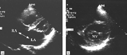
Apical four-chamber view of echocardiography showed a large atrial septal defect (double headed arrow) and side-by-side relationship of the atrioventricular valves with less than usual offsetting of the hinge points (a). Parasternal short axis view showed a bileaflet mitral valve within the left ventricular cavity (b). RA: right atrium; LA: left atrium; MV: mitral valve.

Surgeon's view of the opened right atrium. Margins of the vestibular defect are described in the text. Asterisk shows the muscular structure between vestibules of the right and left atrioventricular valves. SCV: superior caval vein; ICV: inferior caval vein; DOF: defect of oval fossa; VD: vestibular defect; TV: tricuspid valve; CS: orifice of a coronary vein.
 You have
Access
- 4
- Cited by