INTRODUCTION
In the past 30 years, all areas of parasitology have been transformed, but perhaps none have emerged from the shadows quite so dramatically as helminth biology. This article presents a personal retrospective of this period, starting from a time before parasite genes or antigens, helper T-cell subsets or cytokines, through to the present day with molecular, biochemical and immunological sciences illuminating many new pathways to control these highly prevalent and damaging diseases.
Helminth parasites include schistosomes (trematodes) and cestodes (such as tapeworms), as well as the nematode roundworms that are the subject of this review. Nematodes include the major gastrointestinal parasites of man (Ascaris, hookworm and Trichuris) which together infect up to 2 billion people in the world today (Hotez et al. Reference Hotez, Brindley, Bethony, King, Pearce and Jacobson2008); in addition, vector-borne filarial nematodes (e.g. Brugia and Onchocerca) still infect over 100 million people. To this toll of human health must also be added the immense load of gastrointestinal nematodes in livestock, as well as zoonotic species such as Toxocara canis that frequently invade human subjects. Why then has this parasite group not been more urgently prioritized for eradication? To some extent, diseases of developing countries (and generally poorer communities within those countries) were deemed in the past to be less important than those of the Western world. Even in the tropical context, because helminth infections cause relatively little direct mortality, priority was given to diseases with a more lethal profile. Only by calculating the cumulative economic and social costs of disability, growth stunting, fevers and nutritional deficits, can the deleterious effects of helminths be truly appreciated, and appropriate attention paid to the challenge they represent (King et al. Reference King, Dickman and Tisch2005; Hotez et al. Reference Hotez, Brindley, Bethony, King, Pearce and Jacobson2008).
The fact that a large proportion of infected individuals may, at any one time, appear asymptomatic is also a reflection of a central issue, that helminths can induce a state of immunological tolerance in the host which pre-empts expulsion and, in all probability, minimizes pathological damage. This, together with many other aspects of the complex and varied life histories of nematodes, provides a unique research landscape that mixes molecular and experimental laboratory sciences, with human epidemiology and the ecology of parasite transmission, to describe a dynamic interaction with the host that extends in many cases to overt manipulation of host immunity. Dissecting these relationships from specific molecular partners, to general outcomes at the population level, is an exciting and rewarding enterprise, as may be appreciated from the sections below.
DEFINING PARASITE MOLECULES
While today we have entire genomes with catalogues of parasite proteins, for many years helminth immunology relied on poorly-defined ‘antigen’ preparations which were generally solubilized whole worm extracts. In comparison, analyses of B and T cell immune responses had already, by the 1970s, progressed to individual epitopes on key pathogenic or model protein antigens (Maizels et al. Reference Maizels, Clarke, Harvey, Miller and Sercarz1980). It has proven a long path to achieve a similar delineation for helminth antigens. Among the very first steps towards the molecular definition of nematode antigens, was made by Bridget Ogilvie who showed that Nippostrongylus brasiliensis altered expression of acetylcholinesterase isoforms in accord with the immune status of the host (Jones and Ogilvie, Reference Jones and Ogilvie1972). This work not only identified individual, functional antigens, but demonstrated that parasites could alter antigen expression in response to environmental conditions within the host. There has since been a direct progression, through tracking surface and secreted antigens, cDNA cloning and now proteomics and genomics, to the modern-day cornucopia of antigens from across the entire range of helminth species.
Surface labelling of nematode antigens
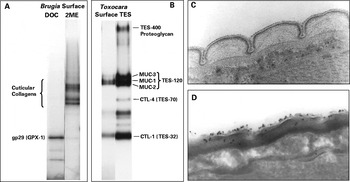
A transformational breakthrough in antigen identification came at the end of the 1970s when Bridget Ogilvie and Michael Parkhouse at NIMR introduced surface labelling as a technique for nematodes. Individual parasite antigens could be visualized for the first time, even if at that time identification at the sequence level was still elusive. It was also startlingly clear that the surface-accessible repertoire was limited to a small number of defined proteins (Philipp et al. Reference Philipp, Parkhouse and Ogilvie1980; Maizels et al. Reference Maizels, Philipp and Ogilvie1982), as illustrated in Fig. 1 A and B. Further, each stage in the life cycle presented a different and unique set of antigens, which only rarely cross-reacted with other stages (Philipp et al. Reference Philipp, Taylor, Parkhouse and Ogilvie1981; Maizels et al. Reference Maizels, Partono, Oemijati, Denham and Ogilvie1983a). In every case, these surface antigens were highly immunogenic in the host, allowing specific serological responses to be mapped, and where tested were also found to stimulate protective immune responses against the parasite (Grencis et al. Reference Grencis, Crawford, Pritchard, Behnke and Wakelin1986).
Surface antigens, secretions and surface coat of parasitic nematodes. (A) Brugia adults, surface labelled with iodogen (a non-permeable reagent which iodinates of tyrosine residues) and solubilized in deoxycholate, a non-ionic detergent (DOC) or with SDS and 2-mercaptoethanol (2ME). The major surface glycoprotein, (gp-29, glutathione peroxidase) is released into detergent, while under reducing conditions, the disulphide-linked cuticular collagens are solublilized (Selkirk et al. Reference Selkirk, Nielsen, Kelly, Partono, Sayers and Maizels1989). (B) T. canis larvae, surface labelled as (A), or ES proteins labelled with same reagent. Identities to antigens discussed in the text are indicated. Note that the mucins are the major carriers of the dominant trisaccharide glycan. (C) Cryo-electron micrograph showing Ruthenium Red-stained polyanionic surface coat exterior to the cuticle of T. canis larvae. (D) Cryo-immunoelectron micrograph localizing binding of monoclonal antibody to the trisaccharide glycan binding to the surface coat (Page et al. Reference Page, Rudin, Fluri, Blaxter and Maizels1992b).

With the advent of cDNA cloning, nematode surface antigens were conclusively identified, such as the gp29 glutathione peroxidase from Brugia malayi (Maizels et al. Reference Maizels, Gregory, Kwan-Lim and Selkirk1989; Cookson et al. Reference Cookson, Blaxter and Selkirk1992), also independently discovered by Eileen Devaney in B. pahangi (Devaney, Reference Devaney1988), and the TES-32 C-type lectin of T. canis (Maizels et al. Reference Maizels, de Savigny and Ogilvie1984; Loukas et al. Reference Loukas, Mullin, Tetteh, Moens and Maizels1999), which has homologues in hookworms and other nematodes (Loukas and Maizels, Reference Loukas and Maizels2000).
In 1981, in collaboration with Felix Partono of the University of Indonesia, we had begun to examine the antigens of human filarial parasites, and the immune responses to them. We were able to show that all the lymphatic filariae, including Brugia timori and Wuchereria bancrofti, expressed highly similar sets of surface antigens which were also strongly cross-reactive in terms of human antibody recognition (Maizels et al. Reference Maizels, Partono, Oemijati, Denham and Ogilvie1983a, Reference Maizels, Partono, Oemijati and Ogilvieb, Reference Maizels, Burke, Sutanto, Purnomo and Partono1986; Morgan et al. Reference Morgan, Sutanto, Purnomo, Sukartono, Partono and Maizels1986). These findings sparked curiosity into why there should be antigenic conservation within this set of parasites, rather than the diversity that might be expected under the pressure of the specific immune response. One possible answer is that filarial parasites induced some form of immunogical tolerance, favouring conservation of ‘tolerised’ specificities (Maizels and Lawrence, Reference Maizels and Lawrence1991).
Labelling experiments also investigated the structure of the nematode surface, which is an unconventional structure with a collagen-rich extracellular matrix bounded by a lipidic epicuticle. Interestingly, in Trichinella spiralis the epicuticle is not a conventional lipid bilayer, but is arranged as cylindrical lipid bundles based on a hexagonal pattern (Gounaris et al. Reference Gounaris, Smith and Selkirk1996). In T. canis, the fact that most surface antigens were also secreted (Maizels et al. Reference Maizels, de Savigny and Ogilvie1984) led to the discovery of a labile surface coat, external to the epicuticle (Fig. 1C and D), involved in parasite immune evasion (Page et al. Reference Page, Rudin, Fluri, Blaxter and Maizels1992b).
Secreted ES antigens
In 1975, Don de Savigny published a simple technique for culturing T. canis larvae, in serum-free medium, which allowed the collection of significant quantities of ‘excretory-secretory’ (ES) antigens (de Savigny, Reference de Savigny1975). In collaboration with Don, we identified the ES proteins (Maizels et al. Reference Maizels, de Savigny and Ogilvie1984), as did the group of Bob Grieve (Badley et al. Reference Badley, Grieve, Bowman, Glickman and Rockey1987b), and once cDNA libraries became available each of these antigens were cloned (Maizels et al. Reference Maizels, Tetteh and Loukas2000). In vitro biosynthetic labelling of secreted proteins with radioactive amino acids (Parkhouse et al. Reference Parkhouse, Clarke, Maizels and Denham1985) allowed identification of ES products from species and stages which are available in only small numbers, such as from the infective larvae of W. bancrofti (Maizels et al. Reference Maizels, Burke, Sutanto, Purnomo and Partono1986). While the technologies of the 1980s allowed Gek-Eng Kwan-Lim to visualize the broad array of adult B. malayi ES antigens (Kwan-Lim et al. Reference Kwan-Lim, Gregory, Selkirk, Partono and Maizels1989), nearly 20 years elapsed before these were matched to genome sequence data by proteomics (Hewitson et al. Reference Hewitson, Harcus, Curwen, Dowle, Atmadja, Ashton, Wilson and Maizels2008). The detection of ES products in the serum of infected humans (Des Moutis et al. Reference Des Moutis, Ouaissi, Grzych, Yarzabal, Haque and Capron1983) and animals (Maizels et al. Reference Maizels, Denham and Sutanto1985), also proved important in developing diagnostic circulating antigen tests (Weil et al. Reference Weil, Lammie and Weiss1997).
T. canis provided an unrivalled model system in which to study the relationship between surface and secreted proteins, because larvae could be recovered from eggs in the laboratory, and cultured at 37°C for many months in serum-free medium. With Malcolm Kennedy and Huw Smith, we generated monoclonal antibodies which demonstrated that most ES components were associated with the parasite surface (Maizels et al. Reference Maizels, Kennedy, Meghji, Robertson and Smith1987b). Tony Page then used these antibodies in immuno-electron microscopy to show that while the C-type lectin TES-32 is integrally distributed in the larval cuticle, the mucin-like glycoproteins (TES-120, see below) are synthesized in internal secretory glands (Page et al. Reference Page, Hamilton and Maizels1992a) and on release form a loosely-attached surface coat which envelops the parasite (Page et al. Reference Page, Rudin, Fluri, Blaxter and Maizels1992b). Immune attack, for example by eosinophils, results in shedding of the surface coat and parasite escape (Fattah et al. Reference Fattah, Maizels, McLaren and Spry1986; Badley et al. Reference Badley, Grieve, Bowman and Glickman1987a). Similar surface coats can be observed in a number of other nematode species (Blaxter et al. Reference Blaxter, Page, Rudin and Maizels1992).
Once identified, the challenge has been to determine the functional properties of ES components. ES material is known to have significant immunological properties; for example, Martin Holland showed that N. brasiliensis ES (NES) is an intrinsically potent Th2-inducing adjuvant (Holland et al. Reference Holland, Harcus, Riches and Maizels2000) while Billy Harnett's laboratory have demonstrated that the major component of ES from the rodent filaria Acanthocheilonema viteae (ES-62) directly interferes with host immune cell signalling, offering therapeutic routes to the treatment of immunological disorders such as arthritis (Harnett et al. Reference Harnett, Goodridge and Harnett2005) and allergy (Melendez et al. Reference Melendez, Harnett, Pushparaj, Wong, Tay, McSharry and Harnett2007). Many ES products are proteases, fulfilling key roles in invasion and nutrition (Williamson et al. Reference Williamson, Lustigman, Oksov, Deumic, Plieskatt, Mendez, Zhan, Bottazzi, Hotez and Loukas2006), but their profile as immunomodulators is less certain. For example, although NES contains proteases (Healer et al. Reference Healer, Ashall and Maizels1991), they are not responsible for its Th2-driving properties (Holland et al. Reference Holland, Harcus, Riches and Maizels2000). Similarly, while ES-62 is a leucine aminopeptidase, its immunoregulatory activity is attributable to phosphorylcholine modifications and not to enzymatic activity (Goodridge et al. Reference Goodridge, McGuiness, Houston, Egan, Al-Riyami, Alcocer, Harnett and Harnett2007).
cDNA cloning and the spliced leader
The second transformational change took place from the mid-1980s with the introduction of molecular cloning of cDNA transcripts (Selkirk et al. Reference Selkirk, Rutherford, Denham, Partono and Maizels1987). Screening B. pahangi expression libraries with monospecific antisera, Murray Selkirk's laboratory isolated clones for key surface-associated antigens of microfilariae (MF22, SHP-1) (Selkirk et al. Reference Selkirk, Yazdanbakhsh, Freedman, Blaxter, Cookson, Jenkins and Williams1991) and adults (gp29 and gp15/400) (Cookson et al. Reference Cookson, Blaxter and Selkirk1992; Tweedie et al. Reference Tweedie, Paxton, Ingram, Maizels, McReynolds and Selkirk1993). A new platform for parasite biology and immunology was created with a rapid succession of collagens, muscle and heat shock proteins, enzymes, inhibitors and lipid-binding proteins being identified by investigators around the world. Pleasingly, the Selkirk laboratory also sequenced the acetylcholinesterases of N. brasiliensis first discovered by Bridget Ogilvie (Blackburn and Selkirk, Reference Blackburn and Selkirk1992; Grigg et al. Reference Grigg, Tang, Hussein and Selkirk1997). By differential screening of an Onchocerca volvulus cDNA library with sera from onchocerciasis and lymphatic filariasis patients, Jan Bradley isolated a set of O. volvulus-specific clones, providing both immunodiagnostic probes and potential immunomodulators (Bradley et al. Reference Bradley, Helm, Lahaise and Maizels1991). Most of the individual parasite proteins studied in the field today were first characterized during this era of discovery.
C. elegans molecular biology was developing rapidly at this time, for example with the discovery of a perfectly-conserved 22-nt spliced leader (SL) sequence which is trans-spliced onto the 5′ end of >50% of mRNA transcripts (Blumenthal and Thomas, Reference Blumenthal and Thomas1988). David Gems exploited this feature to amplify mRNA from T. canis by PCR using SL and oligo-dT (complementary to the poly-A tail at the 3′ end of all mRNAs) primers. He found 2 abundant mRNAs, one of which was a phosphatidylethanolamine (PE)-binding protein (Gems et al. Reference Gems, Ferguson, Robertson, Page, Blaxter and Maizels1995), and the other a mucin (Gems and Maizels, Reference Gems and Maizels1996), 1 of 3 associated with the surface-coat (Loukas et al. Reference Loukas, Hintz, Tetteh, Mullin and Maizels2000b). The SL ‘trick’ was then used to great effect on B. malayi by Bill Gregory to discover the abundant larval transcript (ALT) proteins (Gregory et al. Reference Gregory, Blaxter and Maizels1997), subsequently shown to be a prominent larval-specific antigen shared by all filarial parasites, and a promising vaccine candidate (Gregory et al. Reference Gregory, Atmadja, Allen and Maizels2000). Returning the compliment, Mark Blaxter applied parasite surface labelling techniques to C. elegans (Blaxter, Reference Blaxter1993).
Era of transcriptomes, genomes and proteomes
The late 1990s saw the development of systematic sequencing of cDNA libraries, termed Expressed Sequence Tags (ESTs). For B. malayi, this analysis confirmed that the SL-bearing alt transcripts were indeed very highly expressed by L3 larvae (Blaxter et al. Reference Blaxter, Raghavan, Ghosh, Guiliano, Lu, Williams, Slatko and Scott1996). More directed cDNA sequencing projects were then aimed to capture major products of N. brasiliensis (Harcus et al. Reference Harcus, Parkinson, Fernández, Daub, Selkirk, Blaxter and Maizels2004) and T. canis (Tetteh et al. Reference Tetteh, Loukas, Tripp and Maizels1999), as well as – foraying into cestodes – Echinococcus granulosus (Fernández et al. Reference Fernández, Gregory, Loke and Maizels2002). In the case of N. brasiliensis, for example, we were able to show that signal sequence-bearing proteins, presumed to include most of the secreted (ES) products, had a greater proportion of novel sequences with no similarity to C. elegans or other database entries; this was interpreted as showing a greater diversification among proteins interacting with the host (Harcus et al. Reference Harcus, Parkinson, Fernández, Daub, Selkirk, Blaxter and Maizels2004). With large-scale transcriptomics now accomplished across many members of the nematode phylum (Parkinson et al. Reference Parkinson, Mitreva, Whitton, Thomson, Daub, Martin, Schmid, Hall, Barrell, Waterston, McCarter and Blaxter2004), a rich new resource for helminth research had been made available.
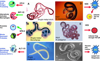
The size of nematode genomes, typically 5–30×107 bp, has proved an obstacle to full genome sequences until recent advances in high-throughput sequencing. In addition, there are varying degrees of polymorphism within each parasite species (Maizels and Kurniawan-Atmadja, Reference Maizels and Kurniawan-Atmadja2002; Redman et al. Reference Redman, Packard, Grillo, Smith, Jackson and Gilleard2008), which can frustrate genome assembly. Nevertheless, a real success in the field has been the publication of the draft sequence of B. malayi in 2007 (Ghedin et al. Reference Ghedin, Wang, Spiro, Caler, Zhao, Crabtree, Allen, Delcher, Guiliano, Miranda-Saavedra, Angiuoli, Creasy, Amedeo, Haas, El-Sayed, Wortman, Feldblyum, Tallon, Schatz, Shumway, Koo, Salzberg, Schobel, Pertea, Pop, White, Barton, Carlow, Crawford, Daub, Dimmic, Estes, Foster, Ganatra, Gregory, Johnson, Jin, Komuniecki, Korf, Kumar, Laney, Li, Li, Lindblom, Lustigman, Ma, Maina, Martin, McCarter, McReynolds, Mitreva, Nutman, Parkinson, Peregrin-Alvarez, Poole, Ren, Saunders, Sluder, Smith, Stanke, Unnasch, Ware, Wei, Weil, Williams, Zhang, Williams, Fraser-Liggett, Slatko, Blaxter and Scott2007). This dataset has allowed us to apply proteomics to nematode ES products; for example, recently James Hewitson and Yvonne Harcus identified some 80 proteins in adult B. malayi ES (BES), including a number detailed below (Hewitson et al. Reference Hewitson, Harcus, Curwen, Dowle, Atmadja, Ashton, Wilson and Maizels2008). For our laboratory, the focus has been on identifying which of these many predicted products and/or known secreted proteins may be immunomodulatory (Maizels et al. Reference Maizels, Blaxter and Scott2001a, Reference Maizels, Gomez-Escobar, Gregory, Murray and Zangb). In Fig. 2, and in a later part of this review, I summarize our findings on candidate modulators which have been obtained from a variety of approaches, ranging from in vitro testing of recombinant proteins, to heterologous expression by transfection into Leishmania for assessment of immunological functions in vivo (Maizels et al. Reference Maizels, Gomez-Escobar, Prieto-Lafuente, Murray and Aebischer2008).
Immunomodulators secreted by nematode parasites and their host target cells. (A) B. malayi adult worms: CPI-2, MIF-1/2. (B) B. malayi microfilariae: SPN-2. (C) B. malayi infective larvae (L3): ALT-1/2. (D) N. brasiliensis adult worms: NES. (E) H. polygyrus adult worms: HES. (F) T. canis adult worms: glycans and lectins. Images are from our own laboratory, with thanks to Bill Gregory (A), Judith Allen and Andrew MacDonald (B), Bill Gregory and Sinclair Stammers (C), Julie Healer and Sinclair Stammers (D), Constance Finney (E) and Tony Page (F). See text for details of all mediators mentioned.

HUMAN FILARIASIS – TOLERANCE AND IMMUNITY
The central immunological feature of filarial infection is antigen-specific hyporesponsiveness, the inverse relationship between the presence of parasites and T-cell reactivity, such that peripheral T cells from microfilaraemic carriers fail to mount a proliferative response to parasite antigen in vitro. Maria Yazdanbakhsh's group in Leiden, collaborating together with colleagues at the University of Indonesia, had shown that unresponsiveness ablated IFN-γ and IL-5 responses (Sartono et al. Reference Sartono, Kruize, Kurniawan-Atmadja, Maizels and Yazdanbakhsh1997), while IL-4 and IL-10 were largely intact. Hence, immunomodulation was relatively selective and targetted ‘effector’ cytokines rather than the regulatory mediators. Maria's group also showed that responsiveness can be regained following drug treatment, demonstrating that hyporesponsiveness is maintained by live parasites (Sartono et al. Reference Sartono, Kruize, Kurniawan, van der Meide, Partono, Maizels and Yazdanbakhsh1995). Hyporesponsiveness can, as first mooted by Eric Ottesen (Ottesen, Reference Ottesen1984) be considered a form of immunological tolerance; these ideas were developed first with Rachel Lawrence, in terms of how tolerance may break down and immunopathology develop (Maizels and Lawrence, Reference Maizels and Lawrence1991) and more recently with Maria Yazdanbakhsh within a wider framework of a regulatory T-cell network (Maizels and Yazdanbakhsh, Reference Maizels and Yazdanbakhsh2003). The key unanswered question is what factors influence the propensity to develop a tolerance-inducing regulatory T-cell response, or an immunity/pathology-conferring effector response.
In addition to hyporesponsiveness, Eric Ottesen's laboratory also highlighted the extraordinarily high levels of parasite-specific IgG4 antibodies present in microfilaraemic patients (Hussain et al. Reference Hussain, Grögl and Ottesen1987). IgG4 is a poorly-researched isotype which represents <5% of normal serum immunoglobulins, and does not bind Fc receptors that activate effector mechanisms. In filariasis, however, the antibody response may be dominated by IgG4, and indeed with Karen Day (then at Imperial College) we showed that there is a significant positive correlation between microfilarial counts and IgG4 titres (Kwan-Lim et al. Reference Kwan-Lim, Forsyth and Maizels1990). Qualitatively, multiple antigenic species are recognized by this isotype (Kurniawan et al. Reference Kurniawan, Sartono, Partono, Yazdanbakhsh and Maizels1998), consistent with a ‘blocking antibody’ hypothesis that in filariasis effector mechanisms may be disabled by the high concentration of IgG4 antibodies. Recently, Achim Hoerauf's laboratory has established that IgG4 is specifically up-regulated by IL-10, which may be produced by regulatory T cells (Satoguina et al. Reference Satoguina, Weyand, Larbi and Hoerauf2005). Hence, IgG4 levels may reflect the degree of immune regulation in a helminth infection, as much as they reflect worm load.
With the development of circulating antigen assays in filariasis, Karen Day applied these to testing epidemiological concepts. Using changes in circulating antigen levels over time to quantify adult worm numbers, and thereby the acquisition of new infections, she showed that adults gained a form of concomitant immunity to incoming larval infections with W. bancrofti (Day et al. Reference Day, Grenfell, Spark, Kazura and Alpers1991b). Together with Bill Gregory, we showed that acquired immunity coincided with non-IgG4 antibody to the surface of infective larvae (Day et al. 1991 a), to a specificity distinct from the major protein antigen components. Hence the antibody profile in a ‘tolerant’ individual is very different from that of an ‘immune’ subject, although there is no evidence as yet from human studies that immunity to filariasis is antibody mediated.
Nematode Antigens and Modulators
These profound effects on the host immune system spurred interest in the molecular products of helminths which may modulate immunity. The following section, and Fig. 2, summarise the major players identified so far.
Cystatins and serpins
Protease inhibitors secreted by nematodes do not simply regulate activity of parasite enzymes, as 2 major ES protease inhibitor families appear to act as immunomodulators by targetting host enzymes. One set, the cystatins (cysteine protease inhibitors) include CPI-2, a 15 kDa surface and secreted protein of B. malayi larvae and adult worms (Gregory et al. Reference Gregory, Blaxter and Maizels1997; Gregory and Maizels, Reference Gregory and Maizels2008), as well as O. volvulus and A. viteae (Hartmann and Lucius, Reference Hartmann and Lucius2003). Although homologues from plants to vertebrates inhibit papain-like cysteine proteinases, only mammals have forms which block asparaginyl endopeptidase (AEP), a key enzyme in antigen processing, through a second inhibitory site. Remarkably, CPI-2 has evolved the same AEP-inhibitory motif, and hence can interfere with antigen processing in human cells (Manoury et al. Reference Manoury, Gregory, Maizels and Watts2001). Consistent with the hypothesis that parasite cystatins have evolved AEP inhibition as an immune interference strategy, Janice Murray showed that C. elegans homologues lacked both the sequence motif and AEP inibition (Murray et al. Reference Murray, Manoury, Balic, Watts and Maizels2005). One unsolved puzzle is how CPI-2, secreted from extracellular Brugia parasites, accesses intracellular antigen-processing compartments of mammalian cells; an untested possibility is that a unique 20-aa N-terminal extension of the filarial CPI-2 protein, absent from other cystatins, is involved in uptake or entry into host APCs.
In the late 1990s, Xingxing Zang started a project to identify T-cell-stimulating antigens from B. malayi. He identified a serine protease inhibitor (serpin) SPN-2, which was also the most abundant transcript from the microfilarial (Mf) stage. In collaboration with a leading serpin laboratory, we found that SPN-2 selectively inhibited 2 neutrophil proteases (Zang et al. Reference Zang, Yazdanbakhsh, Kiang, Kanost and Maizels1999), but not a range of other serine proteases tested. Subsequently, another group noted that the SPN-2 sequence varied from the consensus for inhibitory proteins, and reported lack of inhibitory activity for SPN-2. Irrespective of this controversy, SPN-2 selectively drives Th1 responses (Zang et al. Reference Zang, Atmadja, Gray, Allen, Gray, Lawrence, Yazdanbakhsh and Maizels2000), an interesting finding in view of the fact that B. malayi Mf are one of the few helminth forms which induce Th1 in vivo (Lawrence et al. Reference Lawrence, Allen, Osborne and Maizels1994). SPN-2 is a major target of antibody in infection, and presence of the IgG4 isotype to this antigen can be considered diagnostic of active infection (Zang et al. Reference Zang, Atmadja, Gray, Allen, Gray, Lawrence, Yazdanbakhsh and Maizels2000), as antibody levels decay rapidly following chemotherapeutic clearance of parasites. Notably, no C. elegans homologues encode a signal sequence, indicating that B. malayi has evolved to secrete the serpin for a new function within the host (Zang and Maizels, Reference Zang and Maizels2001).
Glycans, mucins and lectins
Carbohydrate specificities play a major role in the immunology of infections. In the case of T. canis ES, we found that it comprised approximately 40% carbohydrate by weight (Meghji and Maizels, Reference Meghji and Maizels1986); subsequent characterization of the glycan structures by mass spectrometry, the first for a parasitic nematode, identified a dominant O-linked trisaccharide similar to mammalian blood group sugars, but modified with 1–2 additional O-methyl groups (Khoo et al. Reference Khoo, Maizels, Page, Taylor, Rendell and Dell1991). These trisaccharides have more recently been synthesized (Amer et al. Reference Amer, Hofinger and Kosma2003) and confirmed as major antigens in infection (Schabussova et al. Reference Schabussova, Amer, van Die, Kosma and Maizels2007). Monoclonal antibodies reactive to the trisaccharides also bind to the mucin glycoproteins produced by the major larval secretory glands (Page et al. Reference Page, Hamilton and Maizels1992a), and bind the labile surface coat of the parasite (Page et al. Reference Page, Rudin, Fluri, Blaxter and Maizels1992b). Whether the mucins and glycans interact further with the immune system once they are secreted seems likely but has yet to be documented.
Alex Loukas subsequently discovered, by protein sequencing, that T. canis TES-32 was a C-type lectin (CTL), with calcium-dependent carbohydrate specificity (Loukas et al. Reference Loukas, Mullin, Tetteh, Moens and Maizels1999), presaging a much wider presence in helminth secretions of this family of carbohydrate binding proteins (Loukas and Maizels, Reference Loukas and Maizels2000; Loukas et al. 2000 a). Because immune system receptors such as DC-SIGN are members of the same gene family, the possibility arose that secreted parasite CTLs interfere or compete with host lectins for ligands, thereby blocking host immunity. If so, it would constitute a fascinating role reversal, as similar CTLs are employed in C. elegans to mediate immunity against bacterial infection. In a further functional twist, the surface-bound CTL of the marine nematode Laxus captures symbiotic bacteria required for sulphur metabolism (Bulgheresi et al. Reference Bulgheresi, Schabussova, Chen, Mullin, Maizels and Ott2006). It is notable that while CTLs are prominent in the secretions of T. canis and hookworms, they are poorly represented among the filarial parasites; however, adult B. malayi secrete an abundant S-type lectin (galectin), which while bearing no structural similarity to CTLs, might serve a similar physiological role in vivo (Hewitson et al. Reference Hewitson, Harcus, Curwen, Dowle, Atmadja, Ashton, Wilson and Maizels2008).
Cytokine homologues
The immunosuppressive cytokine TGF-β belongs to an evolutionarily ancient extracellular signalling family that also includes bone morphogenetic proteins (BMPs). From observations of filarial parasite hyporesponsiveness, and with identification in C. elegans of the TGF-β homologue DAF-7, we hypothesized that B. malayi may express a functionally active mimic of TGF-β. Natalia Gomez-Escobar used degenerate PCR to isolate a B. malayi homologue (TGH-1), with a 7-cysteine active domain as found in the BMPs, which is primarily expressed in developing larvae (Gomez-Escobar et al. Reference Gomez-Escobar, Lewis and Maizels1998). A second homologue more like DAF-7 and human TGF-β in containing a 9-cysteine active domain emerged from ESTs; moreover, transcription of this gene (TGH-2) was maximal in the developmentally-arrested Mf and mature adult stages, which were found to secrete the protein (Gomez-Escobar et al. Reference Gomez-Escobar, Gregory and Maizels2000). More recently, we have found ES from Heligmosomoides polygyrus contains an active TGF-β mimic able to induce immunosuppressive regulatory T cells (Grainger et al., manuscript in preparation). Currently we are testing whether this biological activity is indeed attributable to a nematode homologue of the mammalian gene so central to immunoregulation.
A second ancient evolutionary family, which encompasses both mammalian cytokines and parasite homologues, is that of the Macrophage Migration Inhibitory Factor, or MIF (Vermeire et al. Reference Vermeire, Cho, Lolis, Bucala and Cappello2008). The first member of this family from B. malayi (MIF-1) was reported by Alan Scott's laboratory (Pastrana et al. Reference Pastrana, Raghavan, FitzGerald, Eisinger, Metz, Bucala, Schleimer, Bickel and Scott1998), and Xingxing Zang isolated a second homologue, MIF-2, which was subsequently crystallized in Edinburgh (Zang et al. Reference Zang, Taylor, Meyer, Wang, Scott, Walkinshaw and Maizels2002). Because of the apparent contradication for a tissue-dwelling parasite to produce a potentially pro-inflammatory mediator, we examined the activity of MIF in more detail. In vivo, MIF was able to induce the production of Ym-1, a key product of alternatively activated macrophages (Falcone et al. Reference Falcone, Loke, Zang, MacDonald, Maizels and Allen2001). Most recently, Lidia Prieto-Lafuente established that in the presence of IL-4, MIF loses ability to induce IL-12 and TNF, and instead promotes the development of these suppressive macrophages (Prieto-Lafuente et al. Reference Prieto-Lafuente, Gregory, Allen and Maizels2009).
Novel proteins – ALTs, VALs and vaccines
Many parasite products involved in establishment in the host will not be recognized or functionally categorized by sequence alone. We were intrigued by novel gene transcripts that are highly expressed in infective larval stages, as judged by their abundance in EST datasets. For B. malayi, 2 such families stood out at levels >1%, the abundant trans-spliced transcripts alt-1 and alt-2 mentioned above, and a Venom Allergen-Like (VAL) homologue similar to Ancylostoma secreted protein (ASP).
ALT-1 and -2 proteins are approximately 80% identical to each other. Homologues exist in all filarial nematodes, for example in O. volvulus in which Ted Bianco's laboratory showed ALT proteins stockpiled in the glandular oesophagus of the vector-borne infective larva (Wu et al. Reference Wu, Egerton, Pappins, Harrison, Wilkinson, Underwood and Bianco2004). ALT proteins are the foremost filarial vaccine candidates, with data from several laboratories showing protection in animal models (Gregory et al. Reference Gregory, Atmadja, Allen and Maizels2000). We wished, however, to test the immunomodulatory properties of ALT proteins, as the dominant parasite products at the first encounter with the immune system. With Natalia Gomez-Escobar, we adopted the strategy of transfection of Leishmania parasites, to allow in vitro effects on macrophages and in vivo outcomes in mice to be evaluated (Maizels et al. Reference Maizels, Gomez-Escobar, Prieto-Lafuente, Murray and Aebischer2008). When either ALT-1 or ALT-2 were expressed as a transgene in L. mexicana, growth in macrophages was enhanced; cellular mRNA analyses showed higher levels of SOCS-1 expression due to ALT transfection. Because SOCS-1 inhibits IFN-γ signalling, the effects of ALT may therefore dampen Th1 effector mechanisms. Consistent with this hypothesis, the virulence of ALT-expressing lines was significantly amplified, with up to 3-fold more parasites in vivo (Gomez-Escobar et al. Reference Gomez-Escobar, Bennett, Prieto-Lafuente, Aebischer, Blackburn and Maizels2005).
Nematode VAL/ASP proteins are similarly elusive in function. Homologues range from pathogenesis-related proteins in plants through to testis-specific proteins in mammals, but have no assigned biological function. It is possible that they represent a conserved structural framework compatible with a variety of functional activities. The first nematode homologue (Ancylostoma secreted protein) was discovered by Peter Hotez' laboratory (Hawdon et al. Reference Hawdon, Jones, Hoffman and Hotez1996), and a closely-related product is now in human vaccine trials. Since then, VAL/ASP proteins have been shown to be dominant ES products of many organisms, such as H. contortus (Yatsuda et al. Reference Yatsuda, Krijgsveld, Cornelissen, Heck and De Vries2003). In our laboratory, Janice Murray isolated the B. malayi VAL protein and showed it to be highly immunogenic although its vaccine efficacy, under the conditions tested, was less potent than that of ALTs (Murray et al. Reference Murray, Gregory, Gomez-Escobar, Atmadja and Maizels2001).
Surprisingly, when we applied the transcript-led approach to T. canis (Tetteh et al. Reference Tetteh, Loukas, Tripp and Maizels1999), we found not only the major secreted lectins and mucin aproprotein, but also a set of 4 abundant novel transcripts (ANTs) with similarity to each other only in their 3′ untranslated regions. No protein corresponding to these transcripts could be identified, but 27 nt in the 3′ UTR bore similarity to the lin-4 suppressive micro-RNA of C. elegans. The possibility of post-transcriptional suppression of protein translation was supported by transfection of C. elegans with GFP reporter constructs containing the ANT 3′ UTR, which resulted in sequence-specific suppression of GFP expression (Callister et al. Reference Callister, Winter, Page and Maizels2008). Hence, empirical proteomic analysis is crucial before interpreting parasite expression profiles based on cDNA alone.
Phosphorylcholine (PC)
The presence of PC in somatic antigens from N. brasiliensis had been discovered in the 1970s (Péry et al. Reference Péry, Luffau, Charley, Petit, Rouze and Bernard1979). Remarkably, many monoclonal antibodies raised to filarial antigens were PC-specific, and reacted with a circulating parasite antigen (Forsyth et al. Reference Forsyth, Spark, Kazura, Broen, Peters, Heywood, Dissanayake and Mitchell1985; Maizels et al. Reference Maizels, Burke and Denham1987a). In human patients, PC is found on a high molecular-weight proteoglycan-like complex, while in the rodent filaria A. viteae, PC occurs on one secreted protein, leucine aminopeptidase (LAP) or ES-62. More recently, James Hewitson identified that in B. malayi, PC is bound not to LAP but to a distinct secreted protein, N-acetylgalactosamine transferase (Hewitson et al. Reference Hewitson, Harcus, Curwen, Dowle, Atmadja, Ashton, Wilson and Maizels2008). Interestingly, in another rodent filarial species (Litomosoides sigmodontis), the PC specificity is replaced by dimethylethanolamine (DMEA), containing one less methyl group (Hintz et al. Reference Hintz, Schares, Taubert, Geyer, Zahner, Stirm and Conraths1998). This microheterogeneity in the presentation of PC-like epitopes may relate to differences in innate recognition systems of the respective host species.
THE METAMORPHOSIS OF NEMATODE IMMUNOLOGY
From the modern perspective, helminth immunology 30 years ago was a blank canvas; infection was associated with eosinophilia, mastocytosis and IgE, but the interconnectedness of these phenomena was not obvious. Mechanisms of immunity were interpreted from in vitro assays or by in vivo transfer of unseparated lymphocyte populations or infection sera likely to have contained cytokines. The transformation of this field came through both technical and conceptual revolutions. The identification of cytokines and their receptors, monoclonal reagents and gene-targetted (knockout) mice, and the development of fluorescent cell staining and fast cell sorting have all provided precise tools to dissect immunity to parasites. Moreover, with the delineation of Th1 and Th2 in 1986 by Coffman and Mosmann (Mosmann et al. Reference Mosmann, Cherwinski, Bond, Giedlin and Coffman1986), helminth immunology also became conceptually alive.
Nematodes clearly provoked strong Th2 responses, including IL-4 and IL-5, stimulating IgE and eosinophilia. Immunity to gut nematodes was shown to be Th2-dependent by Kathryn Else and Richard Grencis in Manchester for Trichuris muris (Else et al. Reference Else, Finkelman, Maliszewski and Grencis1994) and by Fred Finkelman, Joe Urban and colleagues for other gastrointestinal species with a range of in vivo cytokine manipulations (Finkelman et al. Reference Finkelman, Shea-Donohue, Goldhill, Sullivan, Morris, Madden, Gause and Urban1997). Interestingly, different Th2-dependent mechanisms were then found to be involved in immunity to each species studied, with N. brasiliensis, for example, being most sensitive to IL-13-induced goblet cell mucin production, and Strongyloides ratti expulsion requiring mast cell activation (Maizels and Holland, Reference Maizels and Holland1998). Whether Th2 immunity is sufficent against tissue-dwelling nematodes, however, was far less clear (Allen and Maizels, Reference Allen and Maizels1997); Rachel Lawrence had shown that B. malayi microfilariae in fact drive Th1 responses (Lawrence et al. Reference Lawrence, Allen, Osborne and Maizels1994), and IL-4-deficient animals are not more susceptible to infection (Lawrence et al. Reference Lawrence, Allen, Gregory, Kopf and Maizels1995). Mice deficient in IL-4R (and also unresponsive to IL-13) are likewise resistant to B. malayi larval infection (McSorley et al. Reference McSorley, Harcus, Murray, Taylor and Maizels2008). Hence, control of tissue helminth infections requires an important non-Th2 component. With very recent recognition of other T-cell subsets in infection, this issue is now being re-examined.
The Th1/Th2 paradigm also seemed, at first, highly applicable to human filariasis, with a unarguable antigen-specific Th2 bias in infected patients (Sartono et al. Reference Sartono, Kruize, Kurniawan, Maizels and Yazdanbakhsh1996). However, the paradox remained that Th2 responses often co-existed with active helminth infections, calling into question their protective effect. Moreover, cytokine responses did not accord exactly with expectations, with intact IL-4 responses in the absence of IL-5 (Sartono et al. Reference Sartono, Kruize, Kurniawan-Atmadja, Maizels and Yazdanbakhsh1997). Two lines of argument could be offered to account for these discrepancies; first, that patients showed a ‘modified Th2’ similar to that observed in desensitized allergic patients (Platts-Mills et al. Reference Platts-Mills, Vaughan, Blumenthal, Pollart Squillace and Sporik2001), in which effector cytokines such as IL-5 are shut down; or that a ‘third force’ of suppressive, regulatory T cells controlled both Th1 and Th2 responses (Sakaguchi, Reference Sakaguchi2000), and were involved in the maintenance of host hyporesponsiveness to parasite infection (Hoerauf and Brattig, Reference Hoerauf and Brattig2002; Maizels and Yazdanbakhsh, Reference Maizels and Yazdanbakhsh2003). As explained below, both models may apply in parallel.
Dendritic cells and macrophages
In the first encounter between pathogens and the innate immune system, dendritic cells (DCs) shape the ensuing response, through recognition of pathogen molecules and selective induction of T-cell differentiation. A central paradigm is that bacterial LPS, ligating TLR4, upregulates surface costimulator molecule expression and induces IL-12 release, driving a Th1 outcome. Adam Balic asked whether DCs exposed to NES would represent the opposite side of the coin, eliciting a Th2 bias, and indeed this was the case (Balic et al. Reference Balic, Harcus, Holland and Maizels2004). Interestingly, the capacity to drive Th2 responsiveness was not accompanied by the upregulation of surface markers observed in LPS stimulation, but by blocking the ability of LPS to induce IL-12 p70, as has also been observed for other helminth products (Cervi et al. Reference Cervi, MacDonald, Kane, Dzierszinski and Pearce2004). Because NES actively promotes Th2 differentiation, even in the presence of Th1-inducing bacterial products, we argued against a ‘default’ hypothesis in which Th2 resulted from an absence of DC stimulation (MacDonald and Maizels, Reference MacDonald and Maizels2008). More recently, with the focus shifting to DC populations in vivo, Katie Smith in our laboratory has shown that nematode infection switches intestinal DCs into a predominantly tolerogenic subset.
An unusual innate immune population was discovered by Judith Allen in the peritoneal cavity of Brugia-infected mice. These were macrophages which exerted a broad and profound anti-proliferative effect on target cells (Allen et al. Reference Allen, Lawrence and Maizels1996), and which required IL-4 but not IL-10 to develop (MacDonald et al. Reference MacDonald, Maizels, Lawrence, Dransfield and Allen1998), While the mechanism of suppression is still unknown, it is cell contact-dependent (Loke et al. Reference Loke, MacDonald, Robb, Maizels and Allen2000). This new Th2-associated macrophage was similar to the ‘alternatively activated’ macrophage identified by Saimon Gordon at Oxford (Gordon, Reference Gordon2003), and was further linked to the synthesis of a chitinase-like molecule Ym1, among other characteristic proteins. From the initial finding in Brugia-infected mice, the study of alternatively activated macrophages has grown into a major field bringing together immunity, wound healing, obesity and innate recognition (Loke et al. Reference Loke, Gallagher, Nair, Zang, Brombacher, Mohrs, Allison and Allen2007; Siracusa et al. Reference Siracusa, Reece, Urban and Scott2008). A functional role for these cells in protective immunity against H. polygyrus, while in its larval phase in the gut wall, has been demonstrated (Anthony et al. Reference Anthony, Urban, Alem, Hamed, Rozo, Boucher, Van Rooijen and Gause2006). A key question is what stimulates the development of this phenotype, and Lidia Prieto-Lafuente has very recently shown that the combination of IL-4 and MIF (whether from host or parasite) can induce suppressive, Ym-1-expressing macrophages in vitro (Prieto-Lafuente et al. Reference Prieto-Lafuente, Gregory, Allen and Maizels2009).
Regulatory T cells and tolerance
The Regulatory T cell (Treg) has represented the latest conceptual tidal wave in immunology, and the concept of a suppressive population inhibiting both Th1 and Th2 was one which resonated immediately in the human filariasis context (Doetze et al. Reference Doetze, Satoguina, Burchard, Rau, Loliger, Fleischer and Hoerauf2000). In 2001 Matthew Taylor started testing whether, in an animal model, Tregs blocked Th2 immunity to L. sigmodontis. Matt showed that ablation of Treg activity, using anti-CD25 and anti-GITR antibody treatment in vivo, significantly reduced worm load (Taylor et al. Reference Taylor, Le Goff, Harris, Malone, Allen and Maizels2005). This was the first immunological intervention able achieve a measure of ‘cure’ in a susceptible infected host. While anti-CD25 alone abated Treg numbers, additional anti-GITR or anti-CTLA-4 (Taylor et al. Reference Taylor, Harris, Babayan, Bain, Culshaw, Allen and Maizels2007) were required to kill parasites. Because infected mice displayed antigen-specific hyporesponsiveness, which was reversed by the combined antibody treatment, we suggested that CD25+ Tregs tolerize the effector T-cell population, which requires re-activation through GITR ligation or CTLA-4 blockade (Taylor et al. Reference Taylor, Harris, Babayan, Bain, Culshaw, Allen and Maizels2007).
Mark Wilson and Constance Finney then studied Tregs in the model system of H. polygyrus, well characterized by Jerzy Behnke and David Pritchard at Nottingham. Mark found a significant rise in Treg numbers during infection, which Constance demonstrated was accompanied by raised expression of CD103 (considered to be a Treg activation marker) and increased suppressive effect per cell (Finney et al. Reference Finney, Taylor, Wilson and Maizels2007). Mark Wilson used H. polygyrus to explore the interaction between airway allergy and helminth infection, because this parasite does not transit the lungs. Infection resulted in a profound suppression of inflammation (measured, for example, by eosinophil infiltration) even in animals rendered fully allergic prior to infection. Most importantly, CD4+CD25+ Tregs from infected, allergen-naive animals can, on transfer to an allergen-sensitized but uninfected recipient, suppress airway allergy (Wilson et al. Reference Wilson, Taylor, Balic, Finney, Lamb and Maizels2005). Thus, helminth-induced Tregs were shown functionally to inhibit bystander reactivities, contributing to our perception of the ‘hygiene hypothesis’ discussed below.
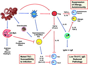
The question remains as to whether helminths stimulate expansion of pre-existing ‘natural’ Tregs, specific for self antigens, or induce the differentiation of new ‘adaptive’ Tregs from naive peripheral cells. We suspect that both may occur (Fig. 3). Matt Taylor showed that L. sigmodontis activates the natural Treg subset early in infection (Taylor et al. Reference Taylor, van der Werf, Harris, Graham, Bain, Allen and Maizels2009), while B. malayi can drive bystander (ovalbumin-specific) T cells into a regulatory phenotype (McSorley et al. Reference McSorley, Harcus, Murray, Taylor and Maizels2008). Most recently, John Grainger has found that H. polygyrus ES contains a TGF-β-like activity which, acting through the TGF-β receptor of mouse T cells, can convert non-regulatory cells into ‘adaptive’ Tregs, confirming that some nematodes at least have evolved to exploit the host regulatory network (Grainger et al., manuscript in preparation).
Immunoregulation in nematode infections and the ‘Hygiene Hypothesis’. Infection generates both activating signals (blue arrows), regulatory signals (red arrows) and inhibitory interactions (red bars). Hence, a Th2 response is generated through innate immune system cells, but in chronic infection adopts a modified or muted phenotype in which proliferative responses are poor (due to either or both alternatively activated macrophages and the effects of regulatory T cells). The Th2 response is sufficient, however, to inhibit pro-pathogenic Th1 and Th17 responses, again in combination with Treg activity. IL-10 switches B cells into IgG4 production, reducing IgE, and minimising overall pathology of infection. The high levels of Tregs (induced via DC, or directly by parasite molecules), together with B cells, create a regulatory environment which inhibits pro-allergic and autoimmune T cells from mounting a response. The observed associations between infections and protection from allergies and autoimmunity may be attributed to a regulatory network such as that depicted here.

IMPACT OF INFECTION ON ALLERGIES AND AUTOIMMUNITY – REVISING THE HYGIENE HYPOTHESIS
The inverse epidemiological association between helminth infections and allergies been has often remarked upon, but was not well substantiated until Anita van den Biggelaar in Maria Yazdanbakhsh's group showed that schistosome-infected Gabonese schoolchildren had lower Th2-dependent allergic reactivity than their uninfected classmates (van den Biggelaar et al. Reference van den Biggelaar, van Ree, Roderigues, Lell, Deelder, Kremsner and Yazdanbakhsh2000). This very significant work intimated a causal link between infection and reduced allergy which demanded more than a simple Th1 vs Th2 model, because schistosomes themselves elicit a potent Th2 response.
Maria Yazdanbakhsh's work was crucial for the reformulation of what had earlier been proposed as the ‘Hygiene Hypothesis’. Although originally conceived in terms of diminished bacterial stimulation of Th1 resulting in stronger pro-allergic Th2 responses, the hypothesis required revision to account for mounting Th1-mediated autoimmune diseases in countries with the more aseptic Western lifestyle, as well as the ability of Th2-inducing helminths to counter allergy. By incorporating a regulatory T cell network, able to dampen both Th1 autoimmunity and Th2 allergy, and dependent on extraneous organisms, a new version of the Hygiene Hypothesis emerged (Yazdanbakhsh et al. Reference Yazdanbakhsh, van den Biggelaar and Maizels2001, Reference Yazdanbakhsh, Kremsner and van Ree2002), which can be integrated with a model of immunoregulation in infection as shown in Fig. 3.
To test this hypothesis, Mark Wilson set up his studies described above and confirmed that nematode-driven Treg expansion could mediate suppression of airway allergy (Wilson et al. Reference Wilson, Taylor, Balic, Finney, Lamb and Maizels2005). A number of other groups have shown reductions in both allergic and autoimmune pathologies in mice infected with helminth parasites (reviewed in Wilson and Maizels, Reference Wilson and Maizels2004), and abated human autoimmune pathologies in helminth infection have also been reported (Correale and Farez, Reference Correale and Farez2007; Elliott et al. Reference Elliott, Summers and Weinstock2007). However, the modifying effects of infection are not limited only to parasites, or even only to pathogenic organisms. For example, among commensal bacteria, particular species are associated with immunoregulatory protection (Mazmanian et al. Reference Mazmanian, Round and Kasper2008). Perhaps there is even a parallel between bacterial commensals and the better-adapted helminth parasites, if both have mastered the art of conditioning the host immune system (Maizels, Reference Maizels2005).
CONCLUSION
While in 30 years our knowledge of helminth parasites has changed beyond recognition, the prevalence of the diseases they cause remains a critical problem. Fortunately, the challenge of disease control has now been embraced by agencies such as the Global Progamme for Elimination of Lymphatic Filariasis and the Bill and Melinda Gates Foundation, but there remains great potential to transform the profile of both parasitic and immunological diseases. In particular, the following areas can be highlighted.
First, the new genomes as well as the availability of C. elegans genetics, should greatly facilitiate elucidation of biochemical pathways and new drug candidates. A notable achievement of 2008 was the description of new anthelmintics targetting the nematode acetylcholine receptor (Kaminsky et al. Reference Kaminsky, Ducray, Jung, Clover, Rufener, Bouvier, Weber, Wenger, Wieland-Berghausen, Goebel, Gauvry, Pautrat, Skripsky, Froelich, Komoin-Oka, Westlund, Sluder and Maser2008). More distant, but attractive, drug targets would be immunomodulatory molecules, if neutralization potentiated the immune response to infection. We may also look forward to exploiting parasite immunomodulators as drugs in their own right to ameliorate inflammatory diseases such as allergy and autoimmunity.
Second, the onward march of cellular immunology is generating a constant stream of new models, reagents and tools which are being applied to helminth infections; just as the regulatory T-cell concept could explain an important aspect of the immunobiology of infection, so new cell types, cytokines or signalling pathways still to be defined will be instrumental in constructing the full picture of the host-nematode interaction. In addition, novel helminth products may lead us to new immune system receptors and pathways which have evolved specifically to combat helminth infections, enriching our knowledge of the immune system itself.
Third, another dividend of the genomic revolution will be the definition of nematode antigens at the level of key epitopes involved in tolerance and immunity. Coupled with the distinction between (for example) parasite-specific and self-specific regulatory T cells, and the identification of nematode-associated molecular patterns that may trigger the innate immune system, vaccines can be fine-tuned to maximize appropriate immune responsiveness and avoid deleterious outcomes (whether excessive pathology or excessive regulation).
It is perhaps overly optimistic to expect that all of these goals will be met in the next 30 years; but with renewed efforts now being placed in treatment of human infections on the global scale, and the speed of scientific advance still accelerating, it is tempting to speculate that with an unrecognizably deeper understanding of helminths, and far stronger array of practical tools for disease control, we will by then be close to eradication of helminths as a major global health problem.
I owe a huge debt of gratitude to all my many colleagues, mentors and students involved in the research described here, first at Mill Hill (1979–1983) then Imperial College London (1983–1995) and more recently at Edinburgh; I have not had room to detail all their many contributions. Space has limited the extent to which I have been able to refer to a great number of contemporary publications in this area. I would also like to thank the MRC and the Wellcome Trust in particular for supporting this research over many years.