

Introduction
The genus Cheirostylis consists of 53 species with a wide distribution in tropical parts of Africa, Asia and Oceania (Seidenfaden, Reference Seidenfaden1978). Cheirostylis sp. can be propagated by seed asymbiotically, but they show a very low germination rate, which may be due to the association with the mycoheterotrophic condition as demonstrated in Cheirostylis montana (Roy et al., Reference Roy, Watthana, Stier, Richard, Vessabutr and Selosse2009). Eight species of Cheirostylis are found in Thailand, namely Cheirostylis didymacantha Seidenf., Cheirostylis macrantha Schltr., Cheirostylis griffithii Lindl., C. montana Blume, Cheirostylis pusilla Lindl., Cheirostylis spathulata J. J. Sm., Cheirostylis thailandica Seidenf. and Cheirostylis yunnanensis Rolfe (Thaithong, Reference Thaithong1999). Of these, C. didymacantha Seidenf. and C. thailandica Seidenf. are classified as the endangered and vulnerable species in Thailand Red Data: Plants (Santisuk et al., Reference Santisuk, Chayamarit, Pooma and Suddee2006). Nowadays, this orchid is under the threat of extinction due to the destruction of forests that leads to its habitat loss and degradation (Salin, Reference Salin2006).
From our survey, Cheirostylis were found in the deciduous or bamboo forests of Thailand's Saraburi and Prachuap Khiri Khan provinces. Genus classification was based on plant morphology according to published descriptions (Rao, Reference Rao2011; Santisuk et al., Reference Santisuk, Chayamarit, Pooma and Suddee2006). C. yunnanensis Rolfe plants were identified by their unique characteristics of oblong petals that are truncated at the apex with hypochile saccate and seven thin terete calli in each half (Rao, Reference Rao2011; Santisuk et al., Reference Santisuk, Chayamarit, Pooma and Suddee2006). In addition, five samples of unknown species of Cheirostylis were found in Buriram (2), Kanchanaburi (2) and Loei (1) provinces, but species identification was not possible as the plants were in the seedling stage with no flowers.
Several types of molecular markers have been developed and applied to the genetic analysis of orchids. Of these, microsatellites or simple sequence repeat (SSR) markers are more powerful than other marker types due to the high level of polymorphism, co-dominance, high transferability and reproducibility (Butcher et al., Reference Butcher, Decroocq, Gray and Moran2000). Therefore, this study aimed to develop microsatellite markers specific to C. yunnanensis Rolfe for genetic diversity analysis of the species.
Experimental
Genomic DNA of C. yunnanensis Rolfe was extracted from young leaf tissue using the Cetyltrimethylammonium bromide method (Doyle and Doyle, Reference Doyle and Doyle1987). A microsatellite-enriched genomic DNA library was constructed as described by Sraphet et al. (Reference Sraphet, Boonchanawiwat, Thanyasiriwat, Boonseng, Tabata, Sasamoto, Shirasawa, Isobe, Lightfoot, Tangphatsornruang and Triwitayakorn2011). Genomic DNA was digested with three restriction enzymes, AluI (Takara, Japan), HaeIII (Takara, Japan) and AfaI (Takara, Japan). The digested products were purified using the Wizard SV gel and the PCR Clean-Up system (Promega, Madison, WI, USA), and then ligated with double-stranded linkers (linker-1; 5′-GTTTAGCCTTGTAGCAGAAGC-3′ and linker-2; 5′-pGCTTCTGCTACAAGGCTAAACAAAA-3′) and hybridized with (AG)20 and (CAG)20 as biotinylated oligonucleotide probes, followed by capture using streptavidin-coated magnetic beads (Invitrogen Dynal AS, Norway) and amplified with linker-1. The amplified fragments of 0.5–1.5 Kbp were purified using a QIAquick Gel Extraction Kit (QIAGEN, Hilden, Germany) before cloning into the pGEM®-T Easy Vector System (Promega, Madison, WI, USA) and transformation of DH5α-competent Escherichia coli cells. Cells were cultured on Luria broth agar plates containing 100 μg/ml of ampicillin, 80 μg/ml of X-gal and 20 mM of Isopropyl-beta-D-thiogalactopyranoside. Positive colonies with 0.5–1.5 Kbp inserts were selected for sequencing. Finally, SSR motifs were detected and primers were designed using the WebSat program (http://wsmartins.net/websat/; Martins et al., Reference Martins, Lucas, Neves and Bertioli2009).
A total of 48 SSR primer pairs were developed and used to analyse 96 samples of C. yunnanensis Rolfe collected from Saraburi (48) and Prachuap Khiri Khan (48) provinces, Thailand. Moreover, five samples of unknown species of Cheirostylis were used for cross-species amplification. Polymerase chain reaction were carried out, as described by Kunkeaw et al. (Reference Kunkeaw, Tangphatsornruang, Smith and Triwitayakorn2010), in a total volume of 15 μl containing 25 ng of genomic DNA, 0.06 μM of each primer, 0.2 mM of Deoxyribonucleotide triphosphates, 1.5 mM of MgCl2, 1 × PCR buffer and 1 U of Taq DNA polymerase (Promega, Madison, WI, USA). PCR cycles were set up as follows: 94°C for 2 min, followed by 35 cycles of 94°C for 30 s, 55°C for 45 s and 72°C for 1 min, and 72°C for 5 min. The PCR products were separated on 5% denaturing polyacrylamide gels and visualized by silver staining (Benbouza et al., Reference Benbouza, Jacquemin, Baudoin and Mergeai2006). The observed heterozygosity (H o), expected heterozygosity (H e), Hardy–Weinberg equilibrium (HWE) and unweighted pair-group method with arithmetic mean (UPGMA) dendrogram were determined using the Tools for Population Genetics Analysis (TFPGA) according to the method described by Miller (Reference Miller1997). Fisher's exact tests for linkage disequilibrium (LD) between pairs of loci were performed using GENEPOP version 4.0.10 (Raymond and Rousset, Reference Raymond and Rousset1995; Rousset, Reference Rousset2008).
Results and discussion
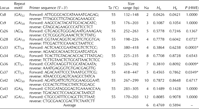
Of the 48 newly developed SSR primer pairs, 13 were able to successfully amplify the genomic DNA of C. yunnanensis Rolfe, and 11 showed polymorphisms among the tested samples. The rest of the primers either showed non-specific products or no PCR products, probably as a result of mutations at the SSR locus (Wang et al., Reference Wang, Liu, Peng, Xu, Yuan, Feng, Yu, Wang and Wei2014). Genotypic data were collected and grouped based on their species and locations, and then subjected to analysis using the TFPGA. The number of alleles detected varied from 2 (Cs8) to 12 (Cs47), with an average of six alleles per locus. The H e value for the individual locus ranged from 0.0421 (Cs8) to 0.9078 (Cs47) with an average of 0.5894, while the H o value ranged from 0.0426 (Cs8) to 0.8085 (Cs47) with an average of 0.4769. A significant deviation from HWE (P< 0.05) was detected at three loci, namely Cs33, Cs36 and Cs37. LD was observed between Cs34 and Cs36. The details of the characteristics of these 11 microsatellite loci are presented in Table 1.
Characteristics of the 11 microsatellite loci derived from Cheirostylis yunnanensis Rolfe, including locus name, repeat motif, primer sequence, annealing temperature (Ta), size range, number of alleles (Na), observed heterozygosity (H o), expected heterozygosity (H e) and significant deviation of P value for Hardy–Weinberg equilibrium (P (HWE))

*P< 0.05.
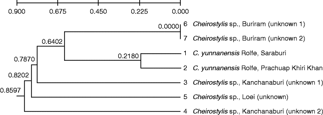
All of the 11 polymorphic primers derived from C. yunnanensis Rolfe were tested for cross-species amplification with five samples of unknown species of Cheirostylis. The results showed that eight primer pairs were able to generate amplification products in all the tested samples, with the number of alleles ranging from one to two. The results of UPGMA analysis showed that C. yunnanensis Rolfe from two locations were classified in the same group, but were separated from the other unknown species (Fig. 1). The genetic information obtained from the analysis using microsatellite markers developed herein will be useful for developing a conservation programme for Cheirostylis sp. The results also indicated that the SSR markers developed in this study are not only useful for the genetic study of C. yunnanensis Rolfe, but may also have application in the identification of Cheirostylis sp.
Unweighted pair-group method with arithmetic mean dendrogram showing the genetic relationship between Cheirostylis yunnanensis Rolfe and unknown Cheirostylis sp.

Acknowledgements
This study was supported by the National Research Council of Thailand, Plant Genetic Conservation Project under the Royal Initiation of Her Royal Highness Princess Maha Chakri Sirindhorn (RSPG), and Mahidol University. The authors thank Professor Dr Wongsatit Chuakul, Faculty of Pharmacy, Mahidol University for help with sample identification.