




No CrossRef data available.
Article contents
Progressive dilation of the diameter of the aortic root in adults with a bicuspid aortic valve
Published online by Cambridge University Press: 24 May 2005
Abstract
Background: To determine the rate of progression of dilation of the aortic root in adults with a bicuspid aortic valve. Methods: We reviewed retrospectively the transthoracic echocardiograms of 50 adults with a bicuspid aortic valve. Each patient had had at least two examinations made 12 months apart. Measurements were taken at four levels: at the basal attachment of the leaflets of the valve within the left ventricular outflow tract, at the widest point of the sinuses of Valsalva, at the sinutubular junction, and in the ascending aorta 1 cm beyond the sinutubular junction. Results: Progressive dilation occurred at all levels, ranging from 0.3 mm/yr at the basal attachment within the left ventricular outflow tract to 1.0 mm/yr, 1 cm beyond sinutubular junction. These rates of dilation were greater than the reported rate of 0.8 mm per decade in the normal population. The rate of dilation found in the ascending aorta 1 cm beyond the sinutubular junction was significantly greater than at the other sites (p = 0.005). The 21 patients with baseline measurements greater than 34 mm had a significantly higher rate of progression (p = 0.007). Sex, age, and the degree of valvar obstruction or regurgitation did not significantly influence the rate of progression of dilation. Conclusion: There is a significantly higher rate of dilation of the aortic root in adults with a bicuspid aortic valve when compared to the normal population. Periodic evaluation of the ascending aorta is essential in these patients, even after replacement of the aortic valve. Other imaging modalities should be considered if the region beyond the sinutubular junction is not well visualized by transthoracic echocardiography.
Information
- Type
- Original Article
- Information
- Copyright
- © 2003 Cambridge University Press
A bicuspid aortic valve is a frequent cardiac malformation that occurs in approximately 1% of the general population.1 Its most common complications are valvar stenosis and regurgitation.1–3 Bicuspid aortic valves can accurately be detected by echocardiography,4 and periodic evaluation by this technique has mainly been focused on the progression of the valvar lesions. Associations with aortic abnormalities, such as coarctation, dilation and dissection of the aortic root, however, have been well described.1, 3, 5–7 A high prevalence of dilation of the aortic root occurring independently of any functional aortic valvar abnormalities has been reported.8, 9 The lack of association between the severity of the valvar lesions and the extent of dilation of the aorta raises the hypothesis that the anomalies might be part of a common developmental defect, with independent rates of progression. Necropsy studies in patients with bicuspid aortic valves have shown that enlargement of the aortic root might be secondary to cystic medial necrosis, a process similar to the one encountered in the Marfan's syndrome.5, 6 No information is available, however, regarding changes in size of the aortic root over time in patients with a bicuspid aortic valve, and how such changes relate to the progression of valvar lesions. The aim of our study, therefore, was to determine the rate of progression, if any, of the diameter of the ascending aorta in patients with a bicuspid aortic valve, and to identify any risk factors for rapid dilation.
Selection of patients
We reviewed retrospectively the charts of all patients with a bicuspid aortic valve followed at the Adult Congenital Heart Clinic of our tertiary centre. Patients were included if they fulfilled the following criteria:
- 18 years and older.
- Echocardiographic confirmation of a bicuspid aortic valve.
- Two or more transthoracic echocardiograms at least 12 months apart with adequate visualization of the ascending aorta.
- Doppler evaluation of the aortic valve available for offline review.
Patients who had undergone replacement of the aortic valve with no intervention on the aortic root were included if the diagnosis of a bicuspid aortic valve was confirmed at surgery, and at least two echocardiograms taken 12 months apart after the operation were available for offline review. Patients with Marfan's syndrome, a history of prior surgery on the aorta, aortic coarctation, or associated congenital cardiac defects were excluded.
In all, we identified 243 patients with a diagnosis of bicuspid aortic valve in the database of the Adult Congenital Heart Centre. Of these, 97 were excluded, because of the presence of aortic coarctation in 34, ventricular septal defect in 19, other associated congenital cardiac defects in 22, intervention on the aortic root in 16, genetic syndromes in 4, and transplantation of the heart in 2. An additional 81 patients had had only one echocardiogram performed at our institution, and were also excluded. Most of these patients had been recently transferred from their pediatric hospitals, and had been seen only once so far at the Adult Congenital Heart Centre. Transthoracic echocardiograms were unavailable, or inadequate for offline analysis, in 15 patients. After these exclusions, 50 patients remained to form our study group.
Methods
A bicuspid aortic valve was diagnosed when only two aortic leaflets were visualized in systole in the short-axis view. Echocardiographic examinations were retrospectively reviewed by one of the same two echocardiographers (MCB or JFB) to confirm the diagnosis, and to measure the diametres of the aortic root in the parasternal long-axis view at end-diastole according to the leading edge technique at four levels:
- The left ventricular outflow tract at the basal attachments of the hinge points of the aortic valvar leaflets.
- The widest point of the sinuses of Valsalva.
- The sinutubular junction.
- The ascending aorta 1 cm beyond sinutubular junction (Fig. 1).

Figure 1. Schematic illustration of the four levels of the aortic root measured by cross-sectional transthoracic echocardiography: the basal attachment of the valvar leaflets within the left ventricular outflow tract (1), the sinuses of Valsalva (2), the sinutubular junction (3), and the ascending aorta 1 cm beyond the sinutubular junction (4).
Adequate measures were obtained at the level of the left ventricular outflow tract in 47 patients, at the widest diameter of the sinuses in 50 patients, at the sinutubular junction in 49 patients, and in the ascending aorta in 38 patients. The degree of aortic regurgitation was assessed by M-mode color Doppler in the parasternal long-axis view by measuring the ratio of the height of the regurgitant jet relative to the height of the left ventricular outflow tract, and by calculating the pressure half-time in the apical five-chamber view. Mild regurgitation was considered present if the ratio in the parasternal long axis was less than 24%, and the pressure half-time was greater than 500 ms moderate regurgitation was defined as a ratio between 25% and 64%, and a pressure half-time between 250 and 500 ms. Severe regurgitation was deemed present with a ratio greater than 64%, and a pressure half-time less than 250 ms. The pattern of flow in the descending aorta on pulsed-wave Doppler was also graded. Holodiastolic reversal of flow in the descending thoracic aorta was considered consistent with significant regurgitation independently of the other criterions. The degree of stenosis was assessed by continuous Doppler evaluation. Stenosis was defined as mild if the mean gradient was between 10 and 29 mmHg, moderate if it was between 30 and 49 mmHg, and severe over 50 mmHg. The aorta was considered to be dilated when its diameter was greater than 34 mm at any level according to the data reported from the cohort examined in the Framingham Heart Study.10 All patients had at least two echocardiograms 12 months apart, with the mean time between the first two echos being 1.96 ± 0.84 years, with 15 patients having three echocardiograms, and two patients having four. The mean follow-up between the first and the last echocardiogram was 2.43 ± 0.98 years.
Variability between observers
The measurements were performed twice in a blinded manner by one observer (MCB) in 10 patients to assess any intraobserver variability. A second observer (JFB) performed the same measurements to determine any interobserver variability. The intra and interobserver variability was assessed by an intraclass correlation coefficient. An intraclass correlation coefficient greater than 0.75 was regarded as excellent concordance. For the intraobserver variability, the intraclass correlation coefficient was 0.97 at the basal level within the left ventricular outflow tract level, 0.99 at the level of the sinuses of Valsalva, 0.99 at the sinutubular junction, and 0.99 within the ascending aorta. The intraclass correlation coefficient for the interobserver variability was 0.71 at the left ventricular outflow tract, 0.95 at the sinuses of Valsalva, 0.98 at the sinutubular junction, and 0.95 within the ascending aorta.
Statistical analysis
Continuous variables are presented as mean plus or minus standard deviation. Comparisons between means were made using Student's t-test, and proportions were compared using the χ2-test. The rate of progression of dilation was defined for each level of the aortic root by taking the difference between the diameters on the last and first echocardiogram divided by the time between these two exams. Comparisons of the rates of progression at the various levels within the aortic root were made using a one-way repeated measures ANOVA with level as the factor. Adding the following covariates in the model to evaluate their influence on the rates of dilation further refined the analysis:
- Age.
- Sex.
- Size of the aorta at baseline.
- Presence of aortic regurgitation.
- Presence of moderate to severe aortic stenosis.
A p value of less than 0.05 was considered statistically significant. All statistical analyses were performed with the Statistical Analysis System, version 8.2 (SAS Institute Inc., Cary, NC, USA).
Results
Valvar lesions
Varying degrees of valvar problems were found in the 50 patients, including 30 males, having a mean age at the first echocardiogram of 30.1 ± 8.1 years, with a range from 18 to 46 years (Table 1). The aortic valve had been replaced in 3 patients with a mechanical prosthesis 1 to 8 years before the initial echocardiogram, with one having a mean residual gradient of 32 mmHg with no regurgitation, and the other two having mild regurgitation, with mean gradients across the prosthesis of 12 and 14 mmHg, respectively. In 13 patients, the gradient across the aortic valve had progressed during the course of the study, changing from no stenosis to mild in 2, to moderate stenosis in another, from mild to moderate stenosis in 2 further patients, and from moderate to severe stenosis in 8 patients. Aortic regurgitation, mild in two and moderate in one, had developed in three patients, while regurgitation had progressed from mild to moderate in 3 further patients, and from mild to severe in 1.
Table 1. Hemodynamic aortic valvar lesions at baseline.

Dimensions of the aortic root
The dimensions at baseline varied from 24 ± 4 to 33 ± 7 mm (Table 2). Men had a significantly larger aortic root compared to women at all levels (p < 0.0003). Only men had aortas wider than 40 mm in diameter. The root was greater than 34 mm at atleast one level in 21 patients, 18 males and 3 females, at their baseline echocardiogram. In one man with a body surface area of 1.89 m2, the dilation, at 35 mm, was limited to the sinuses of Valsalva. In six additional patients, the dilation was limited to the ascending aorta. The other 14 patients all had dilation at more than one level. Patients with aortas wider than 34 mm were older than the others, at 34 versus 27 years (p = 0.003), but this difference was not statistically significant when only patients with aortas wider than 40 mm were compared to the others. For these patients, the differences in age were 32 versus 30 years (p = 0.5).
Table 2. Diametres of the aorta at baseline.

Relationship with degree of valvar hemodynamic lesion
At baseline, there was no significant difference in mean gradient between patients with aortic dimensions greater or less than 34 mm, the values being 27.9 as opposed to 27.6 mmHg (p = 0.9). Similarly, no differences were found in patients with an aorta larger than 40 mm compared to the others, values being 31.2 versus 27.2 mmHg (p = 0.5). In patients with an aorta wider than 34 mm, regurgitation was mild in 16 and moderate in 3, compared to regurgitation being mild in 15 and moderate in 2 in those with aorta smaller than 34 mm (p = 0.5).
Progressive dilation of the aortic root
Progression in widening of the root occured at all levels, ranging from 0.3 to 1.0 mm/yr. The rate of progression 1 cm above the sinutubular junction was greater than at the level of the basal hinge points within the left ventricular outflow tract, at 1.0 versus 0.3 mm/yr (p = 0.0004). Dilation in the ascending aorta was also greater than at the sinuses of Valsalva, at 1.0 versus 0.6 mm/yr (p = 0.005), and at the sinutubular junction, the latter diameter increasing at 0.6 mm/yr (p = 0.005). The rate of progression was also greater when at least one level of the aortic root was wider than 34 mm. In these patients, dilation ranged from 0.6 mm/yr at the level of the left ventricular outflow tract to 1.5 mm/yr in the ascending aorta 1 cm beyond the sinutubular junction (p = 0.007). Sex, age, presence of moderate to severe aortic stenosis, or presence of aortic regurgitation did not significantly influence the progression of dilation.
Discussion
Our study has demonstrated a significant rate of progression of dilation of the aortic root in adults with a bicuspid aortic valve. Our measurements showed progressive dilation that was four times that reported in the normal population at the level of the outflow tract,10 and ten times that reported in the normal population in the ascending aorta beyond the sinutubular junction.11 The progression of dilation was more rapid in patients with aorta wider than 34 mm, but was not associated with age, sex, or the degree of aortic valvar stenosis or regurgitation.
Many authors have studied the normal range of aortic root dimensions using M-mode and cross-sectional echocardiography. Using data collected from the Framingham Heart Study, Vasan et al.10 measured the size of the aortic root size by M-mode at the level of the basal attachments of the valvar leaflets in 1849 healthy men and 2152 healthy women. The dimensions increased gradually with age, the rate of change being 0.8 mm per decade in men, and 0.9 mm per decade in women. The upper limit of normal for the size of the aortic root in men and women younger than 60 years was considered as 34 and 30 mm, respectively. The main limitation of this study, however, was that M-mode echocardiography did not permit measurements within the ascending aorta or at the sinutubular junction.
Roman et al.12 measured the aortic root at four levels using cross-sectional echocardiography in 135 healthy adults. They reported larger measurements at the level of the sinuses of Valsalva level compared to the basal attachments of the leaflets within the left ventricular outflow tract, and concluded that the use of M-mode nomograms falsely diagnosed dilation of the root in almost one-fifth of the adults. In this series, depending on body surface area, the upper limit of normal for an aorta at the level of the sinuses of Valsalva in adults younger than 40 years old ranged from 30 to 37 mm. At the sinutubular junction, the values ranged from 26 to 31 mm.
Hager et al.11 used helical computed tomography to determine normal diametres in 70 healthy subjects aged from 17 to 89 years. They reported mean diametres of 29.8 ± 4.6 mm at the level of the aortic sinuses, and 29.4 ± 4.2 mm within the ascending aorta beyond the sinutubular junction. They also found a rate of progression of 0.1 mm/yr at both the sinuses and the portion beyond sinutubular junction in these normal subjects. We chose 34 mm as our upper limit of normal for the aortic root based on the data reported in the Framingham Heart Study, knowing however that normal dimensions in healthy adults have not been extensively defined by transthoracic echocardiography at all four levels, and vary according to the level measured, the age, and the body size. Most of our patients were young adults in their 2nd or 3rd decade of life, with no patient aged over 46 years, so age was not a factor in explaining the increased diametres we encountered.
Associations between a bicuspid aortic valve and aortopathy including coarctation, hypoplasia of the aortic arch, aneurysm of the ascending aorta, aortic dilation, and dissection have been well described.1, 3, 5–7 Pachulski et al.13 found almost three-fifths of their patients with a bicuspid aortic valve to have an aortic root wider than 34 mm, compared to only one-fifth of those with a trifoliate aortic valve. The proportion in our series was two-fifths. Taking the upper limits found in the normal controls as a reference for normal dimensions, Nistri et al.14 found that the prevalence of enlargement of the root in patients with a bicuspid aortic valve was 7.5% at the level of outflow tract, 19.6% at the sinuses of Valsalva, 15% at the sinutubular junction, and 43.9% beyond the sinutubular junction. We also found that the aorta tends to dilate more in its ascending portion than at the other levels.
The fact that the aortic root seems to dilate independently of any hemodynamic valvar lesions in patients with a bicuspid aortic valve8, 9 could probably be explained by the structural abnormalities found in the aortic wall of these patients. In 1972, McKusick15 reported for the first time an association between the congenitally bicuspid aortic valve and cystic medial necrosis of the aorta. More recently, de Sa et al.16 reported that the severity of medial abnormalities found in the ascending aorta of patients who underwent pulmonary autograft replacement was significantly more severe in patients with bicuspid aortic valve, with three-quarters of these patients having severe changes, than in those with trifoliate aortic valves, of whom only one-sixth had severe changes. These findings were confirmed by Niwa et al.,17 who analysed the aortic wall using light and electron microscopy in patients with different congenital cardiac disease including bicuspid aortic valve. They found that the media of the aorta above a bicuspid valve was consistently abnormal, with fragmentation of the elastic fibres, along with changes in the orientation of the smooth muscle cells and the content of collagen and glycoproteins, a process similar to the one encountered in the population with Marfan's syndrome. These changes were identical whether the valve was stenotic or incompetent, but were lacking in the media of patients with calcific stenosis of a normal trifoliate aortic valve.
Such abnormalities have been well described in the aorta of patients with Marfan's syndrome,18 in whom the clinical implications of progressive aortic dilation, with increasing risk of aortic dissection, are well defined.19 Using echocardiography, Kornbluth et al.20 retrospectively calculated the rate of progression of dilation of the aortic root in 34 patients with Marfan's syndrome. Dilation progressed at 2.0 mm/yr, being maximal at the level of the sinuses of Valsalva. No relationship was found between the rate of progression and the size of the aorta at the time of the initial examination. In our study, the maximal rate of dilation was only half that reported in the population with Marfan's syndrome, but 10 times greater than that reported for the general population distal to the sinutubular junction. A strong association was found between larger size of the aorta at baseline and rate of progression, suggesting that, when the aorta reaches a certain degree of dilation, it tends to dilate more quickly.
Our results underline the importance of periodic evaluation, at least every 2 to 3 years, of the ascending aorta in patients with a bicuspid aortic valve, even should the valve have been replaced. As it is the ascending aorta immediately above the sinutubular junction that has the potential to dilate more rapidly, other imaging modalities, such as magnetic resonance imaging, should be considered if this region is not adequately visualized by transthoracic echocardiography. Larger studies will be needed to confirm the impact of valvar regurgitation or stenosis on the rate of progression of dilation of the ascending aorta. Further trials will also be needed to establish the best cost-effective approach for the periodic evaluation of the entire ascending aorta in these patients, and to determine if β-adrenergic blockade may retard the rate of increase in size of the aortic root, as it does in the population with Marfan's syndrome.
Limitations of the study
Because of the retrospective nature of our study, several patients had to be excluded due to incomplete echocardiographic examinations. There are variations in the echocardiographic measurements depending on the echocardiographic window, and with the site and the timing within the cardiac cycle. To insure reproducibility, the same two echocardiographers reviewed the echocardiographic exams, producing excellent intra and interobserver variability at all levels except at the basal level of the left ventricular outflow tract. It was possible to measure the ascending aorta immediately beyond the sinutubular junction in only three-quarters of our patients confirming that this segment of the aorta is sometimes overlooked, or difficult to measure by transthoracic echocardiography.
References
Roberts WC. The congenitally bicuspid aortic valve: a study of 85 autopsy cases. Am J Cardiol 1970; 26: 72–83.Google Scholar
Fenoglio JJ, McAllister HA, DeCastro CM, Davia JE, Cheitlin MD. Congenital bicuspid aortic valve after age 20. Am J Cardiol 1977; 39: 164–169.Google Scholar
Sabet HY, Edwards WD, Tazelaar HD, Daly RC. Congenitally bicuspid aortic valves: a surgical pathology study of 542 cases (1991 through 1996) and a literature review of 2715 additional cases. Mayo Clin Proc 1999; 74: 14–26.Google Scholar
Brandenburg RO, Tajik AJ, Edwards WD, Reeder GS, Shub C, Seward JB. Accuracy of 2-dimensional echocardiographic diagnosis of congenitally bicuspid aortic valve: echocardiographic–anatomic correlation in 115 patients. Am J Cardiol 1983; 51: 1469–1473.Google Scholar
Roberts CS, Roberts WC. Dissection of the aorta with congenital malformation of the aortic valve. J Am Coll Cardiol 1991; 17: 712–716.Google Scholar
Burks JM, Illes RW, Keating EC, Lubbe WJ. Ascending aortic aneurysm and dissection in young adults with bicuspid aortic valve: implications for echocardiographic surveillance. Clin Cardiol 1998; 21: 439–443.Google Scholar
Larson EW, Edwards WD. Risk factors for aortic dissection: a necropsy study of 161 cases. Am J Cardiol 1984; 53: 849–855.Google Scholar
Hahn RT, Roman MJ, Mogtader AH, Devereux RB. Association of aortic dilation with regurgitant, stenotic and functionally normal bicuspid aortic valves. J Am Coll Cardiol 1992; 19: 283–288.Google Scholar
Keane MG, Wiegers SE, Plappert T, Pochettino A, Bavaria JE, St. John Sutton MG. Bicuspid aortic valves are associated with aortic dilatation out of proportion to coexistent valvular lesions. Circulation 2000; 102 (Suppl III): III35–III39.Google Scholar
Vasan RS, Larson MG, Levy D. Determinants of echocardiographic aortic root size: the Framingham Heart Study. Circulation 1995; 91: 734–740.Google Scholar
Hager A, Kaemmerer H, Rapp-Bernhardt U, et al. Diameters of the thoracic aorta throughout life as measured with helical computed tomography. J Thorac Cardiovasc Surg 2002; 123: 1060–1066.Google Scholar
Roman MJ, Devereux RB, Kramer-Fox R, O'Loughlin J, Spitzer M, Robins J. Two-dimensional echocardiographic aortic root dimensions in normal children and adults. Am J Cardiol 1989; 64: 507–512.Google Scholar
Pachulski RT, Weinberg AL, Chan KL. Aortic aneurysm in patients with functionally normal or minimally stenotic bicuspid aortic valve. Am J Cardiol 1991; 67: 781–782.Google Scholar
Nistri S, Sorbo MD, Marin M, Palisi M, Scognamiglio R, Thiene G. Aortic root dilatation in young men with normally functioning bicuspid aortic valves. Heart 1999; 82: 19–22.Google Scholar
McKusick VA. Association of congenital bicuspid aortic valve and Erdheim's cystic medial necrosis. Lancet 1972; 1: 1026–1027.Google Scholar
de Sa M, Moshkovitz Y, Butany J, David TE. Histologic abnormalities of the ascending aorta and pulmonary trunk in patients with bicuspid aortic valve disease: clinical relevance to the Ross procedure. J Thorac Cardiovasc Surg 1999; 118: 588–596.Google Scholar
Niwa K, Perloff JK, Bhuta SM, et al. Structural abnormalities of great arterial walls in congenital heart disease. Circulation 2001; 103: 393–400.Google Scholar
Fleisher KJ, Nousari HC, Anhalt GJ, Stone CD, Laschinger JC. Immunohistochemical abnormalities in fibrillin in cardiovascular tissues in Marfan's syndrome. Ann Thorac Surg 1997; 63: 1012–1017.Google Scholar
Gott VL, Greene PS, Alejo DE, et al. Replacement of the aortic root in patients with Marfan's syndrome. N Engl J Med 1999; 340: 1307–1313.Google Scholar
Kornbluth M, Schnittger I, Eyngorina I, Gasner C, Liang DH. Clinical outcome in the Marfan Syndrome with ascending aortic dilatation followed annually by echocardiography. Am J Cardiol 1999; 84: 753–755.Google Scholar

Schematic illustration of the four levels of the aortic root measured by cross-sectional transthoracic echocardiography: the basal attachment of the valvar leaflets within the left ventricular outflow tract (1), the sinuses of Valsalva (2), the sinutubular junction (3), and the ascending aorta 1 cm beyond the sinutubular junction (4).

Table 1.
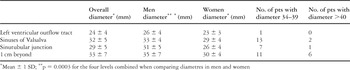
Table 2.
 You have
Access
- 24
- Cited by