Mitral atresia is a rare cardiac malformation that is commonly seen as a part of the spectrum of hypoplastic left heart syndrome. The atretic segment at the position of the left atrioventricular connection may either be formed by fibro-adipose tissue or by a thin imperforate membrane.Reference Anderson and Thiene 1 , Reference Rigby, Gibson and Joseph 3 It is usually associated with an underdeveloped left ventricle and obstructed systemic blood flow in the form of subaortic stenosis, aortic atresia, interrupted aortic arch, or coarctation of the aorta. Rarely, mitral atresia may be associated with transposed great arteries. This is usually associated with left ventricular hypoplasia and aortic or pulmonary outflow tract obstructions. In this case report, we present a rare combination of mitral atresia, transposed great arteries, and unobstructed systemic and pulmonary outflow tracts.
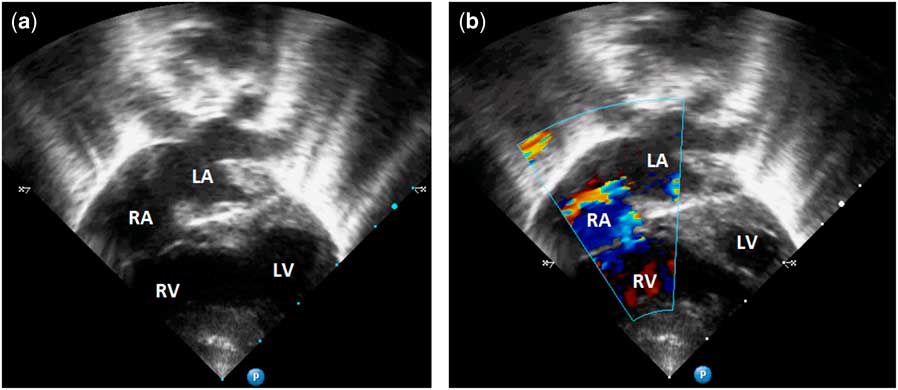
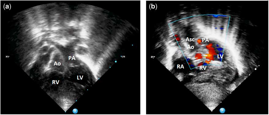
A 1-month-old male baby was referred for failure to thrive. On clinical examination, there was cardiomegaly, a loud second heart sound, and a harsh parasternal pansystolic murmur. The electrocardiogram showed right axis deviation, right atrial enlargement, and biventricular hypertrophy. His four-limb oxygen saturation was 96% on room air; two-dimensional echocardiography showed situs solitus, levocardia, atretic left atrioventricular connection, an atrial septal defect, concordant atrioventricular connections, a large ventricular septal defect, and discordant ventriculo-arterial connections (Supplementary videos 1 and 2). Figure 1 shows an absent left atrioventricular connection – atretic mitral valve – and a normally positioned right atrioventricular valve, which connects to the right ventricle. There was left-to-right shunting across the atrial septal defect. The ventricular septal defect allowed shunting from the systemic ventricle – that is, the morphologically right ventricle – to the left-sided ventricle – that is, the morphologically left ventricle. The ventricular morphologies were established by the smoother endocardial borders of the left ventricle versus the more trabeculated endocardium of the right ventricle. In addition, it was noted that the thick chordae tendinae of the right atrioventricular valve was attached to the interventricular septum, thus identifying it as the tricuspid valve (Supplementary video 1). A full sweep from the four-chamber short-axis view showed the ventricular septum extending into the crux, which established the normal relations of the morphologically right and left ventricles. Figure 2 shows the transposed great arteries and the well-formed left and right ventricular outflow tracts. The aorta and the pulmonary artery had an almost side-by-side relationship. A small patent ductus arteriosus was additionally noted. The left ventricular outflow tract and the ascending aorta were seen to be well developed. Both the right and left ventricles were hypertrophied and normally functioning. There were no anomalous venous connections. The left-to-right shunt across the atrial septal defect in the presence of a large ventricular septal defect with well-developed ventricles and concordant atrioventricular connections and discordant ventriculo-arterial connections created a physiological state of pulmonary over-circulation; however, after 1 week, he developed severe cyanosis due to the development of progressive restriction of the atrial septal defect for which he underwent a successful balloon atrial septostomy. The baby was referred to the cardiothoracic wing for pulmonary artery banding followed by staged Fontan repair.
( a ) Two-dimensional transthoracic echocardiography in the apical four-chamber view showing the absent left atrioventricular connection with the fibro-adipose tissue separating the left atrium and the left ventricle (“mitral atresia”). The tricuspid valve is normal. An atrial septal defect and ventricular septal defect can be seen additionally. ( b ) Left-to-right flow across the atrial septal defect is demonstrated by colour Doppler echocardiography in the same view. LA=left atrium; LV=left ventricle; RA=right atrium; RV=right ventricle.

( a ) Two-dimensional transthoracic echocardiography in a modified sub-costal view showing the well-developed biventricular heart and transposition of the great arteries. The semilunar valves, aorta, and the main pulmonary artery are morphologically normal. ( b ) Colour Doppler echocardiography in a modified apical four-chamber view demonstrating the well-developed ascending aorta and outflow tracts. Right-to-left shunt across the interventricular septum is identified. A small jet due to the patent ductus arteriosus is seen entering the pulmonary artery from the aorta. Ao=aorta; Asc Ao=ascending aorta; LV=left ventricle; PA=pulmonary artery; RA=right atrium; RV=right ventricle.

The survival of a patient with mitral atresia and a normal aortic valve, although better than the survival of patients with mitral atresia and aortic atresia, is still <6 months.Reference David, Richard and Patrick 2 , Reference Rigby, Gibson and Joseph 3 This combination is frequently associated with other cardiac defects such as patent ductus arteriosus, atrial septal defect, functionally univentricular heart, transposed great arteries, abnormal systemic and pulmonary venous connections, straddling of the tricuspid valve, and additional extra-cardiac defects such as asplenia. Eliot et al in 1965 proposed a classification system where mitral atresia was divided into two groups. Group I patients had normally related great arteries and Group II patients had transposed great arteries.Reference Summerell, Miller and Persaud 4 , Reference Eliot, Shone and Kanjuh 5 Group I was further sub-classified based on the presence of hypoplastic left-sided structures–aortic atresia, aortic arch hypoplasia, coarctation – and based on the presence or absence of a ventricular septal defect. Group II was sub-classified based on the presence of one or two functional ventricular chambers. These numeric groupings, however, rarely convey useful descriptive morphological information and, as such, are of limited value compared with creating an accurate verbal description of the cardiac morphology. The patient in our case had mitral atresia, transposed great arteries, and two well-developed ventricles.
In an extensive review of the literature by Moreno et alReference Moreno, Quero and Diaz 6 involving 82 cases of mitral atresia with normal aortic valves, the presence of transposition of the great arteries was mostly associated with left ventricular hypoplasia. In this cohort, well-developed ventricles were seen only in four of the total 82 patients, and a functionally univentricular heart with transposed great arteries and mitral atresia was extremely rare.Reference Moreno, Quero and Diaz 6 Mickell et alReference Mickell, Mathews and Anderson 7 in a series of 35 patients with mitral atresia elucidated the marked heterogeneity in ventricular morphology and ventriculo-arterial connections, especially when associated with a patent aortic valve. Ventriculo-arterial discordance in the presence of two well-formed morphological ventricles, as in our case, was distinctly rare. In addition, the combination of mitral atresia, normal aortic valve, and transposed great arteries is usually associated with pulmonary stenosis or atresia.Reference David, Richard and Patrick 2 , Reference Rigby, Gibson and Joseph 3 Thus, the combination seen in the present case with a well-developed left ventricle and a normal pulmonary valve is particularly rare. This also provides a physiologically favourable haemodynamic state with a near-normal systemic saturation so as to allow relatively prolonged survival compared with other forms of mitral atresia.
A similar combination of anomalies may also be found with a left-handed topology – that is, anatomically corrected malposition – and this needs to be carefully ruled out echocardiographically. In our patient, the morphological characteristics of the ventricular endocardium, septal insertion of the tricuspid valve chordae tendinae, and identification of the extension of the ventricular septum to the crux established the right-handed topology.
Although associated with dismal outcomes in the past, progress in neonatal critical care and advanced surgical methods for treating complex CHD have improved survival. In this case, we initiated a three-stage palliative scheme repair, where an initial pulmonary artery banding procedure will be followed by staged Fontan palliation.
Acknowledgements
G.N.R. and H.V. were primarily involved in diagnosis and imaging. S.M. was involved in preparing the manuscript.
Financial Support
This research received no specific grant from any funding agency, commercial, or not-for-profit sectors.
Conflicts of Interest
None.
Ethical Standards
All ethical standards relevant to the national guidelines of care were maintained while evaluating the case.
Supplementary Material
To view supplementary material for this article, please visit http://dx.doi.org/10.1017/S1047951115001596