Introduction
The petrous apex is a complex region between the middle and the posterior cranial fossa. It encloses various vital structures, including the internal auditory canal, the internal carotid artery, the labyrinth and the facial nerve.
Lesions at the petrous apex can present with various symptoms and signs, depending on the size, location and nature of the pathological process. Cholesterol granuloma is the most common lesion of the petrous apex, while other lesions, like epidermoid cyst, mucocele, benign tumours, fibrous dysplasias, vascular lesions and metastasis, are uncommon.Reference Razek and Huang1,Reference Isaacson, Kutz and Roland2 Radiographic imaging, such as computed tomography (CT) and magnetic resonance imaging (MRI), is of utmost importance in narrowing down the differential diagnosis of petrous apex lesions.Reference Isaacson3
Endoscopic endonasal approaches to the petrous apex are less invasive than other approaches and have gained popularity over the last two decades. Here, we present cases of two different petrous apex pathologies managed with a similar endoscopic approach.
Case reports
Case one
A 39-year-old man presented in late 2019 with complaints of persistent non-pulsatile tinnitus in the left ear, difficulty in hearing within a noisy environment, and headache for the last year. He reported that he had undergone a neurosurgical procedure for similar complaints in 2012, and then two skull base procedures in 2013 and 2014 for recurrence of a brain lesion. The details of this lesion and these procedures were not available.
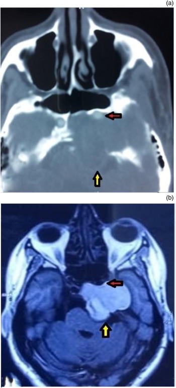
On otoscopic examination, there was a dull tympanic membrane on the left side. Audiometric evaluation showed high-frequency hearing loss on the left side. All cranial nerve examination findings were normal. A CT scan showed a 4.1 × 3.2 × 4.1 cm lesion in the left petrous apex, with extension into the left cavernous sinus, Meckle's cave and the left internal auditory canal, compressing the left cerebellopontine angle and displacing the left internal carotid artery (ICA) inferolaterally (Figure 1a). The MRI scans showed a T1-weighted and T2-weighted hyperintense lesion in the left petrous apex, with extensions similar to those shown on CT (Figure 1b). The lesion did not show fat suppression or contrast enhancement.
(a) Axial computed tomography scan of case one showing a lesion in the left petrous apex compressing the left cerebellopontine angle (yellow arrow) and displacing the left internal carotid artery (red arrow). (b) Axial magnetic resonance imaging scan showing the lesion in the left petrous apex with similar extensions (yellow and red arrows).

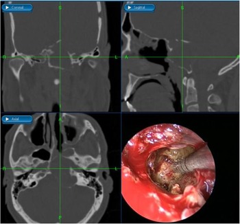
A provisional diagnosis of recurrent (third recurrence) left petrous apex cholesterol granuloma was reached, and the patient was scheduled to undergo trans-sphenoid excision. Initially, excision and drainage were performed using a unilateral approach, and a neo-ostium was created with a temporary stent in situ. However, on follow up, the patient developed stenosis of the opening and had partial recurrence. Subsequently, a bi-nostril approach (instrumentation from the contralateral right nostril) with removal of the posterior and intersphenoid septum was performed under image guidance (Figure 2). A Hadad–Bassagasteguy flapReference Brunworth, Lin, Keschner, Garg and Lee4 was used to line the neo-ostium.
Image-guided excision of cholesterol granuloma through trans-sphenoid corridor in case one. S = superior; R = right; L = left; I = inferior; A = anterior; P = posterior

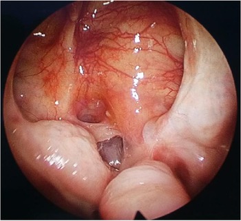
Post-operative nasal endoscopy after four weeks showed a well-mucosalised cavity (Figure 3).
Post-operative photography of nasal endoscopy showing a well-mucosalised sphenoid cavity in case one.

Case two
A 34-year-old man presented in September 2019 with a history of double vision for the last year and persistent headache for 6 months. He had occasional fullness in the left ear, but there were no other ear symptoms.
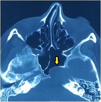
Otoscopic ear examination and audiometric evaluation findings were normal. Extraocular movements were free and full in all directions, and all cranial nerve examination findings were normal. The CT scan showed a well-defined homogeneous mass lesion in the left petrous apex encroaching on the sphenoid sinus and the region of the left ICA (Figure 4). The MRI scan showed a 2.7 × 2 × 1.7 cm T1-weighted hypointense and T2-weighted hyperintense lesion in the left petrous apex. It was non-enhancing on contrast administration and was in close proximity to the horizontal and cavernous ICA.
Axial computed tomography scan of case two showing a well-defined homogeneous mass lesion in the left petrous apex encroaching the sphenoid sinus (yellow arrow).

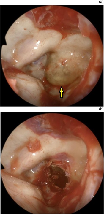
Left petrous apex cholesteatoma was diagnosed, and the patient underwent trans-sphenoid drainage for the same in November 2019. Intra-operatively, a yellowish-brown bulge was observed in the posterior wall of the sphenoid sinus after wide sphenoidotomy (Figure 5a). The bulge was punctured in its medial-most part (Figure 5b), and a cheesy, thick fluid was drained. The opening was widened maximally, and saline irrigation was performed after the removal of contents.
(a) Intra-operative endoscopic image of case two showing a bulge in the posterior wall of the sphenoid sinus (yellow arrow) after wide sphenoidotomy. (b) Site of petrous apex cholesteatoma after removal of contents.

The post-operative period was uneventful, and symptoms subsided within a week of the surgery.
Patient follow up
Both patients were under regular follow up involving nasal endoscopic examination until the travel restrictions imposed because of the coronavirus disease 2019 (Covid-19) pandemic, from March 2020 in India. Currently, both patients are receiving teleconsultation follow up and are asymptomatic. Both patients underwent post-operative imaging at their respective local private centres. Currently, the patients are not able to attend our institution because of ongoing Covid-19 travel restrictions, and we were not able to collect their post-operative images in their absence.
Discussion
Cholesterol granulomas often occur in a pneumatised petrous apex because of the aggressive pneumatisation of the petrous apex that leads to haemorrhage in exposed marrow. An epidermoid cyst (cholesteatoma) of the petrous apex can be either congenital or acquired (following trauma or the result of direct extension from the mastoid cavity), with congenital cases being more common. Congenital cholesteatoma of the petrous apex arises from aberrant ectoderm trapped during embryogenesis.
Both CT and MRI play important roles in the diagnosis of petrous apex lesions. The CT scan shows the detailed anatomy of the osseous structure of the temporal bone and skull base, and can be used intra-operatively for image guidance, as in our case. The MRI scan, when combined with patient history and physical examination findings, helps in reaching a diagnosis, without the need for histopathological examination in most cases, and the same was seen in our cases. Cholesterol granuloma appears as a non-enhancing, T1- and T2-weighted hyperintense lesion on MRI, and as an expansile bony lesion with smooth bone destruction on CT. Epidermoid cyst (cholesteatoma) on MRI is non-enhancing, T1-weighted hypointense to isointense, and hyperintense on T2-weighted imaging, and shows diffusion restriction on diffusion-weighted imaging. On CT, it appears as a non-enhancing, expansile lesion, with variable degrees of bone destruction.Reference Razek and Huang1,Reference Isaacson, Kutz and Roland2,Reference Isaacson3,Reference Le and Roehm5
Observation by serial imaging is the most appropriate management for asymptomatic and incidentally discovered petrous apex cholesterol granulomas, because of the relatively slow clinical development of the lesion and the possible morbidity resulting from surgical intervention. Surgery is considered for lesions that are causing, or threatening, neurological dysfunction because of mass effect or erosion of critical structures.Reference Isaacson3,Reference Sweeney, Osetinsky, Carlson, Valenzuela, Frisch and Netterville6 Spontaneous resolution of cholesterol granuloma is very rare; there is only one such reported case in the literature.Reference Yawn, Sweeney, Carlson and Wanna7 A study comparing surgery versus observation for symptomatic petrous apex cholesterol granuloma found that symptom-specific outcomes were better in patients who were managed surgically.Reference Stevens, Manning, Pensak and Samy8
Excision of the petrous apex cholesteatoma lining and removal of the sac contents is the ideal procedure.Reference Sekhar, Kalfas, Ramanathan, Mai and Schwartz9,Reference Casazza, McCrary, Shelton and Gurgel10 However, its adherence to surrounding structures and its friable nature often make this ideal unattainable, and recurrence is not infrequent.Reference Omran, De Denato, Piccirillo, Leone and Sanna11,Reference Moffat, Jones and Smith12
The surgical approaches for petrous apex lesions can be categorised according to: anatomy (i.e. lateral approaches (translabyrinthine, transcochlear, transcanal-infracochlear, middle cranial fossa) and anterior approaches (medial trans-sphenoidal, transpterygoid infra-petrous, endoscopic translacerum, endoscopic anterior petrosectomy)); or hearing status (i.e. hearing-preserving approaches (transcanal-infracochlear, supracochlear, intralabyrinthine, subarcuate) and non-hearing-preserving approaches (transotic, transcochlear, transtemporal)).Reference Isaacson, Kutz and Roland2,Reference Isaacson3,Reference Gore, Zanation, Ebert and Senior13,Reference Li, Agarwal, Moskowitz and Abuzeid14 Given the disease's benign nature, and the potential for partial petrosectomy to result in significant hearing loss and complete facial nerve paralysis, there has been a gradual shift towards function-preservation surgery.Reference Sharma, Panda, Thakar and Devaraja15 Serviceable hearing can be defined as pure tone audiometry thresholds of lower than 30 dB and a speech discrimination score of higher than 70 per cent.Reference Grinblat, Vashishth, Galetti, Caruso and Sanna16 The anterior endoscopic approach provides an alternate hearing-preserving approach.
Endonasal approaches have gained popularity over the last two decades. These include: (1) a medial approach (trans-sphenoidal and trans-nasopharyngeal) angled medial to the internal carotid artery (ICA); (2) a medial approach with ICA lateralisation; and (3) a transpterygoid infra-petrous approach. The choice of a surgical approach depends on: the relationship of the lesion to the ICA (medial or inferior), the degree of medial expansion, and pathology. The ideal scenario for using the trans-sphenoid approach is when the petrous apex lesion is medial to the carotid and there is a well-pneumatised sphenoid sinus, which was present in both of our cases. If the lesion extends laterally to the ICA, or if the sphenoid sinus is poorly pneumatised, ICA lateralisation can be performed to create more operating space, or the transpterygoid infra-petrous approach could be used.Reference Gore, Zanation, Ebert and Senior13,Reference Zanation, Snyderman, Carrau, Gardner, Prevedello and Kassam17,Reference Kohanski, Palmer and Adappa18
A systematic review comparing surgical outcomes after endoscopic management of cholesterol granulomas of the petrous apex concluded that endoscopic endonasal approaches resulted in higher rates of symptom improvement and lower rates of complications than with open approaches. A comparative meta-analysis of endonasal endoscopic versus open approaches to the petrous apex also concluded the same. However, the recurrence rate was higher in the endonasal approach as compared with the open approach, which may indicate a higher necessity for stenting during endonasal approaches.Reference Eytan, Kshettry, Sindwani, Woodard and Recinos19,Reference Tabet, Saydy and Saliba20
The advantages of the endoscopic approach include hearing and facial nerve preservation, a wide-angled endoscopic view, the avoidance of craniotomy, simple post-operative endoscopic follow up in an out-patient clinic, and faster recovery.Reference Zanation, Snyderman, Carrau, Gardner, Prevedello and Kassam17,Reference Verma, Kumar, Sagar and Kumar21–Reference Sugimoto, Hatano, Noda, Hasegawa and Yoshizaki25 The close proximity to the cavernous ICA, however, leads to an increased risk of vascular injury in inexperienced hands, and it is therefore important to evaluate the anatomical course of the ICA and its variations prior to planning surgery. Other risks include: post-operative epistaxis, injury to the abducens nerve, cerebrospinal fluid leak, restenosis of the drainage pathway, and a recurrence rate of 7.5–15 per cent.Reference Kohanski, Palmer and Adappa18,Reference Park, Wong, Stephens and Saleh26
• Various surgical approaches have been described for petrous apex lesions
• Endoscopic trans-sphenoid approach is used in patients with a well-pneumatised sphenoid sinus and with lesions medial to the internal carotid artery
• This approach is advantageous for the patient as it results in hearing and facial nerve preservation, and avoids craniotomy
• It is also associated with simple post-operative endoscopic follow up in the out-patient clinic and faster recovery
A combined microscopic and endoscopic technique has been used to manage petrous apex lesions; it was found that the introduction of endoscopy in addition to microscopic excision resulted in a less destructive procedure and the preservation of neurovascular structures.Reference Presutti, Alicandri-Ciufelli, Rubini, Gioacchini and Marchioni27–Reference Cazzador, Favaretto, Zanoletti and Martini29
Conclusion
The petrous apex is a complex anatomical area adjoining important neurovascular structures. The use of a nasal endoscope via the trans-sphenoid corridor can provide better access for certain lesions, which can be used for the excision or drainage of petrous apex pathologies.
Competing interests
None declared