Introduction
Abstinence once addicted to nicotine is so difficult that around one out of six women smoke at some point during their pregnanciesReference Egebjerg, Jensen, Nohr and Kruger 1 , 2 despite well documented and disseminated information regarding the detrimental health consequences for the fetus, which includes decreased birth weight, still birth and sudden infant death syndrome.Reference Jaddoe, Troe and Hofman 3 , Reference Mitchell, Ford and Stewart 4 To aid smoking cessation nicotine replacement therapy (NRT) is often used, however, prenatal exposure to nicotine in this way, while perhaps preferable than continued exposure to the myriad of components comprising cigarettes, is also associated with health consequences.Reference Bruin, Gerstein and Holloway 5 In addition to the immediate health consequences for the fetus, exposure to nicotine in utero is associated with a greater risk of becoming addicted to nicotine, especially if nicotine exposure occurs during the adolescent period with the highest risk occurring if exposure occurs during pre-pubescent ages.Reference Chen and Millar 6 – Reference Fidler, Wardle, Brodersen, Jarvis and West 12 Although nicotine use in adolescence can be driven by social factors such as peer pressure, studies using animals models of addiction, which show that rodents exposed prenatally to nicotine will self-administer nicotine to a higher degree than drug naive animals,Reference Chistyakov, Patkina and Tammimaki 13 – Reference Levin, Lawrence and Petro 15 suggest that exposure to nicotine during the prenatal period induces changes in the neurobiological circuitry associated with addiction.
Nicotine has been shown to readily cross the placentaReference Pastrakuljic, Schwartz, Simone, Derewlany, Knie and Koren 16 and accumulate in both the placenta and amniotic fluid, thereby exposing the fetus to a high level of nicotine when compared with the exposure level found in the mother.Reference Luck, Nau, Hansen and Steldinger 17 , Reference Suzuki, Horiguchi, Comas-Urrutia, Mueller-Heubach, Morishima and Adamsons 18 Such exposure to nicotine has been associated with a large array of neurobiological changes in the offspring of the neurotransmitter system at which nicotine acts. Nicotine activates nicotinic acetylcholine receptors (nAChR) in the brain and prenatal and neonatal nicotine exposure have been shown to produce an alternate expression of nAChR mRNA and changes in the numbers of nAChR binding sites across the whole brain, which suggests that prenatal nicotine exposure (PNE) can up- and down-regulate functional nAChRs in multiple regions of the rodent brain. These changes have been shown to occur in areas involved in reward mediation including the ventral tegmental area (VTA) and the nucleus accumbens (NAcc).Reference Huang, Abbott and Winzer-Serhan 19 – Reference Chen, Parker, Matta and Sharp 24 Other PNE-induced effects include a heightened activity of choline acetyltransferase, the enzyme responsible for acetylcholine synthesis, and a decrease in the number of choline transporters, which shuttle choline into acetylcholine-synthesizing cells, suggesting that PNE induces alterations in cholinergic tone.Reference Abreu-Villaca, Seidler, Tate, Cousins and Slotkin 22 In addition, PNE has been shown to change specific membrane channels mediating currents contributing to cellular excitability.Reference Good, Bay, Buchanan, McKeon, Skinner and Garcia-Rill 25 , Reference Pilarski, Wakefield, Fuglevand, Levine and Fregosi 26
The specific PNE-induced alterations underlying the heightened susceptibility to addict to nicotine have not yet been determined; however, as reward mediation is an important component of addiction, changes induced in neuronal groups associated with reward are likely to contribute to the PNE-associated addiction liability. One recently identified critical mediator in the induction of reward is the laterodorsal tegmental nucleus (LDT) (for review, seeReference Kohlmeier, Christensen, Kristensen and Kristiansen 27 , Reference Kohlmeier 28 ) that sends cholinergic, glutamatergic and possibly GABAergic projections to dopaminergic-VTA (DA-VTA) neurons. Through these projections, the LDT controls burst firing of DA-VTA cells,Reference Lodge and Grace 29 which is a firing pattern shown to elicit release of large quantities of dopamine in the NAcc when compared with regular tonic firing.Reference Floresco, West, Ash, Moore and Grace 30 Inactivation of these projections dramatically decreases DA-VTA burst firing and also decreases dopamine efflux in the NAcc induced by stimulation of the LDT.Reference Lodge and Grace 29 , Reference Forster and Blaha 31 , Reference Blaha, Allen and Das 32 Increases in dopamine in the NAcc are observed with the administration of all addictive drugs and have been associated with the rewarding properties of the drugsReference Drevets, Gautier and Price 33 – Reference Pontieri, Tanda and Di 35 suggesting that the LDT may influence reward related behaviours via enhancing DA-VTA excitability. Indeed, optogenetic stimulation of LDT cells that comprise the LDT-VTA pathway results in development of addiction associated behaviours, even in absence of a triggering drug.Reference Lammel, Lim and Ran 36 While glutamatergic projections to the VTA were suggested to be involved in generating this behaviour,Reference Lammel, Lim and Ran 36 cholinergic projections from the LDT have also been shown to terminate on DA-VTA cells belonging to the mesoaccumbal pathway.Reference Omelchenko and Sesack 37 , Reference Omelchenko and Sesack 38 As acetylcholine receptor antagonists microinfused into the VTA decrease dopamine efflux elicited by stimulation of the LDT, these cholinergic projections are believed to play a role in addiction behaviours.Reference Forster and Blaha 31 , Reference Lester, Miller and Blaha 39 , Reference Laviolette, Priebe and Yeomans 40
PNE could induce changes in cellular functioning of LDT neurons which, given the role played by the LDT in addiction processes, would be expected to result in alterations in processing of rewarding stimuli. At this time, it is unknown whether PNE leads to neuronal changes within the LDT. However, the possibility of PNE-induced changes in the LDT are supported by the finding of PNE-induced changes in putative cholinergic neurons located in the pedunculopontine tegmental nucleus (PPT).Reference Good, Bay, Buchanan, McKeon, Skinner and Garcia-Rill 25 Although the PPT is situated adjacent to the LDT, cholinergic neurons of this nucleus primarily send excitatory projections to the DA cells in the substantia nigra and are not thought to participate in direct activation of VTA cells.Reference Forster and Blaha 41 , Reference Oakman, Faris, Kerr, Cozzari and Hartman 42
To test the hypothesis that PNE induces changes in LDT cellular functioning, we examined nicotine-mediated rises in intracellular calcium and nicotine-induced currents as well as changes in membrane currents associated with cellular excitability, using calcium imaging and whole-cell patch clamp, in LDT cells in brain slices from mice exposed to nicotine through the entire gestational period. When compared with responses elicited in non-exposed controls, our findings suggest that PNE induces age-dependent alterations in cholinergic LDT cells. These changes would likely result in a differential functioning of LDT cells, which could alter cholinergic outflow to target regions, including DA cells within the VTA. The PNE-induced changes in cholinergic LDT cells could represent one neuroadaptation to early exposure to nicotine that contributes along with changes occurring in other brain regions to the heightened susceptibility to addict to nicotine observed in these individuals, especially during adolescence.Reference Kandel, Wu and Davies 9 , Reference Lieb, Schreier, Pfister and Wittchen 10
Methods
Animals
All animal studies were conducted in accordance with the European Communities Council Directive of 24 November 1986 (86/609/EEC) as well as with Danish legislations. The Animal Welfare Committee, appointed by the Danish Ministry of Justice, approved the animal study after determining that efforts to diminish and explore alternatives to animal experiments and to minimize animal suffering had been made.
Experiments were conducted using brain slices from mice exposed prenatally to nicotine and control mice. PNE was achieved using an oral exposure model shown to dose dependently increase blood nicotine concentration in the fetus.Reference Pauly, Sparks, Hauser and Pauly 43 NMRI (Taconic, Denmark or Harlan, The Netherlands) dams were housed separately in a cage containing environmental enrichment and nesting material and allowed ad libitum access to food and water. A single male was introduced to the cage and at the same time the drinking water was substituted for a 300 μg/ml nicotine (Nicotine hydrogen tartrate salt, Sigma, USA) solution flavored with 2% saccharine (Saccharin Sodium salt hydrate) to increase consumption.Reference Nesil, Kanit, Collins and Pogun 44 , Reference Matta, Balfour and Benowitz 45 This concentration of nicotine was chosen as it had been shown to result in a plasma concentration of nicotine sufficient to induce dependency and tolerance in mice in other studies.Reference Matta, Balfour and Benowitz 45 Addition of nicotine to the 2% saccharine solution caused the solution to become more acidic, therefore, the pH value of the nicotine solution was readjusted to pH=7.8 using sodium hydroxide. Control animals received a 2% saccharine solution, which was also pH balanced. While some dams receiving nicotine solutions reduced their fluid consumption to half, which is a common phenomenon in mice undergoing this protocol,Reference Pauly, Sparks, Hauser and Pauly 43 animals maintained healthy appearances and grooming and water consumption was within normal limits for mice.Reference Bachmanov, Reed, Beauchamp and Tordoff 46 As reduction in water intake was not consistent across PNE litters, and in other studies, which have also seen a reduction in fluid intake, changes in behavior noted were not believed to be because of reduced intake,Reference Pauly, Sparks, Hauser and Pauly 43 no attempt was made to reduce water intake in control animals. When dams were visibly pregnant, males were removed from the cage. Upon delivery of the litter, the nicotine solution was substituted with solution free water. There was no apparent difference in litter size between control and nicotine conditions and across treatments, pups appeared to be of equal size at birth. However, as animals were not handled until postnatal day (P) 7, to reduce stress responses in both the mother and the pups, physiological differences within this period were not examined by more invasive measures then visual inspection. In another cohort of mice undergoing the same PNE procedure in which handling of the pups was permissible, weight of the pups was monitored 2 days after birth and no differences were apparent between the treatment and control groups (weight at P2; saccharine 2.15±0.14 g, n=4 litters, with ~10 animals in each litter; nicotine 2.15±0.15 g, n=5 litters, with ~10 animals in each litter; two-way t-test P>0.05), supporting our conclusions made from visual observations of animals from this study, that maternal nursing and the continued appearance and weight gain of the pups did not appear to be different between the two treatment groups. While cross-fostering have been used in models of gestational drug exposure to eliminate drug-induced changes in maternal behaviour, which may influence nursing, cross-fostering have been found to significantly decrease rises in NAcc dopamine levels in response to an acute nicotine injection in PNE rats.Reference Kane, Fu, Matta and Sharp 47 Subsequently, cross-fostering was not performed. Data collected from the prenatal saccharine-treated control group were also used in a co-running study examining differences in LDT neurons across development.Reference Christensen, Ishibashi, Nielsen, Leonard and Kohlmeier 48 Animals were kept in a 12 h light/dark cycle, with lights on at 6:00 and room temperature was maintained around 22ºC.
Tissue preparation
Brain slices (250 µm coronal) from control and PNE pups (age P7–P34) containing the LDT were prepared on a Leica vibrotome (VT 1200 S) in ice-cold artificial cerebrospinal fluid (ACSF in mM: NaCl 124.03, KCl 4.99, Na2HPO4 1.12, CaCl2 2.69, MgSO4 (anhydrous) 1.12, Dextrose 9.99, NaHCO3 25.95, oxygenated in 95% oxygen/5% carbogen). Slices were incubated in oxygenated ACSF for 15 min at 37°C and stored at room temperature for a minimum of 1 h before being transferred to the recording chamber, which was continuously perfused with room temperature ACSF with a flow rate of 3–6 ml/min.
Calcium imaging
‘Bulk-load,’ calcium imaging, conducted with the calcium indicator dye fura 2-AM (15 μM in DMSO; Molecular Probes, Invitrogen, Denmark)Reference Tsien 49 can be used as an indirect indicator of changes in intracellular calcium levels in neurons of the LDTReference Kohlmeier, Inoue and Leonard 50 and was used in the present study. Brain slices from three different age groups, group A (P8–P10), group B (P11–P15) and group C (P16–P21), previously shown to exhibit differential rises in intracellular calcium levels in response to nicotine application,Reference Christensen, Ishibashi, Nielsen, Leonard and Kohlmeier 48 were incubated at 31ºC with oxygenated fura 2-AM for 10 min plus 1 min for every day over the age of 10. When fura 2-AM crosses the cell membrane the molecule can undergo de-esterification, which traps the dye intracellularly. The quality of dye loading, however, decreases with age and animals above the age of 21 days were therefore not included in this series of studies. Following incubation with fura 2-AM, slices were transferred to the recording chamber and a continuous flow of ACSF was allowed to washout any leftover dye (30 min) before imaging commenced. As the fluorescent emission of fura 2-AM changes when calcium is bound,Reference Grynkiewicz, Poenie and Tsien 51 changes in intracellular calcium levels were inferred by alterations in fluorescent intensity detected by using appropriate fluorescence microscopy. Fluorescent measurements were performed with a frame transfer, cooled 12 bit CCD camera system (Sensicam, PCO Instruments, Germany) using TILL-VISION software (Till Photonics, Germany), which was mounted on a fixed stage microscope (Olympus BX50WI, USA). Under fluorescent imaging, regions of interest (ROI) incorporating dye loaded cells were selected and changes in fluorescence were monitored within these regions. Pixels within the ROIs were summed according to binning parameters chosen to provide the best balance of the temporal and spatial resolution (2×2). Fura 2-AM is a ratiometric dye and at excitation wavelengths of 340 and 380 nm rises in intracellular calcium will result in an increase or, a decrease, of fluorescence, respectively. Therefore, changes in fluorescence were measured at both excitation wavelengths (340 and 380 nm) and the two values were rationed (F=F 340/F 380) after autofluorescence, which was measured from an area devoid of any fluorescent cells, had been subtracted. By using ratiometric measurement, errors introduced by dye leakage, bleaching and dye loading were minimized. The spatial and temporal alterations in fluorescence are presented using the equation ΔF/F where the difference in the fluorescence at baseline (F) was subtracted from the maximum change in fluorescence (ΔF), which was then divided by baseline fluorescence. An upward going deflection in fluorescence indicates a rise in intracellular calcium, and cells exhibiting a rise exceeding a 2% change in ΔF/F were considered as responders. For each wavelength, exposure times were selected as those maintaining the brightest pixel in the field between 7–10% of the upper limit of the dynamic range, which minimized bleaching of the dye and phototoxicity owing to overexposure.
Whole-cell patch clamping
Thin-walled borosilicate recording electrodes (3–7 MΩ) were produced on a Sutter P-97 horizontal puller (Sutter Instruments, USA) and filled with patch solution containing (in mM) K-gluconate 144, KCl 2, HEPES 10, EGTA (tetraacetic acid) 0.2, Mg-ATP 5 and Na-GTP 0.3. To identify recovered cells post-hoc, biotinylated Alexa-594 (25 µM, Molecular Probes) was also included in the patch solution and allowed to passively fill the cell during the recording. Under differential interference contrast optics, the LDT was located and neurons were visualized with a 40× water immersion objective (NA 0.8, Olympus) using the same imaging system as that utilized for calcium imaging measurements. Under visual control, seals exceeding 1 GΩ was obtained between the pipette and the cell membrane with a patch clamp EPC9 amplifier (HEKA, Germany) operated in voltage clamp mode controlled by Pulse version 8.8 (HEKA). Continuous current traces from the recorded cells were acquired using AxoScope 10.2 (Molecular Devices Corporation, USA) and Axon Digidata 1440 A digitizer (Molecular Devices Corporation) at a sampling frequency of 10 kHz. Recordings were discarded if the holding current exceeded 50 pA or if the series resistance exceeded 30 MΩ. We did not correct for the liquid junction potential (~15 mV). Within the LDT, cholinergic cells are on average larger than GABA-containing cells, although cell size is not a definite marker of cell phenotypeReference Boucetta and Jones 52 and co-localization between GABA and acetylcholine (ACh) has been suggested.Reference Mieda, Hasegawa, Kisanuki, Sinton, Yanagisawa and Sakurai 53 , Reference Jia, Yamuy, Sampogna, Morales and Chase 54 Regardless, we targeted larger multipolar neurons in order to maximize the number of cholinergic cells recorded, with definitive phenotype determined using immunohistochemistry after the recordings were finished.
PNE has been shown to induce an upregulation of nAChR in the brainstem, which includes the LDT. The upregulation appeared to be transient and present from at least P7 until ~P15.Reference Slotkin, Orband-Miller and Queen 21 , Reference Miao, Liu, Bishop, Gong, Nordberg and Zhang 23 As we were interested in examining differential responses to nicotine in PNE animals, which may vary across ontogeny, we therefore divided the recorded cells into two groups: P7–P15 days (group I) and P16–P34 day (group II) previously shown to exhibit ontogenetically differentiated nAChR-mediated responses.Reference Christensen, Ishibashi, Nielsen, Leonard and Kohlmeier 48
Drugs
Nicotine [(-)-nicotine ditartrate, Tocris, UK, 10 µM] was prepared under minimum light exposure from frozen aliquots each day and, for whole-cell patch clamp experiments, applied with a picospritzer III (Parker Hannifin Corporation, USA) with a pressure of 15–20 PSI for 10 s using borosilicate pipettes (3–7 MΩ). The application pipette was situated just above the tissue surface ~60–80 μm from the recorded cell to achieve a relatively local application. For calcium imaging studies, nicotine (100 mM) was picospritzed for 2 s with a lower pressure of 6–10 psi to diminish tissue movement, which created artefacts in the fluorescence recordings. In addition, to maximize nicotine exposure of the entire area under investigation, the application pipette was located further above tissue just outside the field of view.
Protocols
Recordings were performed with a membrane potential maintained at −60 mV with the injection of appropriate holding currents. To examine the shape of the action potential and afterhyperpolarization (AHP), cells were recorded in current clamp mode and current was injected for the minimal duration of time to elicit a single action potential uncontaminated by transients of the pulse. Characteristics of the action potential were measured using custom macros designed for Igor Pro software (Wavemetrics, USA). The firing threshold of the action potential was defined as the potential at which the second derivative of the voltage waveform exceeded three times its standard deviation in the period preceding the spike onset. The spike amplitude was measured as the difference between the spike peak and the firing threshold and the spike width1/2 max was measured at 50% of the maximum spike amplitude. The maximum speed of the rise slope to the action potential peak and the slope of the decay from the action potential peak to baseline were measured as the maximum and minimum of the smoothed first derivative of the voltage waveform. Both the afterhyperpolarization (AHP) amplitude and minimum were determined and the AHP amplitude was measured as the voltage difference between the AHP minimum and the firing threshold. Ih currents were detected in whole cell, voltage clamp mode, following hyperpolarization of the cell from −60 to −110 mV for a duration of 500 ms. Amplitude of the Ih current was defined as the difference between the minimum current required to reach −110 mV and the current applied at the end of the 500 ms hyperpolarization step to maintain a membrane potential of −110 mV. The input resistance (R i ) was determined using a 100 ms hyperpolarization to −70 mV. The current required to induce this change in membrane potential was measured and R i was calculated using ohms law.
Analysis of data
The amplitude of nicotine-induced currents was analysed using Igor Pro software. The number, frequency and amplitude of spontaneous excitatory postsynaptic currents (sEPSCs) were measured using MiniAnalysis (Synaptosoft, USA). For analysis, a period of 30 s before (control) and 30 s after nicotine application, was examined and Kolmogorov–Smirnov (K–S test) statistics were used to determine if a significant difference between the cumulative distribution of sEPSCs of control and drug period was present within individual cells using a significance level of 0.05. Results are presented as mean values±standard error of mean and statistical comparisons were done using t-tests, χ² test and ANOVAs with a significance level of 0.05. All figures were made using Igor Pro software.
Immunohistochemistry
Brain nitric oxide synthase (bnos) is a known and reliable marker for cholinergic neurons within the LDTReference Vincent, Satoh, Armstrong and Fibiger 55 , Reference Vincent and Kimura 56 and remains robust in pontine brain slices maintained for several hours. LDT neuron from which recordings sourced can, therefore, be identified as either cholinergic or non-cholinergic based on the presence or absence of bnos. To definitively identify recorded cells as cholinergic or non-cholinergic, immunohistochemistry was performed on cells filled with the fluorescent dye Alexa-594 during recordings. Accordingly, after whole-cell recordings were completed, brain slices were fixated in 4% paraformaldehyde for at least 4 h before being transferred to a 30% sucrose in phosphate buffer saline solution. Using a Leica CM 3050 S cryostat, slices were resectioned to 40 µm. Slices were then incubated overnight with a primary antibody (anti-bNOS, rabbit polyclonal, cat N7280; Sigma-Aldrich, Denmark A/S) and following washout, incubated in a secondary antibody for half an hour (anti-rabbit, goat, Cat A11008; Molecular probes, Denmark). The excitation and emission bandwidths of Alexa-594 and the secondary antibody were distinguishable under fluorescent microscopy thus allowing the determination of the recorded cells phenotype as cholinergic or non-cholinergic depending upon the presence or absence of staining for bnos, respectively.
Results
PNE is associated with smaller nicotine-induced rises in calcium
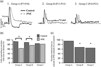
To initially determine whether PNE induces changes in cells within the LDT, we examined nicotine-mediated increases in intracellular calcium using ‘bulk-load’ calcium imaging, which allows the simultaneous monitoring of a large number of neurons for drug-induced rises in calciumReference Kohlmeier, Inoue and Leonard 50 without disrupting the intracellular environment. We conducted our studies in three age groups, as we had previously found differences in nicotine-induced calcium between these ranges of agesReference Christensen, Ishibashi, Nielsen, Leonard and Kohlmeier 48 and we wished to determine whether these differences were altered by PNE. In LDT cells from PNE animals, divided into three age groups (group A, P7–P10; group B, P11–P15; group C, P16–P21), we found an ontogenetic decrease in the amplitude of nicotine-induced rises in ΔF/F, which suggests a decrease in intracellular calcium rises (one-way ANOVA: group A v. group B P<0.05; group A v. group C P<0.05; group B v. group C P<0.05; Fig. 1a). These data collaborate earlier findings of an age-related decrease in the amplitude of nicotine-induced calcium risesReference Christensen, Ishibashi, Nielsen, Leonard and Kohlmeier 48 and extend them to animals exposed to nicotine prenatally.
Prenatal nicotine exposure (PNE) decreases the proportion of cells responding with nicotine-induced calcium rises in younger animals and decreases the amplitude of calcium rises in older cells. (a) Representative traces of ΔF/F showing nicotine-mediated increases in fluorescence for laterodorsal tegmental nucleus cells from control (black, full line) and PNE (grey, dotted line) animals across three age groups, group A, group B and group C. Across both treatment groups there is a decrease in the amplitude of the nicotine-induced rise in calcium across age. Vertical line indicates nicotine application. (b) Histogram showing the percentage of cells responding to nicotine with an increase in the level of intracellular calcium. In PNE animals (striped columns) a smaller proportion of cells responded to nicotine with rises in calcium in group A and group B as compared with the proportion of responding cells from control animals (solid columns). (c) The nicotine-induced increase in intracellular calcium levels for PNE relative to control animals are shown in a histogram and while no significant difference in calcium rises was seen in group A, a significantly smaller rise in calcium was found in group B and group C in PNE animals when compared with the amplitude of rises detected in control animals. Asterisks indicate significance (P<0.05).Reference Christensen, Ishibashi, Nielsen, Leonard and Kohlmeier 48

We subsequently compared differences in nicotine-induced calcium between PNE and control animals across these three age groups. In PNE animals, in the youngest age group examined (group A, P7–P10), we found a significantly smaller proportion of cells responding to nicotine with a rise in intracellular calcium, when compared with the number of responsive cells found in age-matched control animals (control: 94.5% responded, n=329; PNE: 63.7%, n=231; χ² test P<0.05; Fig. 1b). While fewer cells exhibited a rise in calcium in PNE animals in this age group, the amplitudes of the rises in calcium in responding cells did not differ between control and PNE animals in group A (control: 0.47±0.02 ΔF/F; PNE: 0.45±0.04 ΔF/F; two-way ANOVA P>0.05; Fig. 1a1 and 1c). In the middle age group, group B (P11–P15), a significantly smaller proportion of cells also responded to nicotine with a rise in intracellular calcium within the PNE group as compared with the control group (control: 89.7%, n=360; PNE: 77%, n=318; χ² test P<0.05; Fig. 1b). However, while the amplitude of the nicotine-induced calcium rise did not differ between treatment groups in the younger age group, rises were found to be significantly smaller in LDT cells from PNE animals when compared with the control animals in this age group (control: 0.38±0.02 ΔF/F; PNE: 0.26±0.02 ΔF/F; two-way ANOVA P<0.05; Fig. 1a2 and 1c). Interestingly, the reduction in numbers of cells responding with a nicotine-induced rise in calcium appeared to be age associated, as we found no difference in the number of cells responding to nicotine in the oldest age group, group C (P16–P21), between PNE and control animals (control: 84.7%, n=340; PNE: 82.5%, n=496; χ² test P>0.05; Fig. 1b). However, while the proportion of the numbers of responding cells were not different, the amplitude of the calcium rises was found to be significantly smaller in PNE animals as compared with the amplitude seen in control animals (control: 0.26±0.02 ΔF/F; PNE: 0.17±0.01 ΔF/F; two-way ANOVA P<0.05; Fig. 1a3 and 1c).
Taken together, the data from our calcium imaging experiments indicate that in PNE animals, a smaller proportion of cells respond to nicotine with a rise in intracellular calcium as compared with control animals, in animals within the age range of P7–P15. Further, in responding cells smaller rises in nicotine-induced intracellular calcium were elicited in cells from PNE animals between the ages of P11–P21. The PNE-associated reduction in nicotine-induced rises in calcium in LDT cells could be via drug-induced alterations of somatically located calcium permeable nAChRs and/or nAChRs located on terminals impinging on imaged cells and/or calcium-entry mechanisms activated secondarily following nicotinic depolarization of the cell membrane.Reference Shen and Yakel 57 Regardless of the underlying neuroadaptation, the PNE-associated reduction in nicotine-mediated calcium in LDT cells would be expected to differentially influence calcium-dependent processes, such as gene expression and synaptic plasticity,Reference Shen and Yakel 57 , Reference Berridge 58 which have been shown to be induced in the LDT by stimulantsReference Kurosawa, Taoka, Shinohara, Minami and Kaneda 59 , Reference Nelson, Wetter, Milovanovic and Wolf 60 and which may also occur in animals exposed prenatally to nicotine.
PNE does not alter nicotine-mediated inward currents
The results from our calcium imaging studies indicated that PNE resulted in alterations affecting nicotine-induced calcium rises which could be indicative of PNE-induced changes in nAChRs. Prenatal and/or neonatal nicotine exposure has been shown in several studies to transiently up- and down-regulate nAChRs.Reference Huang, Abbott and Winzer-Serhan 19 , Reference Slotkin, Orband-Miller and Queen 21 – Reference Chen, Parker, Matta and Sharp 24 , Reference Huang and Winzer-Serhan 61 Although not specifically examined in the LDT, these PNE-induced alterations in nAChRs were found in several brain regions, including the brainstem where the LDT is locatedReference Slotkin, Orband-Miller and Queen 21 , Reference Miao, Liu, Bishop, Gong, Nordberg and Zhang 23 indicating that functional changes in nAChRs within the LDT in response to PNE might occur. We have previously shown that nicotine induces inward currents in LDT neurons following stimulation of postsynaptic nAChRsReference Ishibashi, Leonard and Kohlmeier 62 and that there is an age related difference in this inward current and we therefore wished to examine whether PNE leads to functional changes in this current. In our earlier study, there was no difference in nicotine-induced inward current amplitude between the two youngest age groups (P7–P10; P11–P15). Therefore, in that study, animals in these two age groups were pooled and accordingly, to examine the effect of PNE on nicotine-induced currents, after establishing in preliminary experiments that there were no differences in this parameter in the PNE animals between P7–P15, we conducted our study across two age groups, group I (P7–P15) and group II (P16–P33), to determine whether the ontogenetic difference persisted in PNE animals.
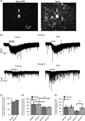
Contrary to our expectations, across PNE and control animals we did not find any significant difference in the number of cells responding to nicotine (puff application, 10 μM, 10 s) with an inward current in either age group (group I: control 94.2%, n=52, PNE 94.3%, n=53, χ² test P>0.05; group II: control 82.6%, n=46, PNE 66.7%, n=51, χ² test P>0.05). The amplitude of the nicotine-induced inward current was also not statistically different between cells from control and PNE animals for either of the examined age groups (group I: control 25.1±2.1 pA, PNE 24.3±2.2 pA, two-tailed t-test P>0.05; group II: control 17.6±2.3 pA, PNE 19.1±2.2 pA, two-tailed t-test P>0.05; Fig. 2b and 2d). The recorded cells were, post-hoc, phenotypically identified as cholinergic or non-cholinergic based on the presence or absence of bnos, respectively (see Fig. 2a), and in cells identified as bnos positive, no difference in the number or amplitude of nicotine-induced inward current was found between control and PNE LDT cells.
Prenatal nicotine exposure (PNE) does not alter the amplitude of nicotine-induced inward currents or the frequency of spontaneous excitatory postsynaptic currents (sEPSCs) elicited by nicotine application. (a) During whole-cell electrophysiological recordings, cells were loaded with the fluorophore Alexa-594 that diffused into the cell from the patch solution. After recording, cells from which data were collected could be located using appropriate fluorescence microscopy (arrows, left panel). Immunohistochemical staining of the slices with an antibody for brain nitric oxide synthase (bnos) (right panel), allowed identification of recorded cells as cholinergic or non-cholinergic based on the presence of both bnos and Alexa-594 (arrow in both panels) or absence of bnos in an Alexa-594 positive cells, respectively. (b) The top two panels are representative whole-cell voltage clamp traces showing the inward current elicited by nicotine application in a cell from a control (left) and PNE (right) animal from the youngest age group, group I. The apparent similarity in the amplitudes of the nicotine-induced current suggests that PNE does not affect this parameter. In the two bottom panels, voltage clamp recordings from laterodorsal tegmental nucleus cells originating from a group II control (left) and PNE animal (right) showing representative nicotine-induced increases in sEPSC frequency and amplitude. Although the frequency of nicotine-induced sEPSCs did not differ significantly across treatment, the amplitude of nicotine-induced sEPSCs is larger in the cell from the PNE animal when compared with the cell from the control animal which suggests that nicotine mediates larger amplitude sEPSCs in cells from PNE animals at this age as compared with cells from control animals. In group I cells no difference was seen in frequency or amplitude between control and PNE animals. (c) Histogram showing the percentage changes in the amplitude of nicotine-induced inward current in cells from PNE as compared with cells from control animals, suggesting that there is no significant difference in nicotine-mediated currents between the control and PNE group. (d) The percentage of nicotine-induced decrease in the interval between sEPSCs for cells from group I and group II are shown in this histogram. The decrease in sEPSC interval between cells from the control and PNE group are not significantly different in either age group, but in both treatment groups a significantly larger decrease in sEPSC interval is seen in the younger as compared with the older age group. (e) The histogram shows the nicotine-induced increase in the amplitude of sEPSCs for the population of recorded cells. While no difference was seen in group I across treatment, the average amplitude of sEPSCs in cells from group II PNE animals was larger than the amplitude of excitatory events in age-matched control animals. Asterisks indicate significance (P<0.05).Reference Christensen, Ishibashi, Nielsen, Leonard and Kohlmeier 48

We have previously shown that the subunit composition of nAChRs in the LDT alters across age and influences the degree of receptor desensitization following nicotine exposure.Reference Christensen and Kohlmeier 63 As a first pass examination of whether PNE induces a change in nAChR subunit composition, which alters the desensitization of receptors,Reference Wooltorton, Pidoplichko, Broide and Dani 64 , Reference Quick and Lester 65 we measured the degree of attenuation of the response of repeat nicotine application since acute receptor desensitization should be indicated by a blunted inward current to second applications of nicotine.Reference Quitadamo, Fabbretti, Lamanauskas and Nistri 66 Responses of membrane current to second applications of nicotine were attenuated in cells from both PNE and control animals and there was no significant difference in the reduction of the inward current induced by the second nicotine application across treatment group (group I: control 43.1±3.8% reduction, n=42, PNE 37.6±3.7% reduction, n=42, two-tailed t-test P>0.05; group II: control 26.9±6.2% reduction, n=29, PNE 29.4±5% reduction, n=24, two-tailed t-test P>0.05). Although this point needs to be confirmed by examining protein expression, our data suggests that exposure prenatally to nicotine does not alter the subunit composition of nAChRs in the LDT. While PNE has been shown to alter nAChR binding in brainstem regions, which is suggestive of changes in nAChRs which would influence functioning, our data do not support the interpretation that PNE has altered functioning of nAChRs in the LDT mediating inward currents.
Nicotine exposure results in an enhancement of spontaneous excitatory postsynaptic currents (sEPSCs) on cholinergic LDT neurons via excitation of nAChRs located on glutamatergic terminals impinging on postsynaptic LDT cells,Reference Ishibashi, Leonard and Kohlmeier 62 and while we did not detect alterations of postsynaptic nAChRs, which mediate inward currents, alterations in nAChRs induced by PNE could extend to receptors located on presynaptic cells mediating sEPSCs. However, we did not find any significant difference in the nicotine-induced increase in frequency of sEPSCs between LDT cells from PNE and control animals (interval between sEPSCs: group I: control 67.7±3% decrease, n=32, PNE 68.1±3.1% decrease, n=47; group II: control 49.3±3.8% decrease, n=28, PNE 49.4±3.6% decrease, n=29; two-tailed t-test P>0.05; Fig. 2c and 2e). In LDT cells from the younger animals, there was no change in the amplitude increase of nicotine-mediated sEPSCs between cells from control and PNE animals (group I: control 25.7±5% increase, n=19, PNE 24.9±6.7% increase, n=20; two-tailed t-test P>0.05). However, the increase in amplitude of sEPSCs elicited by nicotine was significantly greater in LDT cells from PNE animals in the older group when compared with age-matched controls (group II: control 12.2±1.8% increase, n=17, PNE 22±3.5% increase, n=17; two-tailed t-test P<0.05; Fig. 2c and 2d). The difference in amplitude increase between PNE and controls did not extend to bnos-positive cells (group II: control 14.6±2.6% increase, n=9, PNE 19.2±4.89% increase, n=9; two-tailed t-test P>0.05). While insufficient bnos negative cells could be successfully recovered to compare results across treatment our data suggest that the nicotine-mediated increase in amplitude occurs in the non-cholinergic population of LDT cells. Taken together, as the frequency of nicotine-induced sEPSCs did not change across treatment, our data do not support a functional PNE-induced increase in nAChRs located on presynaptic glutamate cells. However, as the amplitude of nicotine-mediated sEPSCs increased in LDT cells from older PNE animals, the results do indicate a late developing alteration in postsynaptic glutamate receptors that could lead to a heightening of nicotine-induced excitation of LDT cells in older animals exposed prenatally to nicotine. As this effect may only extend to non-cholinergic neurons, it would be of interest to determine whether glutamatergic or GABAergic LDT cells are selectively affected.
PNE changes the spike shape and afterhyperpolarization
Although our experiments did not detect evidence that PNE is associated with a detectable change in the numbers, or properties, of functional nAChRs on LDT cells, nicotine exposure in the prenatal period as well as in adulthood has been reported to affect membrane conductances, which could affect cell excitability via alterations of the firing threshold, shape of the action potential and afterhyperpolarization (AHP).Reference Good, Bay, Buchanan, McKeon, Skinner and Garcia-Rill 25 , Reference Ma, Wu and Guo 67 , Reference Wang, Shi and Zhang 68 Changes in these parameters have also been noted to be associated with ontogeny.Reference Christensen, Ishibashi, Nielsen, Leonard and Kohlmeier 48 Therefore, the effects of PNE on the shape of the action potential and AHP were examined across the two age groups.
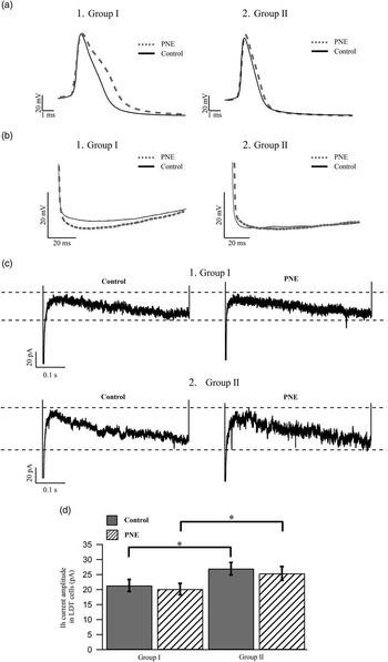
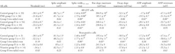
We did not detect a significant difference in the firing threshold between LDT cells from control and PNE animals in either group I or group II (Table 1, Fig. 3a) for the population of cells examined, as well as in a subpopulation of cells identified as bnos positive. This is in contrast to findings in cholinergic neurons of the PPT, in which a more depolarized firing threshold was reported in animals exposed prenatally to cigarette smoke. However, while a more depolarized firing threshold suggested a reduction in excitation of PPT cells in prenatally smoke exposed animals as it would require greater current input to trigger firing, in the same study alterations in conductances were detected which could be expected to heighten excitability.Reference Good, Bay, Buchanan, McKeon, Skinner and Garcia-Rill 25 To examine whether cellular excitability was different in LDT cells following PNE, we compared the minimal current necessary to elicit a single action potential (rheobase). Rheobase was significantly larger in group I PNE animals when compared with the amount of current required to elicit a spike in cells from age-matched controls (group I: control 9.5±1.7, n=4, PNE 17.9±1.7, n=3; two-tailed t-test P<0.05). This is contrary to changes seen in hypoglossal motorneurons in which rheobase was significantly lower in P0–P6 PNE rats, however, this discrepancy could be attributable to differences in species, nuclei and age.Reference Pilarski, Wakefield, Fuglevand, Levine and Fregosi 26 There was no significant difference in rheobase in LDT cells from the older age group between treatment (group II: control 17.6±3.9, n=5, PNE 23.5±7, n=3; two-tailed t-test P>0.05). Although the firing threshold did not seem altered by PNE in LDT cells, our data do indicate there is a decrease in LDT excitability in cells from young animals exposed prenatally to nicotine, indicating that a net larger excitatory input would be required to elicit firing in LDT cells from PNE animals.
Parameters governing the action potential spike shape and afterhyperpolarization (AHP) are altered in prenatal nicotine exposure (PNE) animals while Ih current amplitudes remain unaffected. (a) Representative current clamp recordings showing the spike shape of action potentials from cholinergic laterodorsal tegmental nucleus (LDT) neurons from a control (black, full line) and a PNE (grey, dotted line) animal from group I (A1) and group II (A2), which suggest that PNE induces transient changes in the spike shape in the youngest age group under study. (b) High gain images of the AHPs of cholinergic LDT cells are shown from both a control (black, full line), and a PNE (grey, dotted line) animal from group I (B1) and group II (B2), indicating that the AHP minimum is more hyperpolarized in cells from younger animals prenatally exposed to nicotine when compared with control. (c) The top two voltage clamp recordings (c1) show the Ih currents elicited by a hyperpolarization to −110 mV from two cells identified as cholinergic. The top left trace is from a cell from a group I control animal and the top right trace is from a cell belonging to a group I PNE animal. The two bottom traces display recordings from cholinergic cells originating from a control (left) and a PNE animal (right) from group II (c2). In both age groups, the amplitude of the Ih currents are similar between cells from control and PNE animals suggesting that PNE does not influence the amplitude of the Ih current. (d) As shown in the histogram, across a population of cells, PNE was not associated with a change in the amplitude of the Ih current in LDT cells, however, as previously reported there is a significant ontogenetic associated increase in the amplitude of this current, which is evident in both treatment groups, indicating that PNE does not influence this ontogenetic process. Asterisks indicate significance (P<0.05).Reference Christensen, Ishibashi, Nielsen, Leonard and Kohlmeier 48

PNE induces alterations in the action potential spike shape and AHP of LDT neuronsReference Christensen, Ishibashi, Nielsen, Leonard and Kohlmeier 48

PNE, prenatal nicotine exposure; AHP, afterhyperpolarization; LDT, laterodorsal tegmental nucleus; bnos, brain nitric oxide synthase.
In the top part of the table, measured values for the examined parameters of the action potential across treatment in group I and group II are shown. Underneath the measured values for group I and group II, P-values for statistical comparisons across treatment in group I and group II are seen. The bottom half of the table shows the values of the examined parameters for bnos-positive cells from group I and group II and the P-values for statistical comparisons across treatment.
# indicates the presence of a significant (P<0.05) ontogenetic change between group I and group II for this parameter within the related treatment group.
* denotes a significance level of P<0.05.
In animals exposed to nicotine prenatally the width of the action potential in LDT cells was significantly broader when compared with the spike width in control animals (group I: spike width1/2 max 20.1% broader; group II spike width1/2 max 13.2% broader). However, this change was only significant in cholinergic cells from group I PNE animals (group I: spike width1/2 max 19.8% broader; group II spike width1/2 max 7.4% broader) (Table 1, Fig. 3a). In bnos-positive cells there was a significant reduction in the slope of the rising phase of the action potential in cells from group I PNE animals when compared with that of the control group I (12% reduction). In group II the significant reduction of the rise slope in bnos-positive cells from PNE animals was no longer present (2.3% reduction). Similarly in PNE animals a significant reduction of 17.3% was seen in the slope of the action potential decay phase of bnos-positive cells in group I, whereas the effect of PNE on this parameter of bnos-positive cells from group II (8.5% decrease) was non-significant, indicating the PNE transiently affects conductances important for both the rise and decay phase of the action potential that could underlie the broadening of the spike width. The spike amplitude was also examined but no difference was found between treatment groups.
Previous studies have shown the chronic nicotine injections decrease the amplitude of the initial fast AHP in NAcc cells. This change could be mediated through a nicotine-induced reduction of large conductance, calcium-activated potassium channels (BK-channels)Reference Ma, Wu and Guo 67 as these channels have been shown to be importantly involved in mediation of this initial part of the AHP in NAcc cells.Reference Ma, Wu and Guo 67 In cholinergic cells of the LDT, BK-channels have also been shown to mediate the initial fast part of the AHP.Reference Kohlmeier, Christensen, Kristensen and Kristiansen 27 Therefore, in order to determine whether PNE affects this channel in LDT neurons, we measured the amplitude of the AHP and the AHP minimum. In cells from PNE animals the amplitude of the AHP was significantly larger in the population of cells from the youngest PNE group when compared with the amplitude in group I controls (14.7% larger). In cells from group II we did not find any significant difference across treatment. The AHP minimum in cells from group I PNE animals was significantly more negative than that seen in control animals (8.9% more hyperpolarized), which also extended to a subpopulation of cells identified as bnos positive (6.8% more hyperpolarized, Fig. 3b). In group II cells there was no significant difference in the AHP minimum across treatment protocols. These data indicate that PNE induces changes in the AHP of cholinergic LDT cells that could reflect alterations in BK channels in these neurons.
Taken together, our results show that PNE alters several parameters of the action potential shape and AHP in cholinergic LDT neurons in younger animals and that these changes appear to be transient and approach control values in older animals. The increase in rheobase found in LDT neurons of younger PNE animals suggests that greater depolarization is required to elicit firing. In addition, a more hyperpolarized AHP and a broader spike width would be expected to further alter the responsiveness of LDT cells from younger PNE animals.
The hyperpolarization activated cationic (Ih) current of LDT neurons is unaffected by PNE
To determine whether PNE-affected currents contributing to resting membrane conductances, which participate in setting cellular excitability, we examined changes in input resistance (R i ) across treatment groups. However, we found no significant differences in the R i between PNE and control animals in either age group (group I: control: 1004.9±67.3 MΩ, n=29, PNE: 1007.3±64.1 MΩ, n=32; group II: control: 786.1±63.1 MΩ, n=33, PNE: 786.6±53.4 MΩ, n=46; two-way ANOVA P>0.05), which suggests that PNE does not alter conductances affecting the resting membrane potential. We also examined the holding current and found no difference across treatment in either of the age groups (group I: control: −25.7±2 pA, n=11, PNE: −22.5±2.4 pA, n=13; group II: control: −19.6±1.7 pA, n=17, PNE: −20.8±1.9 pA, n=19; two-way t-test P>0.05) indicating that PNE has not altered the resting membrane potential.
While conductances active at rest did not appear to be affected by PNE, PNE could cause alterations in conductances activated by membrane depolarisations or hyperpolarisations. Ih current is a mixed cationic current activated by a significant hyperpolarization of the cell. We have previously reported that the amplitude of Ih current increases across development in LDT cellsReference Christensen, Ishibashi, Nielsen, Leonard and Kohlmeier 48 , Reference Kristensen, Tyler, Kohlmeier and Leonard 69 and that nicotine decreases the Ih current in cholinergic LDT neuronsReference Christensen, Ishibashi, Nielsen, Leonard and Kohlmeier 48 likely through direct interaction with Ih channels.Reference Griguoli, Maul, Nguyen, Giorgetti, Carloni and Cherubini 70 We therefore investigated the effect of PNE on the Ih current within group I and group II. While we did find age-related differences in the Ih current amplitude across both treatment groups (two-tailed t-test P<0.05), we did not find any significant difference in the Ih current amplitude between LDT neurons from control and PNE animals for either group I or group II (group I: control 21.4±2 pA, n=29, PNE 20.2±2 pA, n=44, two-tailed t-test P>0.05; group II: control 27±2.1 pA, n=50, PNE 25.4±2.3 pA, n=45; two-tailed t-test P>0.05, Fig. 3c and 3d). Differences in Ih current amplitude were also not detected in cells identified as bnos positive (group I: control 19.7±1.6 pA, n=21, PNE 22.3±2.5 pA, n=25, two-tailed t-test P>0.05; group II: control 28.1±2.3 pA, n=33, PNE 26.9±2.2 pA, n=18; two-tailed t-test P>0.05). The absence of alteration in Ih current amplitude suggests that PNE does not affect the number and/or properties of Ih channels. These findings are in contrast to the effect of prenatal tobacco smoke exposure on Ih current in cholinergic PPT neurons, which results in a larger Ih current amplitude when compared with the amplitude of this current in control animals.Reference Good, Bay, Buchanan, McKeon, Skinner and Garcia-Rill 25 The difference in the findings between the two studies suggest that PNE has a differential effect on Ih currents in cholinergic LDT cells from that elicited in PPT cells, and/or, that exposure to non-nicotine bioactive chemicals present in smoke leads to the detected alterations in Ih and accordingly, changes in Ih noted in the PPT were not due directly to nicotine. In the PPT, changes in the Ih current associated with prenatal smoke exposure were proposed to underlie changes found in the resting membrane potentialReference Good, Bay, Buchanan, McKeon, Skinner and Garcia-Rill 25 , which is a conclusion that correlates well with our findings of an absence of both PNE-induced changes in holding current and Ih current in the LDT. Regardless, from our data, we conclude that prenatal exposure to nicotine does not affect the amplitude of Ih present in cholinergic LDT cells and does not prevent the ontogenetic increase in this current across the range of ages studied. Accordingly, PNE does not induce changes in cellular excitability via alterations in this current.
Discussion
In the present study, the proportion of cells responding to nicotine with a rise in intracellular calcium was reduced in LDT cells from younger animals exposed prenatally to nicotine when compared with the proportion of cells responding from control animals. While the amplitudes of nicotine-induced calcium responses across treatment were not different in the youngest age group examined, in cells from PNE animals, within the age range of P11–P21, the amplitude of calcium responses elicited by nicotine was smaller when compared with responses elicited in cells from age-matched control animals. Across the range of ages studied, the action potential spike width of cholinergic LDT cells from animals exposed prenatally to nicotine was transiently increased when compared with cholinergic cells from control animals. The increased action potential spike width in PNE animals correlated with decreases in both the rise and decay slopes of the spike. The AHP minimum was more hyperpolarized in cholinergic LDT cells from younger PNE animals as compared with control animals. The amplitude of nAChR-mediated inward currents as well as the frequency of nicotine-induced sEPSCs was not different between control and PNE animals. However, the amplitude of nicotine-elicited sEPSCs in older PNE animals was significantly larger. Examinations of the R i and the amplitude of Ih current did not reveal any significant PNE-induced changes. Taken together, our data indicate that PNE alters the trajectory of neuronal development of LDT cells. These changes could result in differential functioning of these cells across ontogeny in processes in which they are engaged.
In this study, animals were exposed prenatally to nicotine via maternal oral consumption of a nicotine solution during gestation, which has been shown to dose dependently increase fetal blood nicotine concentration.Reference Pauly, Sparks, Hauser and Pauly 43 Concentrations achieved appear to be sufficient to influence neural development since studies using this application method have shown increases in self-administration in the offspringReference Chistyakov, Patkina and Tammimaki 13 , Reference Klein, Stine, Pfaff and Vandenbergh 14 suggesting an enhanced sensitivity to the rewarding effects of nicotine. While reductions in fluid intake was noted in some of our PNE mothers as has been noted in other studies with a similar exposure paradigm, fluid intake was not compensated for in control animals in our study.Reference Pauly, Sparks, Hauser and Pauly 43 However, we do not believe changes noted in PNE offspring were because of reduced fluid intake as in other studies specifically examining this issue, behavioural actions were not attributed to this reduction, suggestive of nicotine specific actions.Reference Pauly, Sparks, Hauser and Pauly 43 Further indicating that nicotine concentrations are pharmacologically effective, changes in locomotor activity, which is a commonly used indicator of drug effects in areas such as arousal, anxiety and dopaminergic stimulation,Reference Faraday, O’Donoghue and Grunberg 71 , Reference Zocchi, Orsini, Cabib and Puglisi-Allegra 72 have also been noted following this method of nicotine exposure.Reference Pauly, Sparks, Hauser and Pauly 43 , Reference LeSage, Gustaf, Dufek and Pentel 73 Further, oral administration of nicotine to pregnant mice resulted in offspring exhibiting several behavioral, anatomical and neurochemical biomarkers associated with deficits in attention present in prenatally exposed humans, which is of interest to the present study in light of the role played by the LDT in attentional processes.Reference Zhu, Zhang, Xu, Spencer, Biederman and Bhide 74 One large drawback to the oral exposure methodology when compared with other exposure paradigms such as nicotine injection or infusion via osmotic minipumps, where the precise concentration of administered nicotine can be controlled, is that exposure concentrations cannot be determined as nicotine exposure through the drinking water is dependent upon the consumed quantity of nicotine solution by the pregnant dam, which can vary depending on the animal. However, countering this drawback, stress induced by more invasive procedures is avoided by this exposure method, thereby limiting activation of hypothalamic and adrenal stress responses that could induce neurobiological changes in the offspring.Reference Diaz, Ogren, Blum and Fuxe 75 , Reference Lordi, Patin, Protais, Mellier and Caston 76 It is worth noting that although exposure of nicotine through the drinking water does not afford the same control of nicotine exposure as application via pumps or injections, clearance of nicotine from the placenta and amniotic fluid is highly reduced compared with the clearance from the maternal blood system. Therefore, regardless of the route of administration, nicotine accumulates in these compartments, inducing a high and longer lived exposure of the fetus that is buffered from nicotine fluctuations in the dams blood.Reference Pastrakuljic, Schwartz, Simone, Derewlany, Knie and Koren 16 – Reference Suzuki, Horiguchi, Comas-Urrutia, Mueller-Heubach, Morishima and Adamsons 18 , Reference Dempsey, Jacob and Benowitz 77 Other studies examining the actions of prenatal exposure to nicotine have used the smoke exposure paradigm. While this method offers the benefit of approximating the rout of administration associated with human cigarette smoking, it does have the confound that the fetus is exposed to non-nicotine chemical compounds found in tobacco smoke.Reference Talhout, Schulz, Florek, van, Wester and Opperhuizen 78 Taken together, as we were interested in the neurobiological actions of nicotine itself, and the oral consumption model had been shown to effect neurobiological alterations in the fetus at the concentrations utilized in our study,Reference Pauly, Sparks, Hauser and Pauly 43 , Reference Matta, Balfour and Benowitz 45 it was deemed the most appropriate animal model of in utero nicotine exposure for the present examinations.
Calcium imaging and nAChRs
Cellular mechanisms underlying nicotine-mediated rises in calcium were affected by PNE. Changes in these mechanisms could involve alterations in the numbers and/or properties of postsynaptically located nAChRs, which could alter calcium influx directly through calcium permeable nAChRs and/or voltage gated calcium channels activated by nicotine-induced depolarization of the cell.Reference Tsuneki, Klink, Lena, Korn and Changeux 79 PNE has been shown to change the numbers of nAChR binding sites in several brain regions suggesting that PNE induces alterations in the availability of functional nAChRs.Reference Shacka and Robinson 20 – Reference Chen, Parker, Matta and Sharp 24 , Reference Slotkin, Seidler and Qiao 80 Decreases in activity induced by nicotine in prenatal nicotine exposed rats suggests that early life exposure alters the numbers or properties of nAChRs resulting in reductions in function of nAChRs.Reference Britton, Vann and Robinson 81 A large proportion of nAChRs within the LDT appear to be high affinity β2-containing nAChRs,Reference Christensen and Kohlmeier 63 which are particularly susceptible to nicotine-mediated upregulation when exposed to nicotine postnatallyReference Gentry and Lukas 82 – Reference Perry and Kellar 85 as well as during the gestational period,Reference Slotkin, Seidler and Qiao 80 which suggests that PNE could lead to functional upregulations of these receptors. Other studies suggest reductions in function of β2-containing receptors following gestational nicotine, raising the concern that dose and duration of exposure and/or exposure paradigm play a large role.Reference Pilarski, Wakefield, Fuglevand, Levine and Fregosi 86 Prenatal exposure to nicotine also could lead to alterations in other nAChR subunits. Arguing against the conclusion that PNE alters the function of nAChR subunits in the LDT is our finding that there were no changes in the inward current mediated by nicotine in LDT cells from PNE animals, nor in current amplitudes upon second nicotine applications, which would be expected if prenatal nicotine was altering receptor subunit compositions. At the present time, we cannot rule out a change in the numbers of nAChRs nor in their subunit composition, as large changes in the density of nAChRs have been shown in the cortex and thalamus, which did not significantly affect functional outcome.Reference Gold, Keller and Perry 87 In addition, more subtle molecular changes leading to alterations in receptor functioning, without changing actual receptor numbers or composition, could also be occurring and lead to differences in calcium permeability, without affecting total current density. Regulatory proteins of the U-PAR/Ly6, (Lynx) superfamily modulate nAChRs, leading to reductions in functionReference Ibanez-Tallon, Miwa and Wang 88 , Reference Miwa, Ibanez-Tallon and Crabtree 89 and PNE has been shown to alter levels of one member of this family in the hippocampus without leading to changes in the frontal cortex (Morten Skøtt Thomsen, personal communication). While it is currently unknown what the effects of PNE are on other members of this family and whether changes are induced in any of these proteins within the LDT, it is a possibility that actions of PNE could extend to alterations of small protein modulators of the nAChR in the laterodorsal tegmental nucleus leading to changes in calcium permeability, without a large action on current amplitudes.
While future studies need to be conducted to examine the mRNA and protein levels for nAChRs as well as determine whether molecular alterations affecting receptor function are occurring to finally determine whether alterations in numbers of receptors, or receptor subtypes, or receptor function is induced by PNE, as our data raises the suggestion that PNE does not lead to a change in numbers or composition of nAChRs located on postsynaptic LDT neurons, it is worth considering whether PNE-associated changes in calcium rises may be owing to the alterations in other nicotine-mediated mechanisms leading to calcium entry. Excitatory input to cells induced by nicotinic activation of presynaptic nAChRsReference Ishibashi, Leonard and Kohlmeier 62 could also elicit calcium rises, however, as the frequency of nicotine-induced sEPSCs was not altered by PNE, results suggest that changes in presynaptically located nAChRs on glutamate-containing neurons have not occurred. The amplitude of nicotine-induced sEPSCs was shown to increase in LDT cells from older PNE animals indicating a late developing change in postsynaptic glutamate receptors leading to heightened excitatory currents. While increases in nicotine-mediated sEPSC amplitudes could be expected to contribute to larger calcium rises in the postsynaptic cell, decreases were seen in the amplitude of calcium rises from older PNE animals suggesting that this mechanism has a limited effect on intracellular calcium levels. As it does not appear from our data that nAChRs located pre- or postsynaptically have been altered by PNE, this indicates that alterations in other mechanisms underlie the decrease in nicotine-mediated rises in calcium. Calcium influx can occur through voltage-dependent calcium channels (VDCC) and nicotine exposure has been shown to alter the activity of certain subtypes of these channels.Reference Damaj 90 – Reference Stevens, Krueger, Fitzsimonds and Picciotto 92 As a host of neuroactive compounds have been shown to influence calcium entry via these channels in LDT cellsReference Kohlmeier, Inoue and Leonard 50 it is a possibility that in utero nicotine exposure alters properties of these conductances. ACh acting at nACh and muscarinic receptors has been shown to both enhance and decrease activity of VDCCs in the ponsReference Ishibashi, Leonard and Kohlmeier 62 , Reference Kezunovic, Hyde, Goitia, Bisagno, Urbano and Garcia-Rill 93 , Reference Kohlmeier and Leonard 94 and presence of cholinergic agonist at inappropriate developmental times could alter activity of these channels following activation postnatally. Further, similar to proteins that modulate nAChR function, proteins which regulate VDCCs have been characterized, and alterations in their expression may be induced by PNE.Reference Yang and Colecraft 95 As nicotine-mediated rises in intracellular calcium levels can stem from calcium release from intracellular stores,Reference Tsuneki, Klink, Lena, Korn and Changeux 79 , Reference Campusano, Su, Jiang, Sicaeros and O’Dowd 96 PNE could induce changes in control of these stores, which would lead to alterations in calcium rises via nicotine activation of this calcium source. While the precise mechanisms remain to be elucidated, the reduction in nicotine-induced calcium in PNE animals would be expected to influence intracellular calcium-dependent mechanisms such as gene expression and synaptic plasticity within these cells which could exert a large impact on processes in which these neurons are involved.
PNE does not alter the Ih current amplitude
While parameters affecting cellular excitability, such as the firing threshold have been shown to be affected by PNE in the putative cholinergic cells of the adjacent PPT,Reference Good, Bay, Buchanan, McKeon, Skinner and Garcia-Rill 25 we did not find any difference in the firing threshold in neurons of the LDT. There was, however, a significant increase in rheobase in younger PNE animals suggesting that cellular excitability is decreased. This is in contrast to findings obtained in rat hypoglossal motoneurons in which PNE was associated with a lower rheobase indicating a difference in developmental responses to gestational nicotine exposure between rat medullary and mouse pontine nuclei, which might reflect differences in species or nuclei.Reference Pilarski, Wakefield, Fuglevand, Levine and Fregosi 26 Another player in cellular excitability is the Ih current. In the present study, we found that PNE did not induce changes in the Ih current amplitude of LDT cells in either of the age groups examined, which is in contrast to findings from the PPT where prenatal tobacco smoke exposure was shown to increase the Ih current in cholinergic neurons from rats aged P12–P21.Reference Good, Bay, Buchanan, McKeon, Skinner and Garcia-Rill 25 While the differences between the findings may be explained by species or nuclei, it is of interest to consider the in utero exposure models utilized. Our data suggests the possibility that alterations in threshold and Ih detected in the PPT may be elicited by some of the ~5000, non-nicotine chemical compounds in cigarette smoke.Reference Talhout, Schulz, Florek, van, Wester and Opperhuizen 78 Accordingly, the results from our study suggest the possibility that a cellular effect attributed to nicotine within the PPT may actually be the result of the other compounds found in tobacco smoke.
However, if not due to differences in experimental conditions and if changes were induced directly by nicotine, disparate results between PNE-induced changes in the PPT and LDT are worth considering in light of the shared functions subserved by the two adjacent nuclei. Both nuclei are part of the reticular activating system, and responsible for generation of cortical activation, and both appear to be involved in addiction processes. However, in part likely due to a demonstrated sparse innervation of the VTA by the PPT, the functional input of the PPT in addiction processes has been suggested to be gated by the LDT that sends a much heavier pathway to the VTA.Reference Lodge and Grace 29 , Reference Omelchenko and Sesack 37 , Reference Omelchenko and Sesack 38 , Reference Dautan, Huerta-Ocampo and Witten 97 This suggests the possibility that a PNE-induced decrease in excitability of projection neurons of the LDT may temper the outcome within the VTA of any PNE-induced enhancements in excitability in the PPT. However, the precise interplay of PPT and LDT activity and resultant effects on VTA neuronal excitability needs to be elucidated before confident predictions can be made regarding how the differential cellular actions of PNE within these nuclei would alter excitatory transmission directed to the VTA.
PNE alters the neural development of the spike shape and AHP of cholinergic LDT neurons
Our results suggest that PNE increases the amplitude of the AHP of LDT cells in younger animals, and this alteration extends to cholinergic cells. We have previously shown that BK channels influence the amplitude of the initial fast AHP in cholinergic LDT cellsReference Kohlmeier, Christensen, Kristensen and Kristiansen 27 and nicotine has been shown in other cell types to have actions on these channels. Nicotine decreases BK channel activity when co-administered with a BK channel opener in cultured human endothelia cells.Reference Kuhlmann, Trumper, Tillmanns, Alexander and Erdogan 98 Furthermore, in adult mice exposed to nicotine for 5 days, nicotine exposure induced a functional downregulation of BK channels.Reference Ma, Wu and Guo 67 Although these data would predict a nicotine-induced reduction in the portion of the AHP mediated by BK channels, exposure in the prenatal period may lead to unpredicted outcomes on BK function. PNE may lead to changes in BK channel expression patterns leading to the observed alterations in the AHP. However, examinations using protein detection methods such as immunohistochemistry or western blot analysis to quantify alterations in BK channel expression patterns in LDT cells from PNE animals should be conducted. As PNE has been shown to enhance expression of KATP channels and levels of expression of KATP and BK channels have shown to interact, another possibility is that PNE is leading to changes in KATP channels underlying larger membrane after hyperpolarizations.Reference Buttigieg, Brown, Holloway and Nurse 99 , Reference Bournaud, Hidalgo, Yu, Girard and Shimahara 100 PNE was observed to decrease the rise and decay slope of the action potential in cholinergic LDT neurons from younger animals likely contributing to the prolongation of the spike width also detected. Nicotine has been shown to alter the spike width via actions on K+ channels as well as voltage gated sodium channels in cardiac and nerve neurons, respectively.Reference Liu, Zhu and Zhang 101 , Reference Wang, Shi and Wang 102 Accordingly, as nicotine could also affect these channels prenatally, functional changes in these channels induced in LDT cells by gestational nicotine exposure could be one mechanism underlying the decrease in the slope of the action potential rise and decay phases. A broadening of the action potential spike width has been shown to result in a larger influx of calcium.Reference Sabatini and Regehr 103 Therefore, action potentials generated in cholinergic LDT cells would be associated in younger PNE animals with a larger influx of calcium than that induced by similar stimuli in cells from non-exposed animals. Such a heightened influx of calcium in response to action potentials could influence calcium-dependent intracellular mechanisms,Reference Berridge 58 including neurotransmitter release at presynaptic terminals as a larger calcium influx associated with an increase in spike duration has been shown to release a greater proportion of neurotransmitter in target areas.Reference Sabatini and Regehr 103 , Reference Augustine 104 Action potential elicitation in cholinergic LDT cells from younger animals that have been exposed to nicotine prenatally could potentially lead to a heightened cholinergic outflow to target regions. This interpretation is interesting in light of our findings that nicotine exposure elicits action potentials in cholinergic LDT cells.Reference Ishibashi, Leonard and Kohlmeier 62
Functional significance
Through cholinergic and glutamatergic projections to DA-VTA neuronsReference Lammel, Lim and Ran 36 – Reference Omelchenko and Sesack 38 the LDT affects DA-VTA neuronal excitability sufficient to result in efflux of dopamine in the NAcc to levels associated with reward.Reference Lodge and Grace 29 , Reference Forster and Blaha 31 , Reference Drevets, Gautier and Price 33 , Reference Lammel, Lim and Ran 36 The LDT-VTA pathway is likely functionally relevant and involved in the neurobiology underlying processing of rewarding stimuli as exposure to rewarding stimuli such as nicotine and amphetamine excites LDT cellsReference Nelson, Wetter, Milovanovic and Wolf 60 , Reference Ishibashi, Leonard and Kohlmeier 62 and, activation of LDT neurons, which send projections to the VTA, elicits addiction-related behaviour, even in the absence of external rewards.Reference Lammel, Lim and Ran 36 Accordingly, exposure wrought changes in LDT neurons occurring during prenatal development, which could lead to alterations in postnatal excitability would be expected to alter LDT outflow to the VTA and could subsequently influence reward assigned to a stimulus and thereby, susceptibility to addict to nicotine and other drugs of abuse. Our findings suggest the possibility that LDT neuronal excitability and cellular functioning, which could influence output, are altered by PNE. In addition, PNE-associated changes within the LDT also suggest that postnatal exposures to drugs of abuse would be shaped by these changes and result in altered LDT processing and output to the mesoaccumbal pathway and, in combination with PNE-induced changes in other reward related areas,Reference Kane, Fu, Matta and Sharp 47 , Reference Richardson and Tizabi 105 may contribute to the increased susceptibility to addict to nicotine. Some of the effects within the LDT that would be expected to alter cellular excitability were transient, which is consistent with other PNE-associated changes as it has been shown in other brain areas that in utero exposure to nicotine is associated with both time-determined and lifelong teratogenic effects including both physical and neurobiological changes.Reference Slotkin, Orband-Miller and Queen 21 , Reference Goksor, Amark, Alm, Gustafsson and Wennergren 106 – Reference Vaglenova, Parameshwaran, Suppiramaniam, Breese, Pandiella and Birru 110 The effects of PNE on spike shape and AHP apparent in younger animals were absent in cholinergic LDT neurons of older PNE animals, suggesting an age-related diminishment of PNE-induced changes of these parameters. Therefore, while other more long lasting PNE-induced effects on LDT neurons could occur, our results suggest that effects on currents involved in action potential formation and AHP are temporary. However, as our recordings were not done in adult animals since obtaining recordings from adult brainstem tissue containing the LDT is extremely challenging,Reference Kurosawa, Taoka, Shinohara, Minami and Kaneda 59 we can not make conclusions about the impact PNE-induced changes might have in the adult in this nucleus. The transient PNE-induced changes may, however, underlie differential nicotine-induced effects across ontogeny when compared with non-exposed individuals and thereby contribute to the heightened susceptibility to addict to nicotine observed in younger individuals exposed to nicotine in utero.Reference Al Mamun, O'Callaghan and Alati 8 , Reference Kandel, Wu and Davies 9 In this light, it is interesting to note that the younger an individual is when exposure to nicotine first occurs, the more likely it is that addiction will develop.Reference DiFranza 11 The converse relationship between addiction liability and age, likely compounds with ontogenetic changes induced by PNE. This potentially additive effect suggests that PNE-individuals are particularly susceptible to addiction at a very young age, which is unfortunate in light of a current increase in exposure to nicotine in our young via electronic cigarettes,Reference Duke, Lee and Kim 111 and increased marketing of cigarettes following privatization of tobacco production in developing nations.Reference Gilmore, Fooks and McKee 112 In addition, as our exposure methodology only utilized nicotine, comparable to the use of NRT during pregnancy in humans, our results suggest that NRT during pregnancy could also affect neuronal development of LDT neurons and thereby possibly heighten the addiction liabilities of the offspring, lending further support to the caution of temperance in suggesting use of this approach to facilitate abstinence in nicotine-addicted pregnant women.Reference Blood-Siegfried and Rende 113 , Reference Slotkin 114
Conclusion
For the first time we have shown that in utero exposure to nicotine induces neuronal changes in the LDT, a nucleus importantly involved in regulating the activity of DA-VTA cells and in the behavioural actions of several drugs of abuse, which engender reward. Overall, we found a transient PNE-induced increase in the action potential spike width and a decrease of the action potential rise and decay slopes as well as a hyperpolarization of the AHP minimum. PNE also, age dependently, decreased the nicotine-induced rise in calcium and the number of cells responding to nicotine with rises in calcium. Changes associated with PNE could alter LDT neuronal properties as well as transmission of this nucleus to target areas and thereby contribute to processes leading to a heightened sensitivity to the rewarding properties of drugs, increasing their addictive potential. The mechanisms underlying these changes could be diverse and more targeted studies must be conducted in the future to fully elucidate the changes underlying the alterations induced by in utero nicotine exposure and their interactions.
Acknowledgment
The authors gratefully acknowledge the technical assistance of Mr Jason Allen Teem for preparation of brain slices for experimental work and conducting the immunohistochemistry presented in this manuscript.
Financial Support
The University of Copenhagen as well as the Philip Morris External Research Program provided funding for this work.
Conflicts of Interest
The authors disclose that they have no conflicts of interest with respect to this manuscript.
Ethical Standards
All animal studies were conducted in accordance with the European Communities Council Directive of November 24, 1986 (86/609/EEC) as well as with Danish legislations. The Animal Welfare Committee, appointed by the Danish Ministry of Justice, approved the animal study after determining that efforts to diminish and explore alternatives to animal experiments and to minimize animal suffering had been made.