Introduction
Copper is an essential metal for cellular physiology with many key roles in respiration, antioxidant activity, tissue integrity and synaptic function. Although much is known about structural and redox-based actions of copper in proteins and enzymatic processes, the rapidly accumulating evidence for copper as an essential modulator of cell signalling has largely been overlooked. Cell signalling pathways control cell responses to stimulation and are often mediated by outside-in transduction of messages from receptors via specific kinases and ultimately modulation of nuclear transcription. This complex process controls most cellular functions in nucleated cells and the breakdown of this signalling system underlies many disorders in animals and humans. Recent research has seen zinc accepted as a critical second messenger performing modulatory roles similar to calcium in cell signalling (Ref. Reference Maret1). However, a largely ignored body of evidence has demonstrated that copper should be considered as a third key modulator after calcium and zinc. Owing to its potent redox actions, copper has been incorporated in all aspects of cell signalling from modulation of receptor–ligand interactions at the cell membrane, to control of kinase and associated phosphatase functions throughout multiple cell pathways and ultimately control of gene expression in the nucleus.
The consequence of abnormal copper homeostasis, whether through genetic mutation, ageing or environmental influences, leads to major pathological outcomes and plays major contributory roles in cancer, inflammation and neurodegeneration. A growing awareness of copper as an essential signal modulator opens up a vast new area for potential therapeutic intervention in a number of major diseases based on either copper supplementation or removal. However, a great deal is still to be discovered, in particular: how does a metal that is so tightly regulated because of its potential toxic actions provide such diverse and widespread control over cell signalling? This review will provide the evidence to date that demonstrates copper as a leading modulator of major cell signalling processes, the mechanisms by which this may be achieved, the consequences of perturbed copper homeostasis and ultimately the potential for control over copper-mediated signalling in disease.
Cell signal transduction pathways
Cell signal transduction typically involves the highly coordinated transfer of information from the cell membrane to various intracellular locations in response to external stimuli. The outcome is regulation of biochemical actions to control specific cellular functions associated with survival, growth, differentiation and specialised actions within diverse tissue settings (Ref. Reference Ciuffreda2). Signal transduction is mediated through a series of pathways or networks from cell membrane receptors and membrane complexes, via signal amplifying kinases, and often ends in control of enzyme function, nuclear transcription, protein expression and a range of additional cellular processes. Specific signals must be highly coordinated both temporally and spatially within a complex network of pathways and therefore are subject to elaborate and multilayered regulatory mechanisms.
Initiation of cell signalling
External stimuli such as proteins and peptides trigger intracellular signal pathways via membrane receptors. These receptor or receptor complexes are linked to downstream signalling mechanisms. Although there are a range of highly specialised receptor actions, the most common mechanisms for mediating outside-in signalling include conformational change, phosphorylation and compartmentalisation of receptors upon ligand/receptor interaction. The receptors are often associated with membrane complexes that recruit and localise specific downstream protein kinases or control secondary messengers to direct and amplify signals internally (Refs Reference Chen and Goeddel3, Reference Ibanez4). Upon delivery of the external signal, receptors and/or interacting complexes are deactivated through a number of processes, such as de-phosphorylation by protein phosphatases or regulatory control by GTPases.
Major signalling pathways and their regulation
Stimulation of membrane receptors leads to internalised signal transduction via protein kinases, secondary messengers such as phospholipases and adenyl cyclases, Ca2+ and more recently, zinc and regulatory GTPases (Refs Reference Maret1, Reference Latek5). The importance of protein kinases to intracellular signalling is demonstrated by the diverse range and number of proteins involved, with approximately 500 different protein kinases identified in the human genome (Ref. Reference Madhani6). Likewise, a large range of protein phosphatases also exists to provide termination of the signal. The protein kinases, including protein kinases A and C, mitogen-activated protein kinases (MAPK) [extracellular signal-regulated kinase (ERK), c-Jun N-terminal kinase (JNK), p38] and janus kinase/signal transducer and activator of transcription (JAK/STAT) often target phosphorylation to Ser/Thr or Tyr residues on additional signal proteins, leading to a sequential movement of signals throughout the cell. On the other hand, secondary messengers are usually released or generated from stores or parent molecules within a localised area and then removed rapidly upon termination of the signal. One of the most important aspects of intracellular signal control is restricted spatial localisation, and this requires interaction of the specific signalling modules with scaffold proteins, adaptors and lipid domains to ensure that the signal is only mediated to the correct subcellular region.
The complexity of intracellular signalling networks has made it difficult to determine how abnormalities in these processes contribute to disease pathogenesis. However, altered protein kinase metabolism has been identified as a factor in a number of prominent diseases including cancer [Akt, cyclin-dependent kinases (CDKs), epidermal growth factor receptor (EGFR), ERK, focal adhesion kinase (FAK), JNK] (Refs Reference Yan-Na, Ren-Zhong and Zhao-Peng7, Reference Kazi, Kabir and Ronnstrand8, Reference Neuzillet9, Reference Bubici and Papa10, Reference Niederst and Engelman11), neurodegeneration [casein kinase, glycogen synthase kinase (GSK3), CDK5, PTEN-induced putative kinase 1] (Refs Reference Chan and Ye12, Reference Shukla, Skuntz and Pant13, Reference Kanaan14, Reference Wang15, Reference Wilhelmus16), developmental disease (fibroblast growth factor receptor) (Ref. Reference Bocharov17), cardiovascular disease (GSK3, rho-associated protein kinase) (Ref. Reference Shi and Wei18) and many more. Moreover, targeting membrane receptors and their associated intracellular signalling pathways is one of the most common therapeutic approaches for treatment of noninfectious diseases-for example Tamoxifen targeting of the estrogen receptor (Ref. Reference Provenzano, Kurian and Abraham19). In this review, we will examine how copper contributes to multiple intracellular signalling processes, how abnormal copper homeostasis may have a key role in disease through altered signalling outcomes and the exciting prospect of harnessing copper drugs as a basis for therapeutic control of signalling processes in a range of disorders.
Copper in cell functions
Copper is an essential trace metal that can readily change between oxidised (CuII) and reduced (CuI) states in biological medium. This rare characteristic has resulted in the evolutionary harnessing of the enigmatic metal as a critical co-factor in many biological processes and an essential metal for all living cells (Refs Reference Banci20, Reference Telianidis21). These processes include energy metabolism (CcO), antioxidative activity [copper zinc superoxide dismutase (SOD1)], melanin synthesis (tyrosinase), dopamine synthesis (dopamine-β-hydroxylase) and tissue synthesis (lysyl oxidase) (Ref. Reference Nevitt, Ohrvik and Thiele22) (Fig. 1). Copper is also an important structural component of many proteins. However, a key chemical process involves copper ions interacting with molecular oxygen in Fenton reactions, which can produce reactive oxygen species (ROS) with particularly damaging effects on cellular lipids, proteins and nucleic acids. Hence copper distribution and homeostasis are tightly regulated, a process achieved by cells through the careful transfer of copper from molecule to molecule, protein to protein, buffering to maintain copper in a reduced state and sequestration to prevent the metal from accessing abundant oxygen molecules (Ref. Reference Telianidis21). Copper is available to cells from blood or cerebrospinal fluid (CSF). The normal adult concentration range for copper in sera is between 5 and 25 µm with approximately 95% of this bound to ceruloplasmin (Cp) and other major copper-binding proteins (Ref. Reference McMillin, Travis and Hunt23). The normal CSF level ranges from 0.1 to 2 µm (Ref. Reference Kodama24), but synaptic copper can reach concentrations of 100 µm or more (Ref. Reference Gaier, Eipper and Mains25). Transport of copper into a cell is mediated via the copper transporter 1 (Ctr1) membrane protein, most likely after external CuII is reduced to CuI by reductases (Ref. Reference Kim, Wu and Lee26). Upon import, copper (CuI) is chaperoned to several central copper stores (see Fig. 1) by one or more proteins including the copper chaperone for SOD1 (CCS), which delivers copper to its namesake; Cox17 to mitochondria; and Atox1, which delivers copper to specific enzymes, molecules, the nucleus and ER (Ref. Reference Hasan and Lutsenko27). Copper stored in the ER and Golgi is loaded into proteins by the copper transporters ATP7a/b. During periods of high copper exposure, these transporters translocate to the cell membrane, and copper is effluxed from the cell, potentially via either vesicles or direct release (Ref. Reference Telianidis21). In neurons, significant levels of copper are also localised to synaptic vesicles, which upon cellular depolarisation, release copper into the synaptic cleft to modulate membrane receptor function. Although poorly understood, it is also believed that a significant proportion of copper is maintained in internal cellular stores via Ctr2 and/or may be recycled from lysosomes (Refs Reference Telianidis21, Reference Hasan and Lutsenko27, Reference Wee28). Despite a growing understanding of basic cellular copper biochemistry, relatively little is known of how changes in copper homeostasis result in disease. Our main understanding of the latter has come from studies on the genetic disorders, Menkes and Wilson diseases involving mutation of ATP7a and ATP7b, respectively, with a broad range of biochemical abnormalities throughout the body and CNS (reviewed in Ref. Reference Telianidis21). More recently, abnormal copper homeostasis is thought to be an essential component of neurodegenerative disorders including Alzheimer's disease (AD) (Refs Reference Squitti and Polimanti29, Reference Greenough, Camakaris and Bush30) and is involved in several key aspects of cancer, particularly angiogenesis and subsequent metastasis (reviewed in Ref. Reference Tisato31). The relationship between copper and cell signalling in some of these disorders will be covered in detail below.
Schematic representing cellular copper trafficking. Copper enters the cell via the Ctr1 and is directed to intracellular sites by copper chaperones. The copper chaperone for CCS transports copper to SOD1. Atox1 copper to the trans-Golgi network (TGN) where it is incorporated in proteins by ATP7a (or ATP7b in liver). Copper can also be transported into the nucleus by unknown chaperones, or Atox1. Copper is transported to the mitochondria for incorporation into CcO by Cox17. Copper can also be sequestered by metallothionein (MT) or into intracellular stores by Ctr2. Export of copper from the cell is largely through secretion of copper-loaded proteins such as Cp or via trafficking of ATP7a/b to the cell membrane. In neurons, copper is also released at the synapse in vesicles. Both prion protein (PrP) and amyloid precursor protein (APP) have additional, but as yet largely uncharacterised roles, in copper cycling between extra- and intra-cellular sites in neurons and potentially other cell types.

One of the major impediments to gaining a greater understanding of copper-mediated cell signalling processes has been a lack of sophisticated copper probes. The traditional methods for measuring cellular copper have depended on atomic absorption spectroscopy (AAS), inductively coupled plasma-mass spectrometry (ICP-MS), laser ablation (LA)-ICP-MS and more recently, synchrotron X-ray microscopy (Refs Reference Bourassa and Miller32, Reference New33). However each of these techniques has significant limitations for examining the role of copper in cell signalling processes. The techniques either require homogenisation of cells with consequent loss of sub-cellular localisation (AAS, ICP-MS), lack sub-cellular resolution (LA-ICP-MS) or cannot distinguish between total and readily exchangeable (often termed ‘labile’) copper pools that are more likely to be involved in signalling processes (Ref. Reference New33). Although fluorescent probes for calcium and more recently, zinc have provided powerful tools to investigate movement of these metal pools within cells and their role in cell signalling mechanisms, such probes for copper are still in their infancy. Probes for readily exchangeable copper are being developed (Refs Reference Miller34, Reference Domaille, Que and Chang35, Reference Dodani36), however, there still remain questions regarding specificity, subcellular targeting, effect of the probes on the copper pools and how various cellular conditions modulate probe fluorescence regardless of copper binding (Refs Reference New33, Reference Price37). Until these limitations can be overcome, determination of copper signalling will depend on utilising a combination of the techniques described above, together with studies that examine the effects of copper on specific signalling pathways.
Copper control of cell signalling
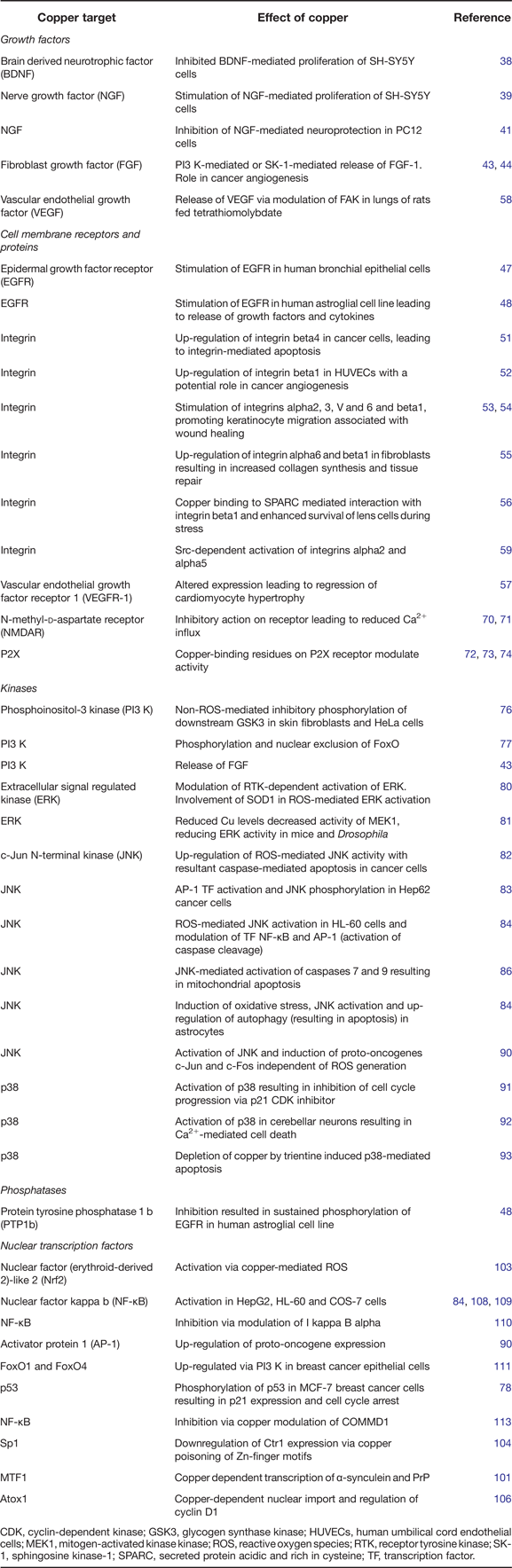
In this review, we will examine the role of copper at each step in the classical cell signalling cascade, beginning with effects of copper on polypeptides targeting membrane receptors, interaction of copper with receptors, copper modulation of kinase and associated phosphatase-based signalling processes, copper as a secondary messenger and finally transcriptional control mediated by copper in the nucleus (Table 1 and Fig. 2).
Schematic representing known sites of copper modulation in cell signal transduction pathways. Copper (Cu) is able to bind to and modulate the interaction of growth factors with cell membrane receptors. Cu can also act in a structural and/or redox role to modify the activation state of membrane receptors for growth factors, either directly or via inhibition of phosphatases that regulate receptor phosphorylation state. Within the cell, Cu can directly regulate kinase activity via structural modification or again via inhibition of phosphatases. Within the nucleus, Cu can modulate action of transcription factors that, in turn, regulate gene expression and subsequently protein synthesis.
Effects of copper on cell signalling pathway targets
CDK, cyclin-dependent kinase; GSK3, glycogen synthase kinase; HUVECs, human umbilical cord endothelial cells; MEK1, mitogen-activated kinase kinase; ROS, reactive oxygen species; RTK, receptor tyrosine kinase; SK-1, sphingosine kinase-1; SPARC, secreted protein acidic and rich in cysteine; TF, transcription factor.
Membrane receptors and associated pathways
Growth factors, receptors and associated tyrosine kinase pathways
Many cell signalling events begin with the binding of a peptide or polypeptide to an external membrane receptor. This process is regulated in a number of ways including through potential structural modification of the interacting protein. Several studies have demonstrated that copper may have an important role in modulating protein binding to target receptors. Copper interacts with the N-terminal domain of brain-derived neurotrophic factor (BDNF) affecting its conformation and subsequent binding to TrkB, the cognate receptor for BDNF on neurons (Ref. Reference Travaglia38). Copper binding occurred within physiological pH within residues 1–12 of BDNF, coordinated through the amino terminus. Copper binding to BDNF peptides or full-length protein inhibited BDNF-induced proliferation of SH-SY5Y neuroblastoma cells in culture (Ref. Reference Travaglia38). Interestingly, this contrasted with the stimulatory effects of copper on nerve growth factor (NGF)-mediated neuronal proliferation (Ref. Reference Travaglia39). Copper coordination by His4 and His8 on one monomer and His84 and Asp105 on the second monomer of the NGF active dimer promoted cell proliferation in SH-SY5Y cell cultures treated with 10 µm copper (Ref. Reference Travaglia39). Binding of copper to NGF is independent of pH, whereas zinc binding is diminished at reduced pH (cerebral acidosis) suggesting that copper may be able prevent zinc-mediated inactivation of NGF during traumatic brain injury (Ref. Reference Ross40). However, these findings contrast with a report from Wang et al. (Ref. Reference Wang41) who reported that both copper and zinc prevented NGF from inducing neuroprotection against oxidative stress in PC12 pheochromocytoma cells. The reason for this discrepancy is not known but could reflect different cell lines, higher copper concentration (100 µm) or contrasting effects of copper on NGF-mediated growth compared with antioxidant action. The association between copper and NGF also appears to be more complex than initially demonstrated. Birkaya and Aletta (Ref. Reference Birkaya and Aletta42) observed that NGF promotes cellular accumulation of copper in PC12 cells with resultant stimulatory action on copper-dependent protein methylation and neurite outgrowth. Potentially, this copper accumulation could adversely interact with ROS resulting in enhanced oxidative stress as reported by Wang et al. (Ref. Reference Wang41).
Copper also has important modulatory effects on other growth factors including fibroblast growth factor (FGF). Copper induces release of FGF-1 from melanoma cells in culture through a mechanism involving activation of the phosphoinositol-3-kinase (PI3 K)-Akt signalling pathway (Ref. Reference Di Serio43). Moreover, copper stimulates FGF-1 release via direct interaction with sphingosine kinase 1 (SK-1). Copper binding promotes release of SK-1, which binds to FGF-1, mediating release of the latter. This mechanism has been suggested to have a role in copper-dependent promotion of angiogenesis (Ref. Reference Soldi44).
In addition to its modulation of ligand conformation and/or growth factor release, copper can also regulate membrane receptor activity through structural modification or phosphorylation. Again, this regulatory action of copper has been demonstrated through its ability to affect activity of growth factor receptors. Wu, Samet and colleagues were among the pioneers of transition metal-mediated cell signalling. Research over several years identified an important role for copper and additional metals in activation of cell signalling pathways in human bronchial epithelial cells (Refs Reference Samet45, Reference Samet46, Reference Wu47). The metal-mediated activation of MAPK pathways is likely to have significant consequences in pulmonary response to combustion-generated airborne metallic particles (Ref. Reference Samet45). Activation of MAPK pathways by copper or zinc involves stimulation (phosphorylation of Tyr845) of the EGFR, leading to downstream up-regulation of mitogen-activated protein kinase (MEK) and ERK activity. The copper/zinc-mediated phosphorylation of EGFR Tyr845 was reported to occur independently of G-protein coupled receptor transactivation and did not involve stimulation of Raf-1 (MAP3 K) (Ref. Reference Wu47). Our own research confirmed the activation of EGFR by copper in a human astroglial cell line, U87MG, over-expressing EGFR. Copper delivered by the cell permeable bis(thiosemicarbazone) complex, CuII(gtsm), induced robust phosphorylation of multiple EGFR tyrosine residues (Tyr1068 and Tyr845) through a cognate-ligand-independent mechanism. This involved inhibition of the protein tyrosine phosphatase, PTP1b, leading to sustained phosphorylation of EGFR (Ref. Reference Price48). This activation resulted in up-regulation of release of cytokines and growth factors with a potential contributory role in the therapeutic action of the complex in vivo (Ref. Reference Crouch49).
Integrins
Another critical mechanism of external sensing and response by cells is via membrane integrin receptors. These transmembrane receptors respond to the extracellular matrix and are activated by common matrix proteins including collagen, fibronectin and vitronectin. Signal transduction is mediated by receptor activation via kinases such as integrin-linked kinase (ILK) and FAK, and results in downstream modulation of MAPK and additional signalling pathways (Ref. Reference Hehlgans, Haase and Cordes50). Cell responses to these signals include migration, tissue invasion, replication or differentiation and are associated with development, tissue repair and angiogenesis (Ref. Reference Hehlgans, Haase and Cordes50). Copper controls integrin-associated signalling via several processes. In cell models of cancer (H322 cells) and wound healing (keratinocytes and fibroblasts), copper complexes have modulated integrin signalling via up-regulation of integrin receptor subunits. Copper-complexed to the salicylaldehydepyrazole hydrazone derivative, (E)-N′-(2-hydroxybenzylidene)-1-(4-tert-butylbenzyl)-3-phenyl-1H-pyrazole-5-carbo hydrazide, (Cu-16) (10 µm), mediated up-regulation of integrin beta4 in cancerous cells, resulting in elevated integrin-dependent apoptosis (Ref. Reference Fan51). Similar effects of copper (10 µm (E)-N′-(2-hydroxybenzylidene)-1-benzyl-3-phenyl-1H-pyrazole-5-carbohydrazide, Cu-15) on integrin beta1 were observed in human umbilical cord endothelial cells (HUVECs) demonstrating a potential role for copper–integrin interactions in angiogenesis (Ref. Reference Fan52). Copper (and zinc) have been found to promote keratinocyte migration necessary for wound healing through stimulation of integrins alpha2, alpha3, alphaV, alpha6 and beta1 (at concentrations of 1–4 µg/ml metal-gluconates or approximately 15–60 µm copper) (Refs Reference Tenaud53, Reference Tenaud54). In contrast, manganese down-regulated alpha3 and beta1 demonstrating that the elevated activity of integrins is not simply metal-dependent. Alternative copper complexes such as Cu-Glycyl-l-histidyl-l-lysine (GHK-CuII) induce up-regulation of integrin alpha6 and beta1 in fibroblasts, resulting in increased collagen synthesis and tissue repair (Ref. Reference Kang55). Copper-mediated changes to integrins in these cells are linked to downstream changes to p63 (a putative stem cell marker of the keratinocytes) and cell nuclear antigen associated with proliferation. Copper-dependent integrin signalling has also been demonstrated in the human lens. The peptide, ‘secreted protein acidic and rich in cysteine’ (SPARC), induces activation of ILK leading to enhanced survival of lens cells. This mechanism involves formation of a complex between SPARC and integrin beta1 during cell stress, a process that requires copper binding to SPARC (Ref. Reference Weaver, Workman and Sage56). Studies have identified a link between integrins and growth factor-mediated responses. Copper (5 µm CuSO4) induces regression of cardiomyocyte hypertrophy through altered expression of vascular endothelial growth factor receptor (VEGFR-1) and subsequent modulation of cGMP-dependent protein kinase 1 signalling (Ref. Reference Zhou, Bourcy and Kang57). In separate studies, depletion of copper led to decreased FAK activity and loss of VEGF expression in lungs of rats fed the copper chelator tetrathiomolybdate (Ref. Reference Mizuno58). Our studies have also shown that cell permeable copper complexes such as clioquinol-copper (CQ-Cu, 10 µm) and pyrrolidine dithiocarbamate-copper (PDTC-Cu) can promote src-dependent phosphorylation of EGFR in U87MG cells via activation of integrins alpha2 and alpha5 (Ref. Reference Price59). The studies demonstrate that copper can modulate multiple sites within a single signalling pathway.
Prion protein and copper signalling
For almost two decades, the membrane-localised cellular prion protein (PrPc) has been recognised as a cuproprotein, capable of binding copper via its octarepeat regions (Ref. Reference Hornshaw60) at physiological extracellular copper concentrations (Ref. Reference Brown61), although the cell signalling roles of copper-bound PrPc have not been thoroughly explored. Experiments from PrPc knockout mice provided the first indications that PrPc-bound copper may participate in physiological synaptic signalling. PrPc knockout mice displayed deficiencies in membrane-bound copper and were unable to elicit appropriate synaptic responses in cerebellar Purkinje cells in response to copper application (Ref. Reference Brown61), suggesting that membrane-bound PrPc may mediate copper import. Nevertheless, substantial controversy still remains regarding whether copper in PrP-binding is physiological or pathological. Several lines of evidence indicate that partial resistance to prion disease onset is correlated to reduced brain copper or reduced copper-binding activity of PrP: disease onset was delayed by (Reference Maret1) a smaller number of octapeptide repeats in PrP (Ref. Reference Stevens62), (Reference Ciuffreda2) treatment with copper chelators (Ref. Reference Sigurdsson63) or (Reference Chen and Goeddel3) a 60% reduced brain copper content, as in hemizygous ATP7a mice (Ref. Reference Siggs64). However, resistance to prion disease was also observed in mice and hamsters fed a high copper diet, indicating a complex relationship between copper and prion disease susceptibility (Ref. Reference Mitteregger65), as PrPc both regulates and is regulated transcriptionally and post-translationally by changes in cellular copper content (reviewed in Ref. Reference Varela-Nallar, Gonzalez and Inestrosa66). Cleavage of PrPc from the cell surface reduced copper content in cerebellar cell membranes in wild-type cells, indicating that membrane-bound copper is primarily associated with PrPc (Ref. Reference Brown61) and likely enters cells in this manner. Copper-dependent transcription of the prion gene (PRNP) (Ref. Reference Armendariz67) is mediated through the copper-responsive transcription factors (TFs) Sp1 and MTF-1 (Ref. Reference Bellingham68) binding to metal response element (MRE) sequences on the PRNP promoter (Ref. Reference Varela-Nallar69).
Additional receptor regulation by copper
In addition to controlling growth factor receptors, membrane integrin function and PrPc, copper has important modulatory effects on cell signalling associated with synaptic transmission. Copper binds to the amino terminal domain of N-methyl, D-aspartate (NMDA) glutamate receptor subunits in the CNS. Copper can induce concentration-dependent inhibitory (greater than 30 µm) or potentiating (less than 30 µm) effects on receptor activation and Ca2+ influx (Ref. Reference Marchetti, Baranowska-Bosiacka and Gavazzo70). Studies have shown that control of copper release into the synapse is critical for modulation of synaptic transmission (Ref. Reference Gaier, Eipper and Mains25) and is controlled by ATP7a at the synapse (Ref. Reference Schlief71). Similarly, copper also regulates P2X ATP-gated purinergic receptors. Huidobro-Toro, Acuna-Castillo and colleagues have identified key copper-binding residues on the P2X receptor that mediate inhibitory activity upon binding (Refs Reference Acuna-Castillo72, Reference Huidobro-Toro, Lorca and Coddou73, Reference Punthambaker and Hume74). Copper may also exert considerable control over synaptic transmission and associated signalling via modulating Ca2+ or zinc binding and regulation of MT expression (Ref. Reference Tamano and Takeda75).
Downstream kinase pathways
PI3 K-Akt
Perhaps the greatest evidence for a key role of copper in modulation of cell signal transduction comes from studies showing activation of MAPKs by copper. Ostrakhovitch and colleagues pioneered this research, demonstrating copper-mediated activation of the well-known cell survival pathway via phosphoinositol 3 kinase (PI3 K) and its immediate downstream partner, protein kinase B (also known as Akt) (Ref. Reference Ostrakhovitch76). Ostrakhovitch et al. reported that copper treatment of skin fibroblasts (10–100 µm CuSO4) or cancerous HeLa epithelial cells (3 µm CuSO4) resulted in up-regulation of phospho-Akt levels and increased inhibitory phosphorylation of its downstream target, glycogen synthase kinase 3 (GSK3) (Ref. Reference Ostrakhovitch76). This elegant publication, and supported by many more, clearly demonstrated that, in contrast to expectation, the signalling activity of copper was not associated with increases in cellular ROS or oxidative stress. While blocking activation of membrane receptor-associated tyrosine kinases or inhibiting ROS formation prevented activation of PI3 K pathway by growth factors, this had no effect on copper-dependent activation. This has been further supported by Eckers et al. (Ref. Reference Eckers, Reimann and Klotz77) who described activation of PI3 K by 10 µm copper (CuSO4) or nickel (NiSO4) (activation was greater with copper than nickel) but without any generation of ROS by the latter. Activation of PI3 K was associated with increased phosphorylation and nuclear exclusion of FoxO TF in HepG2 cells. Further studies by Ostrakhovitch and colleagues have shown that PI3 K-Akt activation, or over-activation in some cases, by copper, can induce cell cycle arrest in some model systems, while in others, provides an insulin-mimetic action (Refs Reference Ostrakhovitch and Cherian78, Reference Barthel79). Copper-mediated Akt activation has also been linked to FGF release in melanoma cells (Ref. Reference Di Serio43). These studies demonstrated the complex pathways that can be modulated by copper through one signalling system in different cellular environments.
ERK
Although the studies described above demonstrated that copper can stimulate kinase activity, more recent investigations of copper and ERK have shown that copper may be essential for kinase activity in certain circumstances. Ctr1 is fundamental for maintenance of copper in cells (Ref. Reference Wee28) (Fig. 1). Modulation of Ctr1 expression has been used elegantly to demonstrate that cellular copper levels control kinase activity. Tsai et al. (Ref. Reference Tsai80) compared kinase activities in normal compared with Ctr1 deficient (Ctr1−/−) cells and reported that the latter revealed greatly reduced activity of ERK after exposure to receptor tyrosine kinase (RTK) ligands, FGF, platelet-derived growth factor (PDGF) and EGF. Similarly, PDGF and EGF increased activation of PI3 K in normal cells but not in Ctr1−/− cells. These findings were supported by copper chelation and supplementation experiments and strongly suggest that copper acts on RTK-mediated cell signalling up-stream of Ras. Interestingly, the authors probed the mechanism of action further and demonstrated that the lack of Ctr1 leads to 17-fold decrease in SOD1 levels. Diminished SOD1 led to reduced H2O2 generation, a known co-factor in ligand-mediated RTK activation, inhibiting activation of the signalling pathway. This indirect role of ROS (H2O2) in activation of signal transduction by copper may help explain the contradictory studies that suggest that copper-dependent activation of signalling does or does not require ROS generation. The studies by Tsai et al. are consistent with similar research reported in Drosophila and mice (Ref. Reference Turski81) in which ablation or mutation of Ctr1 (or copper chelation) resulted in a decrease in activity of MEK1, leading to reduced ERK phosphorylation. Studies in cardiac tissue of Ctr1−/− mice revealed lower phospho-ERK levels and the authors demonstrated that recombinant MEK1 bound two copper atoms at high affinity. Copper (2.5 µm CuSO4) enhanced MEK1–controlled phosphorylation of ERK in a dose-dependent manner (Ref. Reference Turski81). The results indicate that copper is important in several key steps of MAPK signalling, including modulation of SOD1-mediated H2O2 for RTK activation and MEK1-mediated phosphorylation of ERK.
JNK
Considerable evidence for copper-mediated control of cell signalling has come from studies on the toxic actions of excess copper. These studies have been driven mostly by a desire to determine the toxicological properties of exposure to excessive copper, often environmental, or harnessing the toxic action of copper as a potential therapeutic strategy for killing cancerous cells. As with the studies above on copper induction of PI3 K, there have been contrasting roles for copper-mediated ROS generation in activation of JNK. Treatment of hepatocytes with up to 10 µm copper (CuSO4) was found to induce up-regulation of ROS and JNK phosphorylation, leading to caspase activation (Ref. Reference Nawaz82). Exposure of the hepatic cell line, Hep62 to copper-nanoparticles similarly induced JNK stimulation leading to AP-1 TF activation and apoptosis (Ref. Reference Piret83). ROS generation by copper was also found to be associated with JNK activation in HL-60 cells by copper delivered using PDTC (EC50 100 µm with 0.1 µm basal copper in the medium) (Ref. Reference Chen84). The increase in JNK activity was associated with modulation of the TF, NF-κB and AP-1 and induced activation of caspase leading to poly-ADP ribose polymerase cleavage. Additional studies on PDTC delivery of copper (0.1–3 µm PDTC and 3 µm CuSO4) to the B-cell line RIN-m5F supported copper-mediated activation of JNK via oxidative stress and this could be reversed by treating with the antioxidant, N-acetyl-cysteine (Ref. Reference Wu85). In lung epithelial cells, PDTC-Cu (0.5 µm PDTC and 1 µm CuCl2) activated JNK (and ERK) resulting in activation of caspases 7 and 9 and mitochondrial-mediated apoptosis. In addition, ER stress was shown to be induced by PDTC-Cu and was reversed by co-treatment with a JNK inhibitor (Ref. Reference Chen86). Disulfiram has also been used to deliver copper to doxorubicin-resistant HL-60 cells (1 µm disulfiram and 1 µm CuCl2) and in glioblastomas resulting in ROS generation, JNK activation and cell death (Refs Reference Liu87, Reference Xu88). Delivery of copper using the antineoplastic compound, CasIIIia-induced oxidative stress, activation of JNK and up-regulation of autophagy, leading to cellular apoptosis. Oxidative stress induced by copper (10 µm CuCl2 and 0.1 µm neurocuproine) has been shown to activate JNK in astrocytes (Ref. Reference Chen89). In contrast, copper treatment of COS-7 cells (400 µm CuSO4) mediated ROS production and lipid peroxidation but this was found to be unrelated to copper-mediated JNK activation and subsequent induction of proto-oncogenes such as c-Jun and c-Fos (Ref. Reference Mattie, McElwee and Freedman90). The mechanism by which copper controls JNK activation, whether involving ROS or not, is not known.
p38
Analogous findings have been presented for activation of p38 by copper. Moon et al. (Ref. Reference Moon91) reported that PDTC (5–10 µm) blocked cell cycle progression in a copper-dependent manner at G1 in vascular smooth muscle cells via activation of p38 and the p21 CDK inhibitor. Interestingly, copper treatment (50 µm CuCl2) of cerebellar granule cells was found to induce ROS-mediated activation of p38 and ERK but not JNK (Ref. Reference Chen92). Blocking Ca2+ influx prevented p38 activation and reduced copper-mediated cell death. In contrast, a study by Kadowaki et al. (Ref. Reference Kadowaki93) found that depleting copper using trientine was capable of inducing activation of p38 and subsequent apoptosis in fibrosarcoma cells, demonstrating that both excess and limiting copper levels can modulate MAPKs with important consequences to cell fate.
Intracellular mechanisms of copper-mediated cell signalling
Phosphatase-kinase dynamics
Although it is evident that copper is involved in many aspects of cell signal transduction, the mechanisms of this activity are less well understood. As indicated above, some of the effects of copper are likely to be due to a direct role for copper binding to kinases and/or receptors with associated effects on structural conformation and hence activity (Ref. Reference Turski81). However, additional important processes are likely to be involved (Fig. 2). One of these is the reported ability of copper to potently inhibit activity of cellular phosphatases that are essential for modulation of kinase activation and de-activation. Copper inhibits PTP1b activity at submicromolar concentrations (Refs Reference Wang94, Reference Yuan95). We have shown that copper delivered into cells with a bis(thiosemicarbazonato) complex (CuII(gtsm)) inhibits PTP1b, resulting in sustained activation of EGFR and subsequent release of growth factors and cytokines (Ref. Reference Price48). We have found that CuII(gtsm) (50 nm) also inhibits general cellular phosphatase activities (phosphatases 2A) and calcineurin (Ref. Reference Bica96). Urquiza et al. (Ref. Reference Urquiza97) have demonstrated that a copper complex of the antithyroid drug methimazole inhibits alkaline phosphatase activity (ED50 42 µm) and additional studies demonstrate that copper can decrease brain cytosolic phosphatase activity (Ref. Reference Hanahisa and Yamaguchi98). This latter study reported that the use of the reducing agent dithiothreitol prevented copper-mediated inhibition of phosphatase activity implicating action of copper on SH thiol groups. This is consistent with our studies that reducing agents abrogate the ability of copper to inhibit PTP1b (Ref. Reference Price48).
Nuclear signalling
A more complex manner in which copper controls cell signalling is via modulation of nuclear TF activity and localisation. TFs bind to DNA and control expression of genes associated with a vast array of key downstream processes including antioxidant response proteins, inflammation and oncogenes (Ref. Reference Zhang and Glass99). There is a large repertoire of copper-sensing TFs in bacteria, yeast, plants, fungi and lower order organisms, such as Drosophila. These factors possess one or more cysteine-rich copper-binding domains, which may be composed of Cys-X-Cys, Cys-X2-Cys, or Cys-X-Cys-X4-Cys-X-Cys-X2-Cys-X2-His motifs, and may control TF function through redox-dependent conformational switching (reviewed in Ref. Reference Rutherford and Bird100). Copper sensors, including Ace1, Mac1, Crr1 and MTF-1 are reported to regulate a plethora of cellular processes, ranging from copper detoxification and iron metabolism to photosynthesis (reviewed in Ref. Reference Nevitt, Ohrvik and Thiele22).
The situation in mammals is much more complex: although a number of TFs, such as MTF-1, are known to be copper-responsive, the lack of a clear cysteine-rich copper-binding domain on mammalian MTF-1 as compared with Drosophila MTF-1 has continued to puzzle researchers (Ref. Reference Radtke101). It is unclear how copper-mediated MTF-1-dependent responses, such as induction of mRNAs encoding alpha-synuclein and PrP, are mounted in the absence of direct copper binding. Similarly, the antioxidant response TF, Nrf2 has recently been implicated in copper-dependent transcription of antioxidant response element and MRE target genes, regulating diverse processes including cell cycle control, inflammation, metabolism, organelle biogenesis and protein folding (Refs Reference Song102, Reference Wang103). However, it is not clear whether Nrf2 activation involves direct copper binding (Ref. Reference Song102).
An alternative possibility in the case of mammalian MTF-1 is that copper may displace zinc from zinc finger motifs present in MTF-1, which may confer metal-dependent transcriptional specificity. A similar mechanism was demonstrated for the copper-responsive TF Sp1, which is inhibited by direct copper binding or ‘poisoning’ of zinc finger motifs, thereby down-regulating transcription of the copper importer, Ctr1 in response to excess copper (Ref. Reference Song104).
Although copper is generally not localised to the nucleus, nuclear MT import in response to diverse cellular stimuli has been described, and nuclear copper-loaded MT has been detected in human fetal liver samples, which may be a mechanism of copper entry into the nucleus to enable copper-nuclear factor interactions (reviewed in Ref. Reference Cherian and Apostolova105) (Fig. 1). Another strategy in nuclear-copper interactions is that of multifunctional copper chaperones such as Atox1, which shuttles between the nucleus and cytoplasm in response to high copper load (Fig. 1). Atox1 has been recently implicated in regulation of cyclin D1 (Ref. Reference Itoh106) and was proposed to regulate 32 other genes based on putative Atox-binding promoter signatures (Ref. Reference Muller and Klomp107). Given the clear role for copper interaction with plasma membrane receptors as outlined above, a potentially important role for copper in modulation of nuclear receptors remains to be explored.
Many studies have shown that copper can modulate the key inflammatory TF, NF-κB. This has been demonstrated in several cell types exposed to copper including HL60 (as indicated above), HepG2 (400 µm CuCl2) and COS-7 (100–600 µm CuCl2) cells as well as in animal tissues (Refs Reference Chen84, Reference McElwee, Song and Freedman108, Reference Song, Li and Freedman109). In contrast, Satake et al. reported that 500 µm CuCl2 can inhibit NF-κB activation via modulating phosphorylation of the inhibitory binding partner I kappa B alpha (Ref. Reference Satake110). Additional TFs reported to be activated by copper exposure include AP-1 [resulting in proto-oncogene expression (Ref. Reference Mattie, McElwee and Freedman90)]; PI3 K-Akt activated FoxO1a and FoxO4 [resulting in nuclear exclusion of these TFs (Ref. Reference Walter111)]; and the important cancer TF, p53 (Refs Reference Ostrakhovitch and Cherian78, Reference Hoti112). The latter was found to be phosphorylated at Ser15 upon copper treatment of MCF-7 breast cancer cells resulting in p21 expression and cell cycle arrest (Ref. Reference Ostrakhovitch and Cherian78). The ability of copper to modulate TF activity may be driven through either direct binding to copper binding sites or via indirect effects on upstream kinases and phosphatases as described above.
Owing to the reducing cellular conditions, copper-binding proteins generally recognise CuI. A curious exception to this rule is copper metabolism (Murr1) domain containing 1 (COMMD1), which is mutated in Bedlington terriers with copper toxicosis and is involved in both direct and indirect inhibition of NF-κB (reviewed in Ref. Reference Bartuzi, Hofker and van de Sluis113). COMMD1 binding is restricted to CuII, which has been suggested to contribute to copper extrusion or detection and detoxification of toxic labile cellular CuII (Ref. Reference Sarkar and Roberts114).
Disease-specific changes to signalling associated with copper
Altered cell signalling in diseases associated with abnormal copper homeostasis is common (Refs Reference Shukla, Skuntz and Pant13, Reference Trempe and Fon115, Reference Clayton, Minogue and Waugh116, Reference Um and Strittmatter117, Reference Russo118, Reference Jellinger119), however, it is extremely difficult to determine whether the changes to signalling are a direct result of impaired copper handling or are a downstream consequence of copper-mediated changes to cell metabolism. In fact, even indirect relationships between copper and altered cell signalling are difficult to interpret in the complex milieu of tissues. Necessarily, most studies on the role of copper in modulation of cell signal transduction have been performed in cell culture as described in the previous sections. There are a few studies that provide indirect evidence that abnormal copper metabolism does indeed mediate altered cell signalling in disease in vivo. We and others have shown that delivery of copper [10 mg/kg/day CuII(gtsm) or 20 mg/kg/day PDTC] to AD model mice (APP/PS1) using metal complexing agents is associated with increased phosphorylation of Akt and GSK3 in the brain (Refs Reference Crouch49, Reference Malm120) and improvement in cognition. Kitazawa has also shown that 250 ppm CuSO4 fed to AD model mice can modulate CDK5/p25 activity in the brain with effects on amyloid and tau (Ref. Reference Kitazawa, Cheng and Laferla121). Exposure of rats to copper (0.9 and 7.7 µg Cu/g diet) resulted in elevated NF-κB activity and altered protein kinase C (PKC) mRNA expression in separate studies (Refs Reference Persichini122, Reference Davis and Johnson123). However, these studies do not provide direct evidence that copper is controlling these signalling events in animals exposed to a normal copper environment. Owing to the complex effects of altered copper homeostasis on many aspects of animal and human metabolism, it is currently not possible to ascertain a clear role for this metal in cell signalling during disease. This will require the development of novel approaches to cell signal monitoring in vivo.
Therapeutic approaches based on regulation of copper signalling
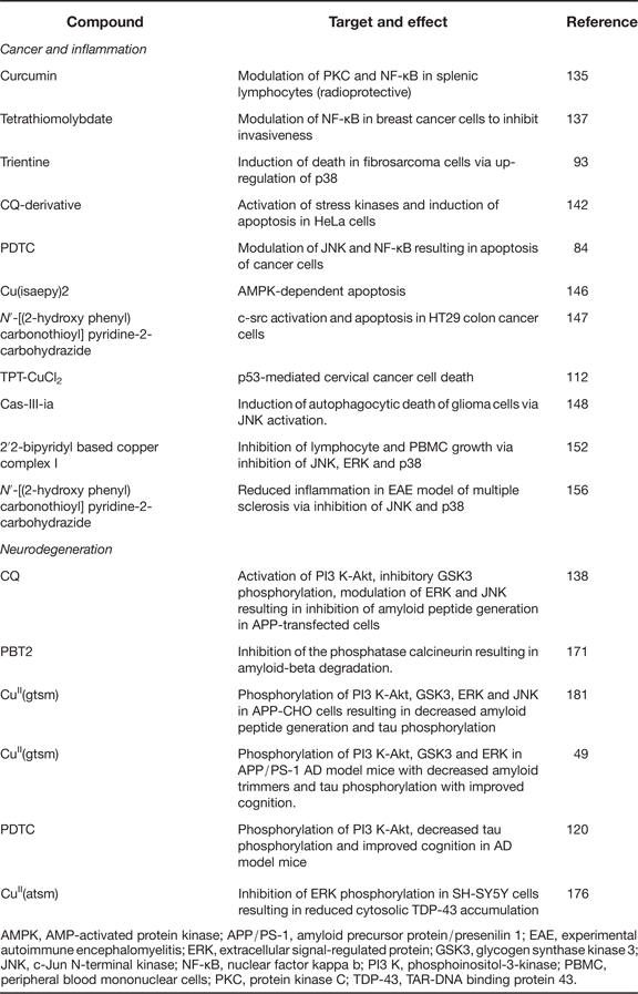
The main purpose in identifying the mechanisms by which copper controls cell signalling is to provide a knowledge base for development of therapeutic treatments for diseases. To this end, there has been a recent focus on development of agents that modulate copper signalling to bring about tumour cell death or promote neuronal survival in cancer and neurodegeneration, respectively (Table 2).
Targeting of signalling pathways with copper-based therapeutics
AMPK, AMP-activated protein kinase; APP/PS-1, amyloid precursor protein/presenilin 1; EAE, experimental autoimmune encephalomyelitis; ERK, extracellular signal-regulated protein; GSK3, glycogen synthase kinase 3; JNK, c-Jun N-terminal kinase; NF-κB, nuclear factor kappa b; PI3 K, phosphoinositol-3-kinase; PBMC, peripheral blood mononuclear cells; PKC, protein kinase C; TDP-43, TAR-DNA binding protein 43.
Cancer and inflammation
In studies aimed at the former, traditional copper chelating compounds have shown promise at inhibiting cancer cell growth, angiogenesis and metastasis. One of the oldest known anticancer agents with copper chelating ability is curcumin, derived from the Rhizome, Curcuma longa. The compound (found in turmeric) has well characterised antioxidant effects in addition to a potential ability to chelate copper (Ref. Reference Leung, Harada and Kee124). The compound has shown protective actions in AD, cancer and inflammatory disease. The antioxidant cellular effects of curcumin are primarily mediated via cellular kinase signalling pathways, including JNK, Akt and Nrf2-dependent mechanisms (Refs Reference Pan125, Reference Jiang126, Reference Liu127, Reference Wang128). Additionally, curcumin also improves numerous indicators of mitochondrial function, from caspase activation to membrane depolarisation (reviewed in Ref. Reference Grubman, White and Liddell129). Aside from direct ROS inactivation as reported for peroxynitrite (Ref. Reference Iwunze and McEwan130), curcumin may also detoxify ROS through its metal-binding and ionophore properties (Refs Reference Dairam131, Reference Garcia132), which can be modulated by alteration of the aromatic ring substituents (Ref. Reference Ferrari133). Copper chelated curcumins and curcuminoids have also found application for treatment of cancer and neurodegeneration via dual-functional pro- and antioxidant effects (reviewed in Ref. Reference Leung, Harada and Kee124). Curcumin/CuII co-treatment-induced apoptosis of human ovarian and breast cancer cells by targeting the NF-κB and mTOR signalling pathways in vitro (Ref. Reference Lou134). Curcumin has also shown radioprotective action as a complex with copper where it protected splenic lymphocytes from irradiation via delaying activation of PKC delta and NF-κB activation (Ref. Reference Kunwar135). Moreover, curcuminoid copper chelates are reported to possess potent antitumour activity in vivo (Ref. Reference John, Kuttan and Krishnankutty136).
Tetrathiomolybdate is a copper chelating agent that has been used in treatment of Wilson disease, a genetic copper overload disorder. Application of tetrathiomolybdate (10 nm) to breast cancer cells has been shown to inhibit cell invasiveness via modulation of NF-κB (Ref. Reference Pan, Bao and Merajver137). Similarly, trientine, a copper chelator also used in Wilson disease therapy induces death of fibrosarcoma cells in a murine transplantation model. As mentioned above, the mechanism is likely due to up-regulation of the stress kinase, p38 by copper chelation (Ref. Reference Kadowaki93). Most recent studies however, have focused on cell permeable copper complexing agents which have the ability to deliver copper into tumour cells and activate cell death pathways. The important caveat with this form of research, however, is that only when compounds are directly and rigorously compared with noncancer cells can copper-dependent cytotoxic effects provide valuable information on therapeutic potential. Delivering enough copper to any cell will be toxic but does not equate to a cancer therapeutic. Unfortunately, this simple but critical point is often lost in the haste to demonstrate therapeutic efficacy of a copper complex. Despite this reservation, there is clear evidence that copper complexes can preferentially kill some cancer cells and this may involve targeting specific cell signalling mechanisms. Considerable evidence supports a potential therapeutic action of the 8-hydroxyquinoline, CQ as a copper delivery agent (Ref. Reference White138) that when applied to tumour cells can promote cell death via inhibition of the proteasome (Refs Reference Daniel139, Reference Ding140, Reference Daniel141). Very little is known regarding the effect CQ or CQ–Cu complexes have on cancer cell signalling, although Chen et al. have demonstrated that a CQ-derivative, 7-methyl-arylsulfonylpiperazine induced ROS and activation of stress kinases, resulting in apoptosis of HeLa cells (1–3 µm compound and 10 µm CuCl2) (Ref. Reference Chen142). As discussed below, more is known of the ability of CQ to modulate cell signalling as a potential therapeutic for neurodegenerative diseases. Similar to CQ, the well-studied copper delivery compound, PDTC, with anticancer cell activity (Ref. Reference Daniel139) has been shown to induce apoptosis via activation of the stress kinase, JNK and modulation of NF-κB as described above (Ref. Reference Chen84).
Many additional copper complexes have been designed specifically as cancer targeting agents (Refs Reference Laine and Passirani143, Reference Duncan and White144, Reference Colotti145), however, again, little is known regarding the mechanisms by which these agents modulate cell signalling as a key aspect of their potential therapeutic activity. Of the few agents where effects on cell signalling have been demonstrated, perhaps unsurprisingly, these are mostly associated with ROS-mediated cell signalling changes leading to activation of apoptosis. Filomeni et al. (Ref. Reference Filomeni146) demonstrated that a copper complex, Cu(isaepy)2 (10–50 µm) targets the mitochondria of a variety of cancer cell types, inducing elevated ROS and AMP-activated protein kinase (AMPK)-dependent apoptosis. Another copper complex, N′-[(2-hydroxy phenyl) carbonothioyl] pyridine-2-carbohydrazide, was shown to inhibit NMT activity in colon cancer cells (HT29) (IC50 12.2 µm). This action led to inhibition of c-src kinase, triggering apoptosis (Ref. Reference Shrivastav147). Several additional copper complexes also mediate cancer cell death, at least in cultured cells via modulation of TFs [TPT-CuCl2 (4 µm) induces p53-dependent cervical cancer death] (Ref. Reference Hoti112), or induction of autophagy [Cas-III-ia (500 nm) induces autophagocytic-mediated death of glioma cells via JNK activation] (Ref. Reference Trejo-Solis148). However, there is still limited understanding of cell signalling modulation by the vast array of anticancer copper complexes currently being developed (Ref. Reference Duncan and White144). This needs to be addressed in order to provide a complete insight into the action of the agents prior to clinical trials.
Interestingly, there have been few advances in the understanding of copper-based therapeutics for modulation of signalling kinases in inflammatory diseases. This is despite the considerable evidence that copper has important modulatory actions on both systemic- and neuro-inflammation (Refs Reference Lutsenko149, Reference Choo150) and the general understanding that targeting copper or using copper-based compounds is an effective strategy to modify the immune system (Ref. Reference Gupte and Mumper151). From the few studies that have been performed on cell signalling by anti-inflammatory copper-based compounds, it is evident that modulation of MAPK pathways is central to their action. Rupesh et al. (Ref. Reference Rupesh152) have shown that 2,2′-bipyridyl-based copper complex I (7 nm) inhibits lymphocyte and peripheral blood mononuclear cell (PBMC) growth through inhibitory action on the MAPKs, ERK, JNK and p38. The copper chelator, N acetylcysteine (NAC), has been investigated for the treatment of diseases ranging from renal and hepatic injury to cystic fibrosis to neurodegeneration. Although NAC has been heralded as a strong antioxidant since the 1980s, the antioxidant effects observed with NAC are likely attributable to GSH synthesis induced by the compound, rather than any direct antioxidant activity (recently reviewed in Ref. Reference Rushworth and Megson153): for instance, NAC is unable to scavenge superoxide (Ref. Reference Aruoma154). There is even evidence that NAC acts synergistically with copper to generate ROS-dependent cell death of cancerous cells (Ref. Reference Zheng155). An NAC-based compound capable of crossing the blood brain barrier has been shown to chelate copper, reducing inflammatory processes in an EAE model of multiple sclerosis also through inhibition of MAPK (JNK and p38) activity, and possibly related to reduced ROS generation (Ref. Reference Offen156). Despite these important findings, considerable research is required to substantially improve our understanding of how copper-based anti-inflammatory therapeutics act via induction of cell signalling changes in immune cells.
Neurodegeneration
Unlike cancer, there has been little focus on modulation of copper-dependent signalling pathways to provide therapeutic benefit in neurodegenerative diseases such as AD, Parkinson's disease (PD) and amyotrophic lateral sclerosis (ALS). This is difficult to reconcile given that abnormal copper metabolism is now well documented in these and related neurodegenerative diseases (Refs Reference Jellinger119, Reference Taba157, Reference Parker158, Reference Ayton, Lei and Bush159, Reference Ward, Dexter and Crichton160, Reference Schrag161) and altered cell signalling is associated to some extent with all molecular pathological changes. Strong potential for achieving valuable therapeutic outcomes from modulation of copper homeostasis was generated from research into the action of copper-complexing compounds in models of neurodegeneration (Refs Reference Crouch49, Reference White138, Reference Bica162). These studies were developed from early successes with the cell-permeable copper chelating compound, CQ, in AD models (Ref. Reference White138). Altered copper metabolism in AD derives from at least two separate processes: the propensity for accumulated extracellular copper to potently drive Aβ aggregation (Ref. Reference Barnham163), and the damaging effects on cuproprotein functions of the consequent intracellular copper deficits (Ref. Reference Adlard164). Even low concentrations of copper (<0.8 µm) can induce Aβ oligomerisation (Refs Reference Atwood165, Reference Curtain166, Reference Huang167) through oxidative modification of a high affinity amino terminal tyrosine residue in Aβ, which not only promotes plaque formation, but may also generate neurotoxic H2O2 via Aβ-mediated reduction of CuII to CuI (Ref. Reference Multhaup168). APP also contains an N-terminal copper binding domain which has been shown to modulate cellular copper homeostasis (Refs Reference Multhaup168, Reference White169), and this may be controlled by phosphorylation of the protein (Ref. Reference Acevedo170).
These important roles for copper in AD pathogenesis led to the examination of copper-targeted therapeutics. The 8-hydroxyquinoline copper chelator, CQ, was found to improve cognitive performance in AD model mice and subsequently improved patient outcomes in a small clinical trial of AD patients (Refs Reference Cherny171, Reference Ritchie172). The method of action was proposed at the time to be due to chelation of copper from aggregated extracellular amyloid beta, reducing the potential toxicity of this accumulating protein in the brains of AD model mice and patients. However, an interesting observation came from these studies. The levels of copper (and zinc) were significantly elevated in the brains of AD mice successfully treated with the CQ. Although CQ was discontinued (as a potential AD therapeutic but still remains in studies as a potential cancer compound), an alternative proprietary 8-hydroxyquinoline, PBT2 is continuing in clinical trials for treatment of AD. Although these initial studies did not examine the copper delivery potential of the compound or ability to modulate cell signalling, we subsequently identified potent kinase regulatory outcomes from CQ and related 8-hydroxyquinoline and phenanthroline compounds, that were dependent upon copper (or zinc delivery) into cells (Refs Reference Filiz173, Reference Caragounis174). Activation of PI3 K-Akt leading to downstream phosphorylation of GSK3 and regulation of ERK and JNK were shown to be key steps in CQ-Cu-dependent inhibition of amyloid accumulation (10 µm CQ-Cu), most likely through up-regulation of amyloid degrading matrix metalloprotease expression (Refs Reference White138, Reference Caragounis174). Subsequent studies have also shown that PBT2 (10 µm) modulates neuronal cell kinase activity, possibly through modulation of the phosphatase calcineurin (Ref. Reference Crouch175) by zinc or copper. Studies also confirm that these compounds have potential neuroprotective actions that are not related to modulation of amyloid (Ref. Reference Parker176) but are copper- or zinc-dependent and involve regulation of kinases and/or phosphatases.
Strong evidence for the potential of copper-mediated modulation of cell signalling as a therapeutic strategy comes from our studies on the bis(thiosemicarbazone) class of metal ligand. CuII(btscs) such as CuII(gtsm) and CuII(atsm) are cell permeable copper delivery compounds with robust therapeutic action in multiple animal models of neurodegeneration including AD, PD and ALS (Refs Reference Crouch49, Reference Soon177, Reference Hung178). The rationale for use of the agents is supported by strong evidence from recent key studies demonstrating conclusively that bioavailable copper is greatly deficient in critical brain regions of several neurodegenerative disorders (Refs Reference Schrag161, Reference Akatsu179). Although several potential therapeutic actions of CuII(btsc)s have been proposed (Refs Reference Hung178, Reference Crouch and Barnham180), growing evidence from our studies is consistent with copper-dependent modulation of cell signalling as the prime therapeutic modality across the diverse models. We initially demonstrated that the CuII(btsc)s (25 µm for 6 h) were able to induce up-regulation of PI3 K-Akt and GSK3 as well as ERK and JNK in neuronal and alternative cell lines, and as with CQ-Cu, this was critical for inhibition of amyloid accumulation (Ref. Reference Donnelly181). Subsequently, our recently published studies have also demonstrated that low concentrations of CuII(gtsm) (50 nm) modulate neurite growth in cultured neurons, likely via control of phosphatase activity (Ref. Reference Bica96). As mentioned above, CuII(gtsm) was also found to activate EGFR via inhibition of PTP1b leading to secretion of growth factors and cytokines, some with therapeutic potential (Ref. Reference Price48).
Importantly, we were able to demonstrate that CuII(gtsm) (10 mg/kg/day) activated the same kinase pathways in AD model mice (Akt, GSK3 and ERK) as we demonstrated in cell culture (Ref. Reference Crouch49). This finding was consistent with the report by Malm et al. (Ref. Reference Malm120) who demonstrated improved cognitive performance of AD model mice treated with PDTC (20 mg/kg/day) and this was associated with elevated brain copper levels and activation of the PI3 K-Akt pathway. Treatment of mice with CuII(gstm) improved cognitive performance in AD model mice. The related compound, CuII(atsm), which has different copper binding properties, has shown robust therapeutic action in four animal models of PD and two models of ALS (Refs Reference Hung178, Reference Crouch and Barnham180, Reference McAllum182). We have shown that 1 µm CuII(atsm) is also capable of inhibiting ERK leading to down-regulation of the toxic protein, TDP-43 associated with ALS neurodegeneration (Ref. Reference Parker176). Interestingly, our studies have shown that these copper complexes are capable of elaborate kinase control, dependent on concentration, time and cell type. Although it remains to be determined the specific cellular localisation of CuII(btsc) action and how this feeds into multiple kinase/phosphatase networks, the early results from these compounds have clearly demonstrated that copper-dependent kinase modulation is an important target for therapeutic intervention in neurodegenerative diseases.
Discussion and the future
This review has attempted to bring together for the first time the large number of studies across broad fields of science and medicine demonstrating that copper is a key mediator of cell signal transduction. It is not surprising that evolution has harnessed the highly ‘pliable’ redox state of copper as a regulator of complex cell signalling systems. It is perhaps more surprising that it has taken a long time for the scientific community to accept copper (and zinc) as bona fide signalling molecules and not just ‘trace elements’. The lack of acceptance of copper as a signal molecule likely stems from its inherent toxicity if mis-regulated and the fact that, as shown here, the studies on copper-mediated control of cell signalling cover a very diverse range of proteins, cells and diseases. There are also a number of additional difficulties in ascertaining the role of copper in signalling. Although the use of fluorescent probes to track intracellular stores of calcium and zinc is well advanced, copper probes are still largely in the developmental stage, with a number of limitations still to be resolved (Ref. Reference New33). Furthermore, as evident from this review, there is a wide range of treatment doses used in studies on copper signalling. These are sometimes outside the physiological range, and it is hard to compare signalling processes triggered by copper complexes that may rapidly increase copper influx, with copper salts that have limited uptake. However, what the studies have shown to date is that copper affects many aspects of cell signalling (Fig. 2) and this modulation leads to major downstream changes, including cell growth, differentiation, death and a multitude of cell functions.
Our review has attempted to detail some of these studies, clearly demonstrating that copper has key regulatory control over cell signalling in many compartments of the cell, including membrane receptors, sub-membrane complexes, MAPKs and tyrosine kinase signalling pathways, modulation of phosphatases and regulation of nuclear TFs. Abnormal handling of copper is prominent in many diseases and as shown here, likely underlies a number of pathological cellular changes. From these observations, obviously, modulation of copper homeostasis can be harnessed as a key therapeutic strategy for cancer, neurodegeneration and potentially additional diseases involving inflammation. For these studies to advance, a great deal of research is still required to determine how and where, within different cell types, copper controls signalling pathways and networks and what is the best mechanism for controlling this as a therapeutic option. This is likely to evolve with advances in sub-cellular detection of copper pools using a new generation of fluorescent copper probes.
Acknowledgements
A.R.W. is the recipient of an Australian Research Council Future Fellowship. Patent protection has previously been sought by the University of Melbourne for the use of bis(thiosemicarbazones) for treatment of diseases. A.R.W. is a co-inventor on this patent application PCT/AU2007/001792, which is the subject of a commercialisation contract between the University and a private company. The company has not funded nor contributed to research described in this manuscript.