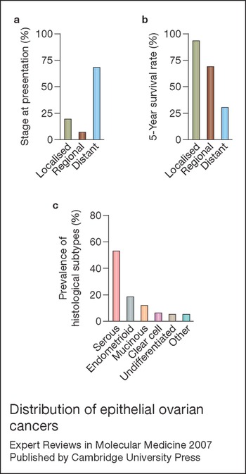
Epithelial ovarian cancer (EOC) is the fifth leading cause of cancer death in women and is the most lethal gynaecological malignancy (Ref. Reference Jemal1). Although 5-year survival rates can approach 90% for women who are diagnosed with tumour confined to the ovaries, approximately 70% of patients present with disseminated disease (Fig. 1a). Early detection of EOC is hampered by the absence of specific clinical symptoms and lack of accurate diagnostic tests. The glycoprotein CA125, the best-characterised serum biomarker for EOC, is used for post-operative monitoring, but alone has insufficient specificity and sensitivity for population screening (Ref. Reference Bast2). For many years, tumour-debulking surgery and taxane–platinum combination therapy have remained the standard-of-care. Despite advances in chemotherapeutic regimens, relapse is frequent and the high initial response rate does not translate to a high cure rate. For women diagnosed with advanced-stage EOC, the average 5-year survival rate is only 30% (Fig. 1b).
Distribution of epithelial ovarian cancers. (a) Distribution of epithelial ovarian cancers by disease stage at time of initial diagnosis. (b) 5-year survival rate by stage. Graphs in (a) and (b) were adapted from figures in Ref. Reference Jemal1 [Jemal, A. et al. (2007) Cancer statistics, 2007. CA Cancer J Clin 57, 43-66]. (c) Distribution of tumours by histological subtype. The graph in (c) was constructed using data pooled from the text of Ref. Reference Clements and Young3 [Clements, P.B. and Young, R.H., eds (2000) Atlas of Gynecologic Surgical Pathology, W.B. Saunders, Philadelphia] and from tabulated data shown in Ref. Reference Seidman4 [Seidman, J.D. et al. (2004) The histologic type and stage distribution of ovarian carcinomas of surface epithelial origin. Int J Gynecol Pathol 23, 41-44].

The heterogeneity of epithelial ovarian cancers
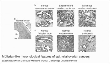
A major challenge to improving early detection of EOC is that it is not a single disease entity, but instead comprises a heterogeneous group of tumours. These tumours are classified according to their patterns of histological differentiation (Refs Reference Clements and Young3, Reference Seidman4, Reference Young and Scully5, Reference Kaku6). The most common histological subtype – serous – constitutes ∼50% of EOCs (Fig. 1c). Serous tumours range from cystic, papillary tumours to solid masses often with papillary surfaces, and histologically are very similar to cancers of the fallopian tube (Fig. 2). By contrast, endometrioid cancers are characterised by their endometrial-like glandular structures, and resemble their counterparts in the uterine corpus. Mucinous tumours are typically composed of glands and cysts lined by cells with mucin-rich cytoplasm that resemble epithelial cells of the endocervix or of the intestine. The prognosis for women with serous EOC is much poorer than for those with endometrioid and mucinous tumours. Serous carcinomas are often high-grade and bilateral, and are more frequently detected at an advanced stage. Endometrioid and mucinous tumours constitute 15–20% and 10% of EOCs, respectively (Fig. 1c). Less common EOC subtypes include clear-cell and transitional-cell (Brenner) tumours (Refs Reference Clements and Young3, Reference Seidman4, Reference Young and Scully5, Reference Kaku6).
Müllerian-like morphological features of epithelial ovarian cancers. (a) Hematoxylin–eosin-stained section of normal human ovary surrounded by monolayered epithelium (arrow) and containing an inclusion cyst (i.c.). (b) Sections of ovarian carcinomas: serous, with papillary features; endometrioid, with glandular features; and mucinous. (c) Sections of normal fallopian tube, endometrium and endocervix. Bar, 50 µm.

The traditional notion that EOCs originate from cells of the simple epithelium that lines the ovarian surface has been controversial. Much of the debate regarding the origin of EOCs stems from their histological complexity that seemingly defies traditional paradigms of tumour histogenesis and progression. Although morphological heterogeneity exists among tumours that arise from other organ sites, it is often observed that some tumours morphologically resemble the tissue of origin while many others appear less differentiated. By contrast, EOCs appear to paradoxically ‘differentiate up’ towards more committed epithelial phenotypes. The ovarian surface epithelium consists of a single layer of flat or cuboidal cells that closely resembles the mesothelium that lines the pelvic cavity (pelvic peritoneum) and the surfaces of abdominal organs (Fig. 2). Cells of the ovarian surface epithelium express several fibroblast markers (vimentin, and collagen types I and III) as well as markers characteristic of simple epithelium (keratin types 7, 8, 18 and 19) (Ref. Reference Auersperg7). These observations have led to the phenotype of the ovarian surface epithelium being described as ‘uncommitted’ (Refs Reference Auersperg8, Reference Feeley and Wells9). Unlike most other normal adult epithelia, the ovarian surface epithelium expresses very little or no E-cadherin (Refs Reference Maines-Bandiera and Auersperg10, Reference Davies, Worsley and Ponder11). However, E-cadherin is expressed in many EOCs, including tumour implants at distant sites (Refs Reference Davies, Worsley and Ponder11, Reference Inoue12, Reference Sundfeldt13). By contrast, loss of E-cadherin expression is widely observed in carcinomas of other organ sites, particularly in advanced-stage disease (Refs Reference Birchmeier and Behrens14, Reference Thiery15).
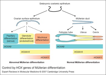
A striking aspect of the apparent ‘differentiating up’ of EOCs is their assumption of specialised morphological features of nonovarian tissue lineages. The epithelium that lines the ovarian surface is a modified mesothelium that derives from the coelomic epithelium (Fig. 3). By contrast, the epithelia of the female reproductive tract – that is, fallopian tubes, endometrium, endocervix and upper segment of the vagina – derive from the Müllerian ducts (Ref. Reference Kobayashi and Behringer16). The Müllerian ducts were originally described in the 19th century and have been thought to derive from the coelomic epithelium (Ref. Reference Auersperg8). A report published this year confirmed the coelomic epithelial origin of the Müllerian ducts by tracing the fate of coelomic epithelial cells that expressed green fluorescent protein (GFP) in developing chick and mouse embryos (Ref. Reference Guioli, Sekido and Lovell-Badge17). In other words, the ovarian surface epithelium and the specialised epithelia of the reproductive tract are of common embryonic origin, but evolve via distinct tissue lineage pathways (Fig. 3). The assumption by the three major subtypes of EOCs of morphological features that resemble those of the Müllerian-duct-derived epithelia seems therefore paradoxical in their misprogramming of differentiation pathways or ‘lineage-infidelity’. The same description can be applied to intestinal-like mucinous EOCs, transitional-cell tumours (showing urothelial-like features), and to clear-cell tumours, which were originally described as mesonephric but are now described as Müllerian (Ref. Reference Clements and Young3).
Control by HOX genes of Müllerian differentiation. The ovarian surface epithelium and Müllerian ducts derive from the embryonic coelomic epithelium. As the Müllerian ducts differentiate to form the fallopian tubes, uterus, cervix and upper vagina, HOX gene expression becomes spatially restricted. These patterns of HOX expression are recapitulated in serous, endometrioid and mucinous epithelial ovarian cancers according to their pattern of Müllerian-like differentiation (Ref. Reference Cheng56).

The disputed cell of origin
The ‘lineage-infidelity’ and ‘differentiating up’ of EOCs has prompted several alternative explanations as to the origin of these cancers. One widely accepted notion, based on histopathological observations, is that serous EOCs arise from cortical inclusion cysts. These inclusion cysts are thought to form from invaginations of the ovarian surface, and are typically lined by tubal epithelium that expresses E-cadherin (Ref. Reference Maines-Bandiera and Auersperg10). There is an increased frequency of cortical invaginations and inclusion cysts in the normal contralateral ovary of women with unilateral disease, as compared with ovaries of age-matched women without disease (Refs Reference Mittal18, Reference Tresserra19). Furthermore, surface epithelial pseudostratification as well as increases in cortical invaginations and inclusion cysts exhibiting papillary projections are frequently observed in overtly healthy ovaries of women with a family history of ovarian cancer (Ref. Reference Salazar20). It is also thought that some serous tumours that were diagnosed as of ovarian origin may actually arise from the fallopian tubes (Refs Reference Piek, Kenemans and Verheijen21, Reference Crum22). This notion largely stems from several histopathological and genetic studies of women with mutations in the BRCA breast and ovarian cancer susceptibility genes who have undergone prophylactic salpingo-oophorectomy, which indicate that cancers of the fallopian tube might not be as rare as originally thought (Refs Reference Colgan23, Reference Paley24, Reference Lee25). Furthermore, some mucinous EOCs are thought to be metastases of gastrointestinal tumours (Refs Reference Seidman4, Reference Young, Gilks and Scully26).
It has also been hypothesised that EOCs arise from the ‘secondary Müllerian system’ (Ref. Reference Dubeau27). This includes structures that exist in the ovarian hilum and in paratubal and paraovarian areas and are thought to represent remnants of the Müllerian ducts (Ref. Reference Feeley and Wells9). The strongest evidence that supports the ‘secondary Müllerian system’ hypothesis comes from studies of endometriosis. Endometriosis is frequently associated with endometrioid and clear-cell carcinomas, but not with serous or mucinous tumours (Refs Reference Sainz de la Cuesta28, Reference Ogawa29). A direct transition from endometriosis to malignancy has been reported in a third of cases (Ref. Reference Sainz de la Cuesta28). In a study of endometrioid and clear-cell carcinomas associated with endometriosis, 63 loss of heterozygosity events were detected in carcinomas and 22 of these were also detected in corresponding endometriosis (Ref. Reference Prowse30). Loss of heterozygosity at the 10q23.3 locus of the tumour suppressor PTEN and somatic PTEN mutations have been detected in endometrial cysts and in endometrial and clear-cell carcinomas (Ref. Reference Sato31). In a mouse genetic model by Dinulescu et al., activation of K-ras in the ovarian surface epithelium was found to induce peritoneal endometriotic lesions, whereas a combination of oncogenic K-ras and Pten loss induced tumours that closely resembled endometrioid carcinoma (Ref. Reference Dinulescu32). Although this study demonstrated a direct molecular link between endometriosis and endometrioid carcinoma, the findings were somewhat surprising since activating KRAS mutations have not been detected in endometriosis (Ref. Reference Vercellini33).
In recent years, three other mouse models have been generated in which tumours were directly induced by introducing specific genetic alterations into the ovarian surface epithelium. In one model, female transgenic mice expressing the transforming region of simian virus 40 under the control of the promoter of the Müllerian inhibitory substance type II receptor gene developed poorly differentiated tumours with some papillary structures at the surface (Ref. Reference Connolly34). Most tumours induced in another model, generated by concurrent inactivation of the tumour suppressors Rb1 and p53, were either undifferentiated or poorly differentiated, but a subset exhibited serous-like features (Ref. Reference Flesken-Nikitin35). Organised papillary projections were seen in some tumours induced in a third mouse model by activation of any two of the oncogenes K-ras, c-myc and Akt in a p53-deficient background (Ref. Reference Orsulic36). These findings have been interpreted as demonstrating the surface epithelial origin of EOCs (Ref. Reference Orsulic36). However, it could be argued that these studies merely demonstrate that ovarian surface epithelial cells are capable of undergoing malignant transformation if specific sets of genetic alterations occur.
Oncogenic events and their subtype specificity
Because EOCs exhibit a diverse spectrum of morphological features, it is not surprising that transcriptional profiles of the different histological subtypes are heterogeneous (Refs Reference Schwartz37, Reference Lu38, Reference Zorn39, Reference Heinzelmann-Schwarz40). It has not yet been determined whether the genes identified as subtype-specific in expression profiling studies are functionally significant to the histological differentiation patterns of the different subtypes. The difference in histopathology of tumours induced in the mouse genetic model of Dinulescu et al. (Ref. Reference Dinulescu32) and the three other models (Refs Reference Connolly34, Reference Flesken-Nikitin35, Reference Orsulic36) described above raises the question as to whether alterations in specific oncogenes and tumour suppressors in ovarian surface epithelial cells could give rise to different tumour subtypes. Several genetic alterations are more prevalent in one tumour subtype than in others. Mutations in p53 are the most frequently detected alteration in high-grade serous ovarian carcinoma, and have been detected in up to 80% of cases (Ref. Reference Shih and Kurman41). Mutations in the oncogenes KRAS and BRAF occur in approximately two-thirds of low-grade serous tumours but are rare in high-grade serous carcinomas (Ref. Reference Singer42). KRAS mutations also frequently occur in mucinous carcinomas (Ref. Reference Enomoto43). Mutations in PTEN and in the CTNNB1 gene that encodes β-catenin are more prevalent in endometrioid carcinomas (Refs Reference Obata44, Reference Palacios and Gamallo45). It is intriguing to note the prevalence of mutations in the histologically similar counterparts that arise from the uterine corpus. As likewise found for EOCs, the frequency of PTEN mutations is higher in the endometrioid subtype of endometrial carcinoma than in uterine papillary serous carcinomas (Ref. Reference Tashiro46). Conversely, p53 mutations are more prevalent in uterine papillary serous carcinomas and have been detected in approximately 90% of these cases (Ref. Reference Lax47).
There are several compelling arguments against the possibility that the tumour histotype might be defined per se by the specific combination of oncogenic events. First, many of the genetic alterations described above occur in histologically diverse cancers that arise from other organ sites. Second, there is considerable evidence that oncogenic transformation of a variety of cell types, including ovarian surface epithelial cells, is accompanied by loss of normal differentiation (Refs Reference Auersperg and Roskelley48, Reference Hoffman49). Third, although papillary structures were observed in a subset of tumours induced in three mouse genetic models of EOC (Refs Reference Connolly34, Reference Flesken-Nikitin35, Reference Orsulic36), the majority of tumours were poorly differentiated. As is the case for malignant EOCs, benign ovarian cystadenomas are categorised by their histological differentiation patterns, with serous and mucinous being the most prevalent subtypes (Ref. Reference Clements and Young3). However, mutations in p53, KRAS and BRAF have not been detected in solitary cystadenomas that are not contiguous to malignant tumour (Refs Reference Zheng50, Reference Cheng51). This strongly implies that the pathways that control malignant transformation are independent of those that control histological differentiation. The morphological heterogeneity of EOCs and their assumption of histological features of tissue lineages distinct from that of the surface epithelium might therefore not be solely explained by alterations in ‘classic’ oncogenic events and could involve other molecular mechanisms.
Determination of morphological identity by homeobox genes
Because the major EOC subtypes exhibit morphological features that resemble those of the Müllerian-duct-derived epithelia, their histogenesis might involve inappropriate activation of genes that normally regulate differentiation of the Müllerian ducts. Homeobox genes encode transcription factors that play essential roles in controlling cell differentiation during embryonic development (Refs Reference Mark, Rijli and Chambon52, Reference Pearson, Lemons and McGinnis53). These genes were first discovered in Drosophila as genes whose mutations caused homeotic transformation – a phenomenon in which body segments form in an inappropriate location or context, such as the formation of additional legs rather than antennae in gain-of-function mutants of the Antennapedia gene (Ref. Reference Gehring and Hiromi54). The 39 genes that constitute the mammalian HOX family of homeobox genes are tandemly arranged in four clusters located on different chromosomes and are aligned in 13 paralogous groups based on their sequence similarities and relative positions in the loci (Refs Reference Mark, Rijli and Chambon52, Reference Pearson, Lemons and McGinnis53). The control of developmental patterning by HOX genes is linked to their colinearity, whereby the spatial and temporal expression patterns of HOX genes along the anterior–posterior body axis is coupled to their relative position in the gene cluster. HOX genes at the 3' end of the clusters are generally expressed early in development and in anterior body regions, whereas those at the 5' end of clusters are expressed later and in more posterior regions (Refs Reference Mark, Rijli and Chambon52, Reference Pearson, Lemons and McGinnis53).
Colinearity is well exemplified in the developing reproductive tract. During early stages of development in the mouse, the tandemly arranged Hoxa9, Hoxa10, Hoxa11 and Hoxa13 genes are uniformly expressed along the axis of the Müllerian duct. As Müllerian duct differentiation progresses, Hoxa9 becomes expressed in the primordia of the fallopian tubes, Hoxa10 in the developing uterus, Hoxa11 in regions destined to become the lower uterine segment and cervix, and Hoxa13 in the future upper vagina (Ref. Reference Taylor, Vanden Heuvel and Igarashi55) (Fig. 3). Expression of these HOX genes remains spatially restricted in the adult reproductive tract in mouse and in humans (Refs Reference Taylor, Vanden Heuvel and Igarashi55, Reference Cheng56). Most of our current understanding of the functions of these HOX genes has come from studies of knock-out mice. Female mice with a targeted disruption of Hoxa10 or of Hoxa11 produce normal embryos, but their uteri exhibit impaired glandular development and are unable to support embryo implantation (Refs Reference Benson57, Reference Gendron58). Moreover, Hoxa10-deficient female mice exhibit homeotic transformation of the anterior segment of the uterus into oviductal-like structures (Ref. Reference Benson57).
In the study of Cheng et al. (Ref. Reference Cheng56), expression of HOXA9, HOXA10 and HOXA11 was not detected in normal human ovarian surface epithelium. However, colinear expression patterns of these HOX genes that normally occur in Müllerian-duct-derived epithelia were found to be recapitulated in serous, endometrioid and mucinous EOCs according to the pattern of Müllerian-like differentiation of these tumours (Fig. 3). Stable expression of the HOX genes in undifferentiated, transformed mouse ovarian surface epithelial cells, and intraperitoneal propagation of these transfected cells in female mice, showed that cells derived from ovarian surface epithelium undergo differentiation along distinct Müllerian lineages when specific HOX genes are aberrantly expressed (Ref. Reference Cheng56). Mouse ovarian surface epithelial cells that stably expressed Hoxa9 formed cystic, papillary tumours that resembled high-grade serous carcinoma; in contrast, expression of Hoxa10 and Hoxa11 induced formation of high-grade endometrioid-like and mucinous-like tumours, respectively. Furthermore, the mouse tumours exhibited patterns of marker expression similar to those of clinical specimens. Mucin 1, claudin-3 and mesothelin, which are more highly expressed in nonmucinous than in mucinous EOCs, were found to be more highly expressed in the papillary tumours derived from Hoxa9-transfected cells than in the mucinous-like tumours derived from Hoxa11-transfected cells. Conversely, mucin 5AC and trefoil factor 1 (TFF1), which are preferentially expressed in mucinous EOCs, were expressed in Hoxa11-transfected cells, but absent from those expressing Hoxa9. In addition, the HOXA7 gene, a neighbour of HOXA9, was found to be expressed in normal tubal, endometrial and endocervical epithelia, but not in normal ovarian surface epithelium (Ref. Reference Cheng56). HOXA7 expression was detected in all three major subtypes of EOC, but its expression was significantly higher in low-grade than in high-grade tumours. Studies in which Hoxa7 was stably expressed in tumourigenic mouse ovarian surface epithelial cells revealed that Hoxa7, while having no Müllerian-lineage-specificity when expressed alone, promoted the abilities of Hoxa9, Hoxa10 and Hoxa11 to induce tumour differentiation along their respective pathways (Ref. Reference Cheng56).
It can be argued that the recapitulation in EOCs of the HOX gene program that normally occurs in Müllerian-duct-derived epithelia might reflect the Müllerian origin of these tumours. However, the demonstration that transformed ovarian surface epithelial cells can differentiate along distinct Müllerian lineage pathways when specific HOX genes are activated (Ref. Reference Cheng56) provides a molecular explanation for the morphological heterogeneity of tumours that derive from surface epithelial cells and their assumption of Müllerian-like features. This study should not be interpreted to prove the surface epithelial origin of EOCs. However, it provides an explanation for the Müllerian-like features of EOCs – the prior lack of which formed the basis of arguments against the surface epithelial origin of these tumours (Ref. Reference Dubeau27).
The finding that colinearity of HOX expression is recapitulated in EOCs might explain why serous tumours are more abundant than the endometrioid and mucinous subtypes (Fig. 1c; Fig. 3). The sequence of HOX expression might also explain why serous tumours can be distinguished from mucinous tumours by their global gene expression profiles, whereas endometrioid tumours show overlap with the other two subtypes (Refs Reference Schwartz37, Reference Heinzelmann-Schwarz40). Although a mechanism for the unique features of clear-cell EOC has not been identified, the observation that clear-cell tumours express HOXA9 and HOXA10 is consistent with the overlapping histological features that this subtype shares with serous and endometrioid tumours (Ref. Reference Cheng56). Gene expression profiles of clear-cell EOCs have been found to be remarkably similar to profiles of clear-cell carcinomas of renal origin (Ref. Reference Zorn39). It is possible that histological differentiation of clear-cell EOCs might involve activation of genes that normally control urogenital patterning. Several studies have reported that CDX2, a homeobox gene that normally controls intestinal differentiation, is expressed in intestinal-like mucinous EOCs, but not in other subtypes (Refs Reference Fraggetta59, Reference Groisman, Meir and Sabo60). The ‘lineage-infidelity’ of EOCs that exhibit intestinal-like features might also be explained by inappropriate activation of CDX2 in cells of ovarian origin.
Aberrant differentiation and stem-cell-like properties
One interpretation of the HOX gene study is that it implies that ovarian surface epithelial cells are pluripotent (Ref. Reference Naora61). Indeed, as discussed earlier, normal adult ovarian surface epithelial cells have been described as having an ‘uncommitted’ phenotype, and these cells exhibit several features that are characteristic of stem cells. The key hallmarks – self-renewal capability and pluripotency – are properties that are important for normal post-ovulatory repair of the ovarian surface (Ref. Reference Auersperg8). It has been speculated by Auersperg and colleagues that the ability of ovarian surface epithelial cells to undergo epithelial–mesenchymal transition enables cells to migrate across the ruptured surface and also enables incorporation of displaced cells into the ovarian stroma (Ref. Reference Auersperg8). Ovarian surface epithelial cells that fail to undergo epithelial–mesenchymal transition and become trapped within the ovarian cortex might form inclusion cysts (Ref. Reference Auersperg8).
Although the morphological heterogeneity of EOCs might be attributed to the intrinsic stem-cell-like properties of adult ovarian surface epithelial cells, it can be also argued that morphological heterogeneity is not a phenomenon unique to EOCs. For example, there exist multiple histological types of breast cancer, with ductal carcinoma accounting for approximately 75% of invasive breast cancers (Ref. Reference Li, Uribe and Daling62). Less common types of breast cancer include lobular, mucinous, tubular and medullary carcinomas (Ref. Reference Li, Uribe and Daling62). It is becoming increasingly recognised that many solid tumours, as well as haematological malignancies, arise from a small population of tumour-initiating cells that have the self-renewal capability and differentiation potential of normal tissue stem cells (Refs Reference Bonnet and Dick63, Reference Al-Hajj64, Reference Singh65). Indeed, a cell population isolated from malignant ascites and expressing markers of multiple ovarian lineages – that is, ovarian surface epithelium, granulosa cells and germ cells – has been identified as putative ‘ovarian cancer stem cells’ (Ref. Reference Bapat66). In another recent study, side populations of cells with stem-cell-like features were isolated from lines derived from two mouse genetic models of EOC (Ref. Reference Szotek67). The notion that EOCs arise from ‘ovarian cancer stem cells’ that are capable of differentiating along different lineages might not only explain the existence of multiple tumour subtypes, but also explain why some tumours exhibit mixed histological features. It is as yet unclear, particularly in solid tumours, as to whether ‘cancer stem cells’ arise by transformation of normal tissue stem cells, or from lineage-restricted progenitors or even mature, differentiated cells. Despite uncertainty as to the actual origin of ‘cancer stem cells’, their discovery has led to a significant paradigmatic shift towards viewing tumours as undergoing a hierarchical development similar to their normal tissue counterparts (Refs Reference Pardal, Clarke and Morrison68, Reference Sell69, Reference Miller, Lavker and Sun70). An implication of this conceptual shift is that the so-called ‘loss’ of differentiation that is histologically observed in many tumours may not be actual loss, but rather reflects incomplete differentiation or maturation arrest of a cell lineage derived from a ‘cancer stem cell’. Therefore, it could be argued that the ‘differentiating up’ of EOCs is not actually a paradox, but rather is a striking example of aberrant recapitulation by tumours of organogenesis.
The advantages of ‘differentiating up’
Although the heterogeneity of EOCs might be rationalised either by the plasticity of adult ovarian surface epithelial cells or by the pluripotency of ‘cancer stem cells’, one unresolved question is the cellular preference for ‘differentiating up’ along the Müllerian lineages. The predominance of Müllerian-like phenotypes in these tumours, rather than phenotypes of other coelomic epithelium derivatives, has been thought to reflect the close primordial relationship between the ovarian surface epithelium and the Müllerian ducts (Ref. Reference Auersperg8) (Fig. 3). By contrast, rarer tumour subtypes have histological features that resemble those of tissues whose embryonic relationship to the ovarian surface epithelium is more distant. One example is the Brenner tumour, a rare form of EOC that is characterised by urothelial-like morphological features (Ref. Reference Clements and Young3). Only 5% or fewer EOCs are classified as undifferentiated (Fig. 1c). This suggests that Müllerian-like differentiation might confer selective advantages on transformed ovarian surface epithelial cells. Unlike the epithelia of the reproductive tract, the normal ovarian surface epithelium has only a tenuous attachment to underlying stromal components. Müllerian differentiation of ovarian surface epithelial cells has been speculated to enhance epithelial–stromal interactions that support malignant growth and to increase responsiveness of cells to growth factors and hormones (Ref. Reference Auersperg8). For example, oestrogen is mitogenic for tubal and endometrial epithelia, but not for normal ovarian surface epithelial cells (Ref. Reference Karlan71). Homophilic cell adhesion may prevent anoikis in EOC cells in the pelvic cavity. Indeed, it has been observed that a significant proportion of EOC cells in ascites of patients exist as multicellular aggregates (Ref. Reference Burleson72).
Although loss of E-cadherin expression and of epithelial differentiation are widely associated with invasive behaviour of most carcinomas, many EOCs, including tumour implants at distant sites, have been found to express E-cadherin (Refs Reference Davies, Worsley and Ponder11, Reference Inoue12, Reference Sundfeldt13). ‘Differentiating up’ might provide growth and survival advantages for EOC cells, but this process seems at odds with well-established paradigms of metastasis. One explanation for this paradox might lie in the differences between dissemination of EOC and metastasis of many other types of epithelial cancers. ‘Classic’ metastasis occurs as a sequence of events involving invasion of surrounding tissues, crossing of the vascular endothelium, survival of tumour cells in the circulation, arrest at a distant site, and extravasation into the surrounding tissue (Refs Reference Fidler73, Reference Pantel and Brackenhoff74). Although EOC can spread by directly extending to adjacent organs, its most widely recognised behaviour is the ‘seeding’ of the pelvic cavity with nests of tumour cells that implant on the pelvic peritoneum and serosal surfaces of abdominal organs (Ref. Reference Naora and Montell75). This mode of dissemination involves transportation of exfoliated tumour cells throughout the pelvic cavity by normal peritoneal fluid and is – in contrast to metastasis of many other epithelial tumours – a somewhat passive process (Ref. Reference Naora and Montell75). EOC cells may not need to undergo epithelial–mesenchymal transition to disseminate by intraperitoneal seeding and, as discussed above, may need to retain epithelial features in order to survive in the pelvic cavity. It is intriguing to note that other types of carcinomas are thought to undergo mesenchymal–epithelial transition once they reach the secondary site in order to establish and grow at this site (Ref. Reference Thiery15). In this regard, EOCs might not mechanistically differ from other types of epithelial tumours in their establishment at secondary sites.
Concluding remarks
Much of our lack of understanding of EOCs has stemmed from the conundrum posed by their apparent ‘differentiating up’ and ‘lineage infidelity’. When viewed in the light of recent molecular studies and emerging paradigms of tumour histogenesis and progression, this baffling class of tumours seems less paradoxical. The different EOC subtypes should be regarded as separate disease entities driven by distinct sets of molecular events. The recognition of the molecular heterogeneity of EOCs is driving current initiatives to develop multiplexed diagnostic assays comprising panels of complementary serum biomarkers. Although improving early detection of EOC will greatly reduce the morbidity and mortality caused by this disease, there is an urgent need for new-generation targeted therapeutic agents. Functional studies to define molecular targets that are the focal points of multiple pathways that drive persistent EOC growth and dissemination therefore represent an important endeavour.
Acknowledgements and funding
The author is currently funded by NIH grant R01 CA101826 and by US Department of the Army grants DAMD 17-03-1-0219 and W81XWH-06-1-0259. The author thanks Dr J. Liu (M.D. Anderson Cancer Center, Houston, TX, USA) for providing tissue specimens shown in Figure 2, and the peer reviewers for their helpful comments.
Note added in proof
In a study recently published in Cancer Cell (Ref. Reference Wu76), Cho and colleagues report that conditional inactivation of the Pten and Apc tumour suppressor genes in the mouse ovarian surface epithelium induces formation of tumours that morphologically resemble human ovarian endometrioid carcinoma.