Dijkerman & de Haan (D&dH) suggest that, analogous to the organisation of the visual system, somatosensory processing for guiding actions (terminating in the posterior parietal cortex [PPC]) can be dissociated from the processing that leads to perception and memory (going through the secondary somatosensory cortex and terminating both in the insula and in the PPC).
In the visual domain, the clinical conditions of optic ataxia and visual form agnosia stand for two separate streams of visual information: dorsal stream for action and ventral stream for perception. Analogously, in the haptic domain a similar dissociation of information processing for external tactile stimuli can be proposed: an action stream represented by tactile apraxia and a perception stream represented by tactile agnosia. As suggested by D&dH, there is also clinical evidence that, despite the parallel processing, the haptic streams for perception and action interact tightly because of functional and anatomical overlaps inside the PPC. Accordingly, both haptic perception of objects and somatosensory guidance of action are processed in the anterior part of the PPC: the human anterior intraparietal area (AIP).
AIP and action (tactile apraxia): Interaction with the ventral premotor cortex. Tactile apraxia is defined as an affection of explorative finger movements in absence of paresis or sensory deficits (Klein Reference Klein1931). The finger movements seem uncoordinated and inadequate to the size and the shape of objects to be explored (Binkofski et al. Reference Binkofski, Kunesch, Classen, Seitz and Freund2001). Typically, the intransitive (not object related) and expressive movements (gestures) are well preserved. Functional imaging studies show that manipulative finger movements involve activation of the AIP, the superior parietal lobule, the secondary somatosensory area, and the ventral premotor cortex (vPMC) (Binkofski et al. Reference Binkofski, Buccino, Posse, Seitz, Rizzolatti and Freund1999a; Jäncke et al. Reference Jäncke, Kleinschmidt, Mirzazade, Shah and Freund2001). Especially the AIP is processing dynamic parameters of ongoing hand actions (Tunik et al. Reference Tunik, Frey and Grafton2005). Electrophysiology in monkeys showed that AIP neurons discharge during object holding and manipulation (Sakata et al. Reference Sakata and Kusunoki1992; Reference Sakata, Taira, Murata and Mine1995) and that those “motor dominant” neurons discharge during hand-related actions in the dark (Murata et al. Reference Murata, Gallese, Kaseda and Sakata1996). The tight interaction between the AIP and the vPMC for coordination of goal-directed hand movements has been already proposed on the basis of monkey data (Jeannerod et al. Reference Jeannerod, Arbib, Rizzolatti and Sakata1995; for review, see also Rizzolatti & Lupino Reference Rizzolatti and Luppino2001; Rizzolatti et al. Reference Rizzolatti, Luppino and Matelli1998). Studies on human lesion provide another argument in favour of this interaction in the haptic domain. Lesions involving the AIP, contralateral to the affected hand, cause tactile apraxia (Binkofski et al. Reference Binkofski, Kunesch, Classen, Seitz and Freund2001; Valenza et al. Reference Valenza, Ptak, Zimine, Badan, Lazeyras and Schnider2001). Furthermore, lesions of the frontal operculum, containing the vPMC, can also impair finely tuned finger movements in a condition resembling tactile apraxia, that is, limb kinetic apraxia (Binkofski & Fink Reference Binkofski and Fink2005; Binkofski et al. Reference Binkofski, Kunesch, Classen, Seitz and Freund2001; Dettmers et al. Reference Dettmers, Liepert, Hamzei, Binkofski and Weiller2003). Thus, there is converging evidence that the AIP constitutes the nodal point of a network for coordination and integration of sensorimotor information for action.
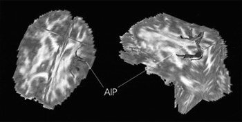
The AIP and perception (tactile agnosia): Links with inferior temporal cortex. For object recognition from tactile stimulation, elementary cutaneous and proprioceptive sensations from the exploratory hand have to be integrated to more complex information reflecting textural and spatial patterns of a specific object. O'Sullivan et al. (Reference O'Sullivan, Roland and Kawashima1994) and Roland et al. (Reference Roland, O'Sullivan and Kawashima1998) found evidence for different information streams, depending on the somatosensory submodalities from positron emission tomographic activation studies of the human brain. Roughness discrimination activated the lateral opercular cortex, but shape and length discrimination activated the AIP. Accordingly, Bohlhalter et al. (Reference Bohlhalter, Fretz and Weder2002) reported two patients with tactile agnosia. One patient had a lesion that affected mostly the anterior parietal lobe and caused a pure aperceptive agnosia, and the second had agnosia for more complex features (like shape) following a lesion of the anterior intraparietal regions (Hoemke et al., in press). This is consistent with neuroimaging data in human patients showing that the AIP is involved in tactile, visual, and transmodal processing of object features (Grefkes et al. Reference Grefkes, Weiss, Zilles and Fink2002). The lateral occipital cortex (part of the inferotemporal cortex) is known to be important not only for visual, but also for tactile, object recognition (Amedi et al. Reference Amedi, Malach, Hendler, Peled and Zohary2001) and maybe even for the supramodal object recognition (Binkofski et al. Reference Binkofski, Buccino, Zilles and Fink2004). Anatomical connections between the intraparietal and inferotemporal areas have been found by the Van Essen group of researchers (see Lewis & Van Essen Reference Lewis and Van Essen2000) in monkeys, and such connections seem also to exist in people (Figure 1).
Diffusion-tensor tractography data showing anatomical connections of the anterior intraparietal area (AIP) with primary sensory, ventral premotor, posterior intraparietal, and inferior temporal areas. (Figure from Pisella et al. Reference Pisella, Binkofski, Lasek, Toni and Rossetti2006, with permission)

AIP the node. Tactile apraxia and tactile agnosia can occur separately, indicating a possible dissociation. Indeed, clinical cases have been reported of patients with tactile agnosia who had intact exploratory hand movements and preserved basic somatosensory functions (Platz Reference Platz1996; Reed & Caselli Reference Reed and Caselli1994; Reed et al. Reference Reed, Caselli and Farah1996). In contrast, as mentioned by D&dH, Valenza et al. (Reference Valenza, Ptak, Zimine, Badan, Lazeyras and Schnider2001) described a patient with pure tactile apraxia who had an intact ability to perceive passively applied stimuli, but showed severe impairments when required to use exploratory finger movements for object identification.
However, Delay (Reference Delay1935), while describing tactile apraxia, had already referred to the association between apractic and agnostic deficits. Accordingly, tactile apraxia is often accompanied by deficits in tactile recognition of objects (or astereognosia) (Binkofski et al. Reference Binkofski, Kunesch, Classen, Seitz and Freund2001; Wernicke Reference Wernicke1876). This underpins the tight interaction of the action and the perception systems. Object characteristics trigger appropriate finger movement patterns that serve the perception of object features (action for perception – “feedback”). At the same time, the object properties determine the movement extent – perception in the service of action (perception for action – “feed forward”) (Lederman & Klatzky Reference Lederman and Klatzky1997). As shown by Bodegard et al. (Reference Bodegard, Geyer, Grefkes, Zilles and Roland2001), the AIP is activated by both passive and active tactile shape discrimination.
Because the AIP receives somatosensory and visual information about objects and is interconnected with the vPMC for planning actions towards the objects and with the inferior temporal cortex for recognising the objects, this area (or its human homologue) seems to constitute one of the most important interfaces between action and perception in the haptic system.