Introduction
Numerous nanoporous materials can be obtained via dealloying. Among these, nanoporous gold (NPG) has stood apart for use in bioanalytical and biomedical applications. This can be attributed to its many desirable properties, including high effective surface area, good electrical conductivity, easy surface modification via thiol-gold linker chemistry, tunable pore morphology, and compatibility with conventional microfabrication. Initial interest in NPG has centered on its use as a versatile material to study structure–property relationships in the context of size-dependent mechanical properties, as well as catalytic and energy-storage applications. Recently, there has been a surge of interest in NPG’s applications in biomedical sciences, as it has offered unique solutions to standing issues in the fields of biosensors and neural interfaces. This article aims to provide a review of NPG-based biosensor and neural interface operation and challenges, as well as opportunities offered by NPG formed by dealloying.
Bioanalytical applications
In most bioanalytical systems, the goal is to sense and extract molecule(s) of interest with high sensitivity and selectivity. We initially describe the considerations for sensing and later extend the discussion to bioseparation applications, as both have significant mechanistic commonalities.
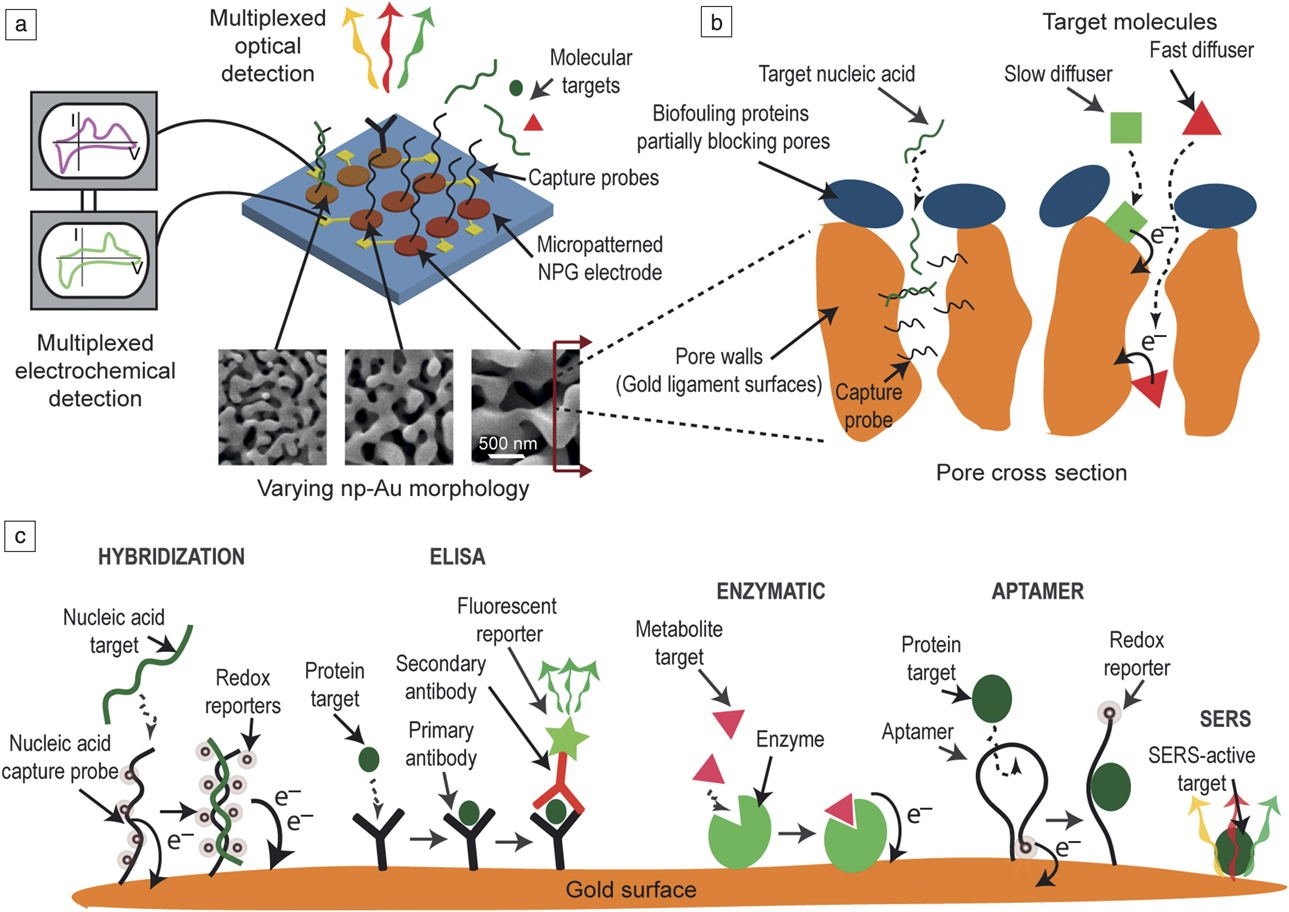
A typical biosensor architecture can be generalized to a capture probe immobilized on a solid support, a target biomarker in liquid or gas medium, and a reporter molecule that mediates the transduction of the capture event to an easily measurable signal (e.g., optical, electrical) (Figure 1a). For example, in the case of a nucleic acid-based sensor, the capture probe could be a short nucleic acid with a sequence complementary to the target nucleic acid biomarker (e.g., DNA or RNA). In this case, the solid support (e.g., nanoporous gold) would be decorated with the capture probe. In order for the sensing event to occur, the target biomarker would need to react with the capture probe, which requires transport of the target biomarker to the capture probe on the electrode from the solution (e.g., blood sample). This capture reaction may be transduced via additional reporter molecules (e.g., double-stranded specific fluorescent markers) that bind to DNA only in its double-stranded form and emit a fluorescence signal that can be captured by an optical detector.
Schematic illustration of nanoporous gold (NPG)-based biosensors. (a) Multiple electrode NPG arrays can be created via microfabrication processes, allowing for both optical and electrochemical detection modalities. (b) The tunable porosity permits biofouling-resilient sensing and enhanced selectivity in electrochemical detection of molecules with different diffusivities and charge-transfer rates. (c) A variety of different capture probes can be immobilized onto the NPG surfaces, allowing for different sensing schemes. Note: SERS, surface-enhanced Raman spectroscopy; ELISA, enzyme-linked immunosorbent assay.
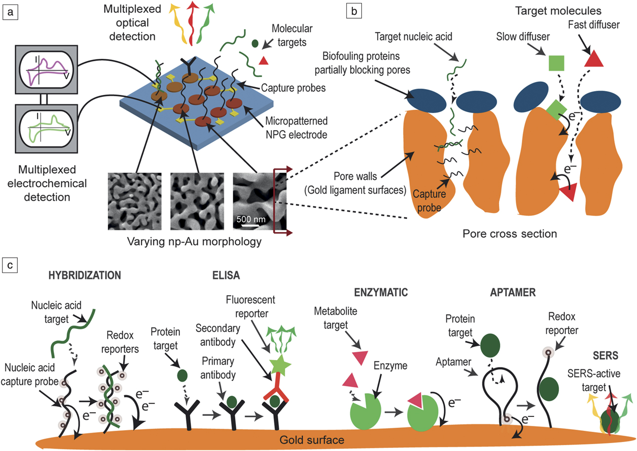
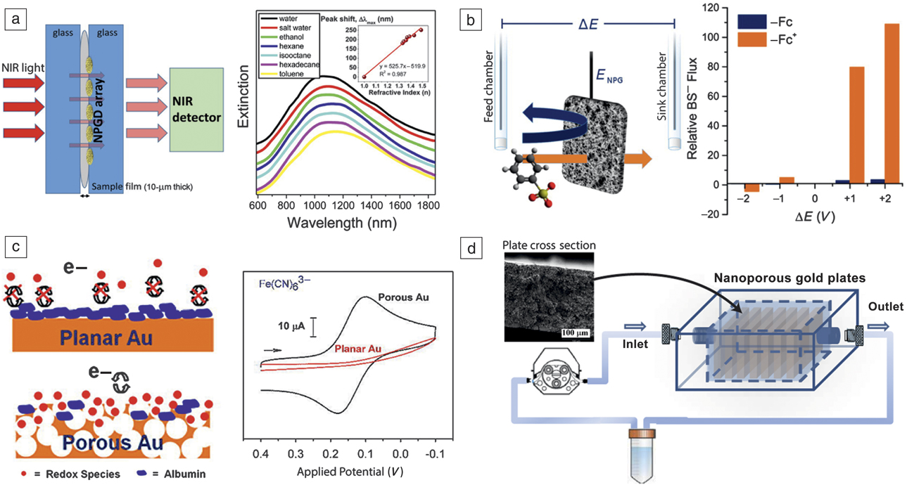
The transport and reaction processes, as well as the effectiveness of the transduction component, constitute the key underlying mechanisms for sensor operation and performance. Much work has been devoted to enhancing various aspects of these processes. There are some performance metrics common to most sensors, with the limit of detection and selectivity/specificity generally being more important ones. In this section, we start by introducing how capture biomolecules are immobilized onto NPG, describe common biosensor architectures, and exemplify transduction modalities, with a focus on enhancing sensor performance (Figure 1 and Figure 2).
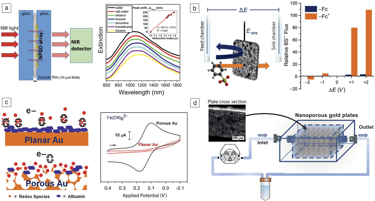
Examples of nanoporous gold (NPG)-based bioanalytical and bioseparation techniques. (a) Normal-incidence extinction spectra of 350-nm diameter NPG disks (NPGD) sandwiched between glass slides and excited with near-infrared (NIR) light for detecting refractive-index change due to various solvents.Reference Shih, Santos, Zhao, Zenasni and Arnob27 (b) Selective permeation of small molecule analytes controlled by modulating the surface charge of NPG membrane via electrochemical modulation of a redox-active self-assembled monolayer (SAM). BS– is benzene sulfonate, Fc is neutral (ferrocenyl)hexanethiol and Fc+ is its oxidized cationic form, ΔE is the applied electrochemical potential difference between feed and sink chambers separated with NPG membrane, and E NPG is the applied potential on NPG to control SAM redox reactions. Reproduced with permission from Reference McCurry and BaileyReference 28. © 2016 American Chemical Society. (c) NPG electrodes exhibit ideal electrochemical signal when immersed in biofouling solutions containing potassium ferricyanide, while significant fouling was evident in the electrochemical response at planar gold. Reproduced with permission from Reference Patel, Radhakrishnan, Zhao, Uppalapati, Daniels, Ward and CollinsonReference 17. © 2013 American Chemical Society. (d) A flow-through system consisting of NPG monolith plates (inset: cross section of a plate) decorated with carbohydrates to assist in the capture of lectins in perfused solution.Reference Tan, Fujikawa, Pornsuriyasak, Alla, Ganesh, Demchenko and Stine29
Probe immobilization and target molecule capture
A wide range of biomolecular capture probes have been immobilized onto NPG, including enzymes, single-stranded DNA, aptamers, antibodies, and lectins (Figure 1c). Immobilization onto NPG has been achieved by simple physisorption within the porous structure,Reference Wu, Sun, Li, Liu, Du, Wang and Xu1 by physisorption/entrapment together with a polymer,Reference Salaj-Kosla, Pöller, Beyl, Scanlon, Beloshapkin, Shleev, Schuhmann and Magner2 cross-linking after physisorption,Reference Sanzó, Taurino, Antiochia, Gorton, Favero, Mazzei, De Micheli and Carrara3 direct thiol–Au interaction,Reference Yan, Wang, Zhao, Zhang, Xu and Ding4 and by conjugation to activated terminal ester groups of self-assembled monolayers (SAMs).Reference Xiao, Li, Wang, Zhang and Si5
Enzymes immobilized on NPG by physisorption have been found to better retain their stability on NPG than when free in solution, as has been reported for laccase, a copper-containing oxidase enzyme.Reference Qiu, Xu, Huang, Ding, Qu and Gao6 While some redox enzymes have been adsorbed alone and reported to display direct electron transfer,Reference Qiu, Xu, Huang, Ding, Qu and Gao6 others have been co-adsorbed together with redox polymers.Reference Salaj-Kosla, Scanlon, Baumeister, Zahma, Ludwig, Conghaile, MacAodha, Leech and Magner7 Enzymes have been physisorbed onto NPG together with cross-linking polymers, or have been cross-linked after physisorption.Reference Siepenkoetter, Salaj-Kosla and Magner8 Enzymes such as laccase have also been covalently linked to SAMs formed from lipoic acid activated to produce reactive esters that form an amide linkage to a lysine residue on the enzyme.Reference Qiu, Xu, Huang, Ding, Qu and Gao9
Thiolated single-stranded DNA oligonucleotides have been directly assembled onto NPG,Reference Daggumati, Appelt, Matharu, Marco and Seker10 as have thiolated aptamers.Reference Zhang, Chang, Hirata, Wu, Xue and Chen11 In the case of DNA and aptamer immobilization, subsequent exposure to a short chain alkanethiol such as mercaptohexanol has been applied to cover exposed sites, limit nonspecific adsorption, and promote better orientation of the DNA strand. Immobilization of antibodies has been achieved by physisorption followed by exposure to bovine serum albumin to block nonspecific adsorption.Reference Fan, Guo, Gao, Zhang, Fan, Ji, Du and Wei12 Antibodies have also been immobilized by conjugation to SAMs presenting activated ester groups for amide bond formation.Reference Li, Wang and Zhang13 Lectins (with specific sugars as targets) have been immobilized onto nanoporous gold modified by activated lipoic acid SAMs.Reference Alla, d’Andrea, Bhattarai, Cooper, Tan, Demchenko and Stine14 Reagents such as dithiobis(succinimidyl propionate) are available for direct formation of an activated SAM without need for the activation step.Reference Ding, Li, Hu and Lin15 The diversity of biomolecular probes that can be attached onto NPG offers the ability to capture a rich set of target biomolecules (e.g., nucleic acids, proteins, carbohydrates, metabolites, small molecules) (Figure 1c).
Once a solution containing the target molecule is introduced, the target molecule needs to reach the capture probe, which is a challenge in nanoporous metals due to the tortuous path the molecule needs to travel to reach the capture probes immobilized within deeper pores. This transport is generally driven by a diffusion-like process hindered by surface–molecule interactions. The transport process can be enhanced by convection (e.g., flow-through cells);Reference Alla, d’Andrea, Bhattarai, Cooper, Tan, Demchenko and Stine14 however, there are large backpressures involved. On the other hand, the nanoporous morphology offers additional attributes that assist with the capture process. For example, tunable morphology allows for introducing cracks in the film so target molecules can permeate the NPG both from top and sides. The transport phenomenon (i.e., diffusion- versus reaction-controlled processes) in NPG electrodes has been shown to enhance sensor selectivity (e.g., detection of dopamine in the presence of interfering ascorbic acid).Reference Qiu, Zhou, Ji, Zhang, Huang and Ding16 Another challenge is the presence of biofouling molecules, such as proteins that can nonspecifically adsorb on electrodes, blocking charge transfer between the electrode and reporter redox molecules. Several groups have reported that NPG films are highly biofouling-resilient and are able to sustain electrochemical function in protein solutions.Reference Daggumati, Appelt, Matharu, Marco and Seker10,Reference Patel, Radhakrishnan, Zhao, Uppalapati, Daniels, Ward and Collinson17 This has been attributed to NPG morphology at a certain pore size (tens of nanometers), which blocks the permeation of large proteins while still allowing the transport of small ions and reporter molecules, effectively serving as a biofouling-resilient electrode (Figure 1b and Figure 2c).
Reporter molecules and transduction modalities
The majority of transduction events involving NPG have been carried out by either optical (e.g., plasmonic signals, fluorescence, color change) or electrochemical outputs (Figure 1a). One general detection strategy is variations of the enzyme-linked immunosorbent assay protocol,Reference Fan, Guo, Gao, Zhang, Fan, Ji, Du and Wei12 where a primary antibody immobilized onto NPG captures the target molecule, followed by a secondary antibody attaching to the target. The secondary antibody is tagged with a reporter molecule such as a fluorescent molecule, quantum dot, or enzyme (e.g., horseradish peroxidase) that leads to a color change in a solution. Similarly, the tag could be an electroactive molecule such as a redox reporter (e.g., methylene blue) where an electrical current is generated at a specific applied potential.Reference Daggumati, Appelt, Matharu, Marco and Seker10
On the plasmonics front, NPG thin films have recently captured intense attention as plasmonic materials with propagating surface plasmon resonance (SPR),Reference Yu, Ahl, Caminade, Majoral, Knoll and Erlebacher18 localized surface plasmon resonance (LSPR),Reference Lang, Qian, Guan, Zi and Chen19 and use as a substrate for surface-enhanced Raman scattering.Reference Biener, Nyce, Hodge, Biener, Hamza and Maier20,Reference Qian, Yan, Fujita, Inoue and Chen21 For each plasmonic modality, the resonant frequency exhibits a strong pore-size dependence, which can be optimized by leveraging the highly tunable morphology of NPG. In order to add an additional length scale to improve coupling between light and plasmons, NPG nanoparticles/nanodisks were fabricated to yield large surface areas, tunable plasmonic resonances, and high-density hot spots.Reference Zhao, Zeng, Parvez Arnob, Sun, Qi, Motwani, Gheewala, Li, Paterson, Strych, Raja, Willson, Wolfe, Lee and Shih22,Reference Wi, Tominaka, Uosaki and Nagao23 These nanoporous disks have been integrated with microfluidic devices for chemical and biological sensing.Reference Zeng, Zhao, Li, Li, Lee and Shih24 A significant advantage of plasmonics-based techniques is that they allow for label-free detection, circumventing the need for reporter molecules. To that end, there are several different label-free detection options available, such as leveraging LSPR’s sensitivity to local refractive index changes near the metal surface (so-called LSPR spectroscopy) due to target molecule adsorption. Beyond refractive index sensing, electromagnetic field localization and enhancement near the nanostructures have been demonstrated to have profound impact on a variety of light–matter interactions, such as surface-enhanced Raman spectroscopy,Reference Biener, Nyce, Hodge, Biener, Hamza and Maier20,Reference Qian, Yan, Fujita, Inoue and Chen21 surface-enhanced infrared absorption,Reference Wang, Kundu and Halas25 surface-enhanced fluorescence,Reference Zhang, Song, Fujita, Zhang, Chen and Wang26 and surface-enhanced near-infrared absorption (Figure 2a).Reference Shih, Santos, Zhao, Zenasni and Arnob27 Each of the transduction modalities described here possess different strengths. For example, electrochemical transduction allows for easy integration with electronics for point-of-care applications, while plasmonics-based methods allow for label-free detection.
Bioseparation modalities
The tunable pore morphology and customizable surface functionalization of NPG has begun to be explored in applications that involve separations. NPG is well suited for flow of solutions through its internal pore structure. NPG monoliths of 0.25-mm thickness have been used for selectively regulating the transport of model analytes across them.Reference McCurry and Bailey28 The transport of methyl viologen (MV2+) or benzene sulfonate (BS–) across NPG monoliths was monitored spectroscopically and found to be strongly dependent on the applied potential and on the ionic strength. The transport behavior was further regulated by modification of the NPG monoliths with alkanethiols with pH-sensitive terminal groups (–NH2 or –COOH). The transport was sensitive to both pH and applied potential. NPG monoliths modified by alkanethiols with terminal mannose (a monosaccharide sugar of the aldose group) units were useful for capture and release of the lectin concanavalin A, a carbohydrate-binding protein originally extracted from the jack-bean, Canavalia ensiformis.Reference Tan, Fujikawa, Pornsuriyasak, Alla, Ganesh, Demchenko and Stine29
The surface coverages of SAMs within the monoliths of bound lectin were determined using thermogravimetric analysis. NPG monoliths modified by lipoic acid and linked to the concanavalin A were suitable for selective capture and release of the mannose-rich glycoprotein ovalbumin.Reference Alla, d’Andrea, Bhattarai, Cooper, Tan, Demchenko and Stine14 In each case, release of protein was achieved by flow of free mannose ligand through the monolith. Solid-phase microextraction experiments using a nanoporous gold fiber combined with analysis by gas chromatography have been reported.Reference Hafez and Wenclawiak30 In this study, the nanoporous fiber more efficiently extracted dodecanethiol from ethanol than the standard polymer fiber. Additionally, the nanoporous gold fiber was used to extract 10 sulfur-bearing compounds from the headspace above an onion sample with the compounds identified by gas chromatography/mass spectrometry. NPG has been modified by thiolated single-stranded DNA and used to capture a 26-mer target DNA from tobacco mosaic virus.Reference Daggumati, Appelt, Matharu, Marco and Seker10 Electrochemical desorption was used to release the double-stranded DNA from within NPG for collection and analysis. The strategy was able to efficiently capture the specific target DNA in the presence of competing nonspecific DNA and serum proteins of various sizes.
Multifunctional biomedical device coatings
Incorporating multiple functions into biomedical device coatings has the potential to improve therapeutic outcomes and prolong/enhance device function. For example, biomedical implants (e.g., vascular stents, orthopedic implants) with the capability to deliver therapeutic soluble factors (e.g., antibiotics, growth factors) in situ have shown that the body tolerates coated implants better. It has been demonstrated that similar benefits are observed due to therapeutic insoluble factors, such as topographical cues (e.g., varied surface roughness) and presentation of immobilized biomolecules.Reference Chapman, Goshi and Seker31
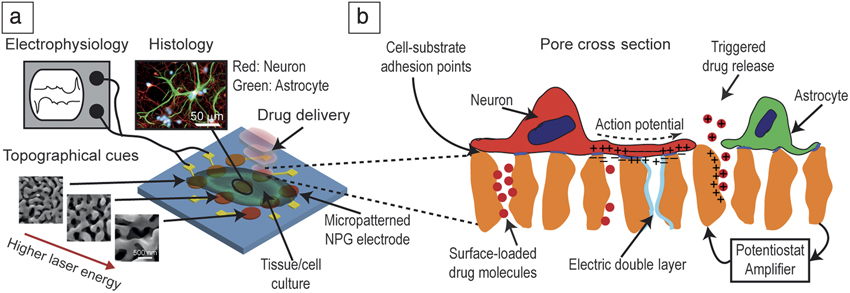
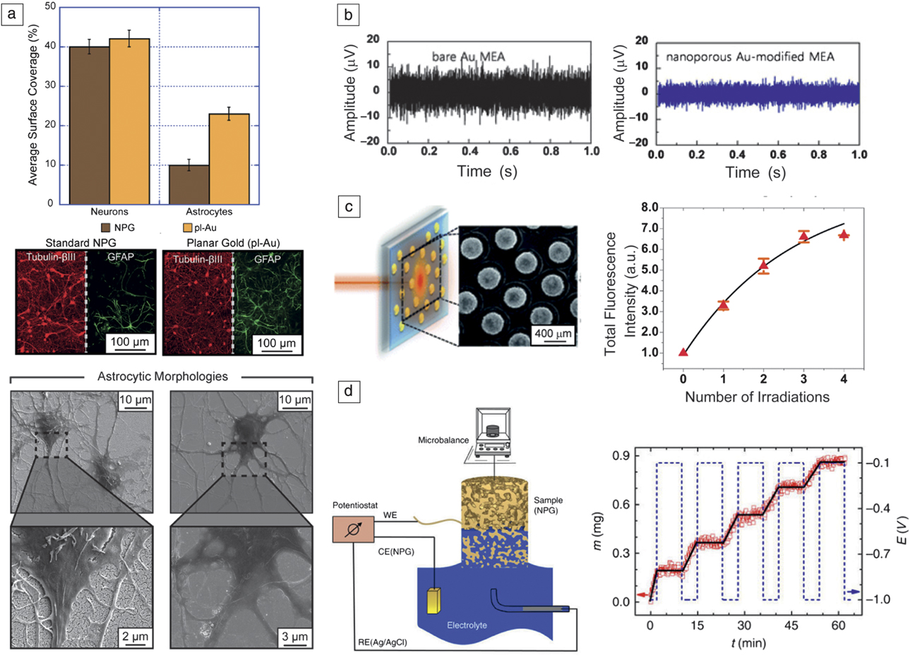
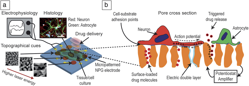
NPG has significant potential to serve as a multifunctional device coating, building on several attributes already discussed and some to be introduced in this section. We specifically focus on NPG as a neural interface for monitoring and modulating neural tissue behavior, which is a promising tool for neuroscience as well as a device for treatment of neurological disorders. The key performance parameters for neural electrodes are to obtain and sustain a high signal-to-noise ratio (SNR) and high spatiotemporal resolution in electrophysiological recordings from neuronal cells. The high spatial resolution generally requires a high-density electrode array with a small individual electrode footprint. The high SNR, on the other hand, requires both low electrode electrical impedance and proximity to the neurons. However, the electrode impedance is inversely proportional to the electrode surface area and neuron-electrode proximity is often prevented by neural cells (activated astrocytes) that cover the electrodes as a part of tissue response to the electrode. Here, we discuss how NPG electrodes are well suited to mitigate some of these challenges (Figure 3 and Figure 4).
Schematic illustration of a multifunctional neural interface. (a) Nanoporous gold (NPG) multiple electrode arrays can support the culture of neural cells and tissue, allowing for electrophysiological recordings with high signal-to-noise ratio. (b) Topographical cues enhance neuron–electrode proximity while reducing astrocyte spreading, which tends to interfere with the neuron–electrode interface. The porous structure and high effective surface area allow for loading and release of pharmaceuticals, thereby modulating neural behavior.
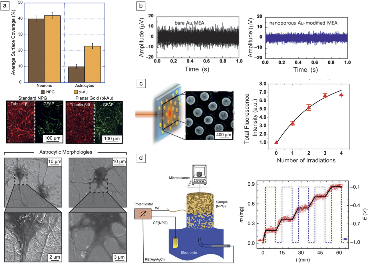
Examples of nanoporous gold (NPG)-based biomedical devices. (a) NPG surfaces reduce astrocyte coverage while maintaining neuronal coverage as illustrated by quantitative immunofluorescence of the cell cultures on NPG and planar gold (pl-Au) surface. Tubulin-βIII is a marker for neurons and glial fibrillary acidic protein (GFAP) is a marker for astrocytes. Scanning electron micrographs display varying response of astrocytes to the two surfaces.Reference Chapman, Wang, Chen, Garrison, Lein and Seker32 (b) NPG-modified multiple electrode arrays (MEA) increase signal-to-noise ratio in electrophysiological recordings of neurons. Reproduced with permission from Reference Kim, Kim, Kim, Han, Chung and JungReference 33. © 2015 IOP Publishing. (c) Photothermal excitation of NPG nanodisks loaded with fluorescent molecules exhibit light-gated release of the molecules.Reference Santos, Zhao, Zeng and Shih36 (d) Electrical modulation of the surface charge triggers wicking of electrolyte into the NPG cylinder. Note: CE, counter electrode; RE, reference electrode; WE, working electrode. Reproduced with permission from Reference Xue, Markmann, Duan, Weissmüller and HuberReference 39. © 2014 Macmillan Publishers Ltd.
Electrode impedance
The first challenge of obtaining high-density electrode arrays with low electrical impedance has been addressed by employing traditional photolithography to fabricate NPG electrodes on glass and silicon substrates.Reference Chapman, Wang, Chen, Garrison, Lein and Seker32,Reference Kim, Kim, Kim, Han, Chung and Jung33 In electrophysiological recordings, the sensing is due to capacitive coupling between the ionic changes (created by neurons) and the electric double layer at the electrode–electrolyte interface. While reduction of electrical impedance due to electrodes with high effective surface area is commonly observed, this reduction was sustained even in biofouling solutions, likely by the aforementioned biofouling resilience (Figure 2c).Reference Daggumati, Appelt, Matharu, Marco and Seker10,Reference Patel, Radhakrishnan, Zhao, Uppalapati, Daniels, Ward and Collinson17
Topographical cues
As discussed earlier, another factor that influences the recording SNR is the proximity of neurons to the electrode. One approach to enhance this has been by introducing micro- and nanoscale features onto electrode surfaces. Cells have the ability to sense the underlying substrate and regulate their adhesion in part as a function of the underlying surface topography (Figure 3b). Studies on the influence of NPG topographical cues on cellular spreading have revealed an interesting phenomenon, where the NPG surface reduced astrocyte (a star-shaped glial cell of the central nervous system) spreading while it did not affect neuronal coverage (Figure 4a).Reference Chapman, Wang, Chen, Garrison, Lein and Seker32 Similarly, the mobility of another important neural cell, microglia, which promotes spreading of astrocytes, was limited on NPG surfaces.Reference Tan, Terrill, Paranjape, Stine and Nichols34 These are important findings that point to NPG’s potential as a neural interface coating for enhanced electrode reliability and SNR for recording and electrical stimulation (Figure 4b).Reference Chapman, Wang, Chen, Garrison, Lein and Seker32,Reference Kim, Kim, Kim, Han, Chung and Jung33
Soluble factor delivery
Another approach to manage tissue response to electrodes is releasing soluble factors, such as drug molecules in situ from the device coatings. This approach, in some ways, is the opposite physical problem of that discussed for biosensing, where here the reaction is the surface–molecule association/dissociation, and transport is the outflux of the molecule through the porous network. In this case, drug molecules are loaded into the porous matrix and retained largely by weak intermolecular interaction between the surface and the molecules. As expected, the large effective surface area of NPG drastically increases the loading capacity.Reference Polat and Seker35 In addition, this surface–molecule interaction largely dictates the molecular release kinetics, extending the release half-lives to almost half of one day from submicron thin films.
The surface–molecule interactions can be modulated by various external stimuli opening the door to on-demand release of drugs. For example, it has been shown that by leveraging the high photothermal-light-harvesting efficiency of NPG nanodisks, molecular release from NPG was triggered by exposure to a near-infrared laser (Figure 4c).Reference Santos, Zhao, Zeng and Shih36 Analogously, by varying the surface charge of NPG via an applied electric potential between NPG and a reference electrode, charged drug molecules were successfully released.Reference Gittard, Pierson, Ha, Wu, Narayan and Robinson37 The molecular release was also gated by the presence of halides that have a higher affinity for the gold surface than physisorbed dye molecules.Reference Polat and Seker35 The biological relevance of the drug delivery capability of NPG was demonstrated by loading NPG-coated glass coverslips with a drug molecule inhibiting cell division. When astrocytes were cultured on the coverslips, the release of the inhibitory drug molecules suspended the spreading of astrocytes compared to control samples.Reference Seker, Berdichevsky, Staley and Yarmush38 Along with surface–molecule interaction-mediated molecular release, it is also possible to modulate the hydrophilicity of NPG by applying electrical potentials and enabling electrically controlled wicking and dispensing of extremely small liquid volumes (Figure 4d).Reference Xue, Markmann, Duan, Weissmüller and Huber39
Device fabrication
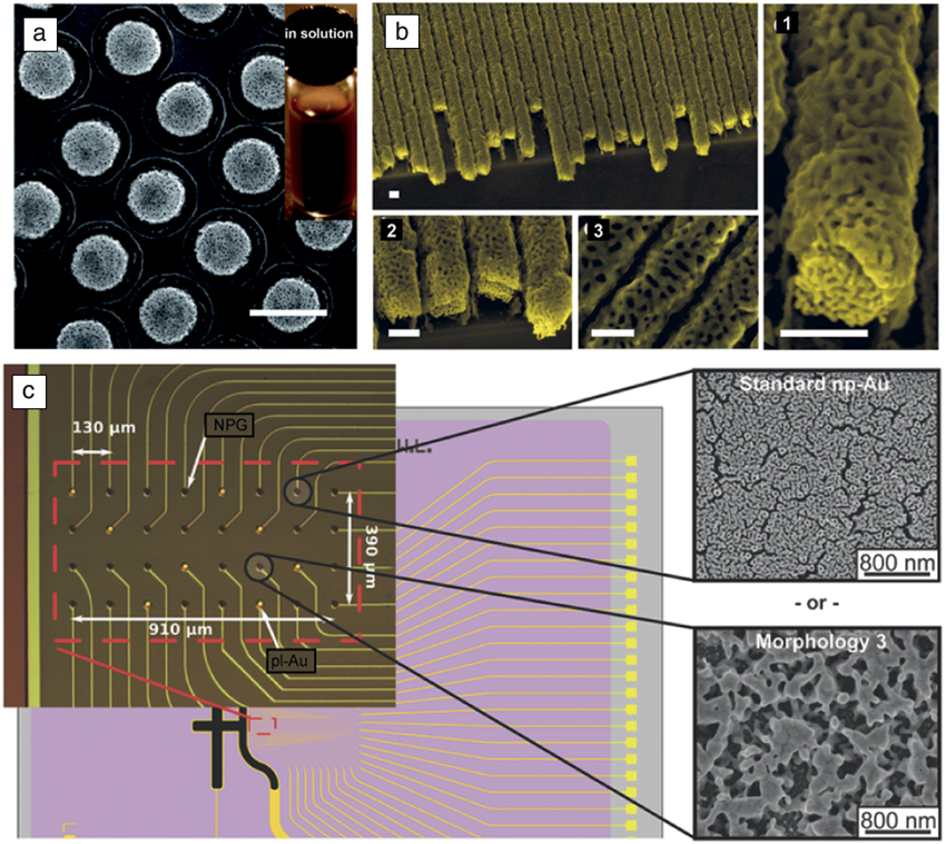
A significant advantage of NPG has been its amenability to being produced in various sizes and geometries (Figure 5), spanning submicron thin-film coatings,Reference Chapman, Wang, Chen, Garrison, Lein and Seker32,Reference Kim, Kim, Kim, Han, Chung and Jung33 nanoparticles,Reference Santos, Zhao, Zeng and Shih36,Reference Liu, Bai, Zhang, Yang, Fan, Zheng, Yin and Gao40,Reference Wang and Schaaf41 and millimeter-sized monoliths.Reference Alla, d’Andrea, Bhattarai, Cooper, Tan, Demchenko and Stine14,Reference McCurry and Bailey28 In its thin-film form, NPG has been deposited via sputtering and evaporation of AuAg alloy constituents. As these methods are compatible with conventional pattern transfer techniques, such as lift-off photolithography and screen printing, micropatterns (e.g., electrode arraysReference Chapman, Wang, Chen, Garrison, Lein and Seker32) have been successfully fabricated on a variety of substrates, including silicon and glass. Thin-film coatings have also been patterned using self-packed polymeric particles as etch masks to create well-defined nanodisk patterns that can either be left as surface coatings or released into solution to produce a colloidal suspension of nanodisks (Figure 5a).Reference Santos, Zhao, Zeng and Shih36 In devices for separation of biomolecules and for capture/release applications, NPG monoliths of 8.00 × 8.00 × 0.25 mm3 dimensions were integrated into a Teflon flow cell and solutions were pumped through using a peristaltic pump (Figure 2d).Reference Alla, d’Andrea, Bhattarai, Cooper, Tan, Demchenko and Stine14 Such setups can be integrated with UV-vis detection capabilities or other sorts of chromatographic detectors. It has been shown that the interior surfaces of NPG monoliths can be fully modified by functional SAMs, such as the reported instance of mannose terminal SAMs for lectin capture and release,Reference Tan, Fujikawa, Pornsuriyasak, Alla, Ganesh, Demchenko and Stine29 and of SAMs conjugated to lectins for glycoprotein capture and release.Reference Alla, d’Andrea, Bhattarai, Cooper, Tan, Demchenko and Stine14 Electroplating of gold-silver alloy is another approach that allows for thicker films and templated growth, such as in the case of producing NPG nanowires by AuAg electroplating in a porous anodic alumina template followed by dissolution of the template,Reference Liu and Searson42 or using advanced lithography techniques to produce arrays of AuAg fibers (Figure 5b)Reference Chauvin, Stephant, Du, Ding, Wathuthanthri, Choi, Tessier and El Mel43 and dealloying of the released AuAg nanowires (Figure 5).
Examples of various nanoporous gold (NPG) structures. (a) Submicron NPG nanodisks patterned on a substrate and subsequently released to create a colloidal suspension. Scale bar = 500 nm.Reference Santos, Zhao, Zeng and Shih36 (b) Large-scale production of NPG nanowires obtained by laser-interferometric patterning of a photoresist layer used as a shadow mask to gold-silver (NPG precursor) evaporation. Scale bars = 200 nm.Reference Chauvin, Stephant, Du, Ding, Wathuthanthri, Choi, Tessier and El Mel43 (c) Photolithographic patterning of NPG multiple electrode arrays and their subsequent thermal modification to display different electrode morphologies used for electrochemical sensing and neural electrophysiology applications.Reference Chapman, Wang, Chen, Garrison, Lein and Seker32
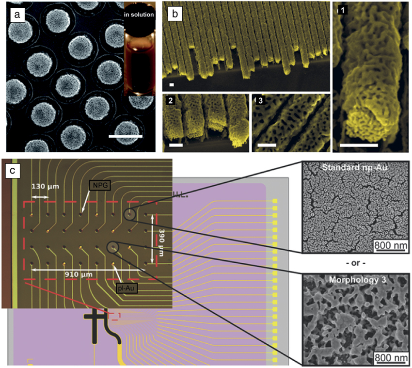
In many applications and fundamental studies of structure–property relationships, it has become crucial to rapidly observe the influence of pore morphology (e.g., porosity and ligament size) on properties (e.g., biocompatibility, biosensor performance, and separation efficiency). To that end, various approaches have been devised to modulate pore morphology via thermal, electrical, and electrochemical means. Some of these techniques have been extended to produce on-chip libraries of NPG with varying morphologies.Reference Chapman, Wang, Chen, Garrison, Lein and Seker32 We expect these libraries to enhance the fundamental studies (e.g., mechanical, optical, biological properties) and applications of NPG films (e.g., catalysis, biosensing).
Summary and outlook
There has been tremendous progress in a little more than five years in bioanalytical and biomedical applications of nanoporous metals obtained by dealloying, particularly of NPG. Some applications, such as detection of biomarkers, have the potential to be used to test patient samples. Further applications, such as integration of NPG monoliths into liquid chromatography columns, are of high interest, while these may require bimodal pore-size distributions with a population of small nanometer-scale pores and larger pores for minimization of backpressure effects. The issue of SAM stability over time in varying solvent environments needs to be investigated if modified monoliths are to be used. The NPG morphology libraries may find use in studying structure–property relationships in a variety of fields, including catalysis.
Acknowledgments
E.S. acknowledges support from the National Science Foundation (CBET-1512745 and CBET&DMR-1454426). W.-C.S. acknowledges support from the National Science Foundation (CBET-1605683, CBET-1151154) and NASA (NNX12AQ44G). K.J.S. acknowledges support from the National Institutes of Health (NIGMS R01-GM111835).

Erkin Şeker is an associate professor at the University of California, Davis, in the Department of Electrical and Computer Engineering and leads the Multifunctional Nanoporous Metals Group. He received his PhD degree in electrical engineering from the University of Virginia (UVA) in 2007. He was a postdoctoral fellow at UVA in the Chemistry Department from 2007 to 2009, and a postdoctoral research associate at the Center for Engineering in Medicine at Harvard Medical School from 2009 to 2011. His research interests focus on structure–property relationships of nanoporous materials in the context of biomedical applications. Şeker can be reached by email at eseker@ucdavis.edu.

Wei-Chuan Shih is an associate professor of electrical and computer engineering, biomedical engineering, materials science and engineering, and chemistry at the University of Houston. He leads the Nanobiophotonics Laboratory and is associate director of the Nanosystem Manufacturing Center. He earned his PhD degree in 2007 from the Massachusetts Institute of Technology Spectroscopy Laboratory/NIH Laser Biomedical Research Center, studying optical spectroscopy techniques for noninvasive chemical/biomedical sensing. He developed optical sensing systems for offshore oil spill monitoring as a Schlumberger Research Fellow from 2007 to 2009. He has been a faculty member at the University of Houston since 2009. Shih can be reached by email at wshih@uh.edu.

Keith J. Stine is a professor of chemistry at the University of Missouri–St. Louis. He received his PhD degree in physical chemistry from the Massachusetts Institute of Technology, working in the field of liquid crystals. He was a postdoctoral associate at the University of California, Los Angeles, working in the field of monolayers. He has been on the faculty in the Department of Chemistry and Biochemistry at the University of Missouri–St. Louis since 1990. Stine can be reached by email at kstine@umsl.edu.