Partial anomalous pulmonary venous connection is an uncommon congenital defect, first described by Winslow in 1739. Reference Winslow1 Incidence of partial anomalous pulmonary venous connection varies between 0.7% at autopsy series Reference Hughes and Rumore2 and 0.1% at retrospective analysis of chest CTs. Reference Ho, Bhalla, Bierhals and Gutierrez3
Partial anomalous pulmonary venous connection is characterized by the failure of one or more pulmonary veins to connect with the left atrium during fetal development. In particular, the anomalous pulmonary veins connect directly to the right atrium or to one or more of its venous tributaries.
The heterogeneous anatomy of partial anomalous pulmonary venous connection results in a left-to-right shunt with a wide spectrum of clinical cases.
Presentation with heart failure occurs rarely. Most often partial anomalous pulmonary venous connection can be apparently asymptomatic and detected only in adulthood. Symptoms may include recurrent pneumonia, wheezing and haemoptysis, typically found in patients with scimitar syndrome. This syndrome includes partial anomalous right pulmonary venous drainage to the inferior vena cava, hypoplastic right lung with displacement of cardiac structures into the right hemithorax, sometimes pulmonary hypertension and anomalous systemic arterial supply to the right lung. Reference Van Praagh, Carrera, Sanders, Mayer and Van Praagh4
The haemodynamic effect of partial anomalous pulmonary venous connection is the result of variable degree of an obligatory left-to-right shunt, which if left unrepaired, can lead to pulmonary vascular remodelling until the development of pulmonary arterial hypertension. Reference El-Kersh, Homsy, Daniels and Smith5
Management of partial anomalous pulmonary venous connection includes close observation, medical therapy, and/or surgical repair, depending on anatomy and clinical presentation.
Surgical repair consists of redirecting the pulmonary venous flow to the left atrium with variable technical complexity, related to the anatomy of partial anomalous pulmonary venous connection.
Considering the wide variety of partial anomalous pulmonary venous connection to different sites (above the heart, the heart, and below the heart), we focused our attention to partial anomalous pulmonary venous connection to below the heart or infradiaphragmatic anomalous connection diagnosed in adulthood.
In the infradiaphragmatic anomalous connection, the pulmonary veins conjoin and form a vertical vessel that passes caudally into the abdomen through the oesophageal orifice of the diaphragm. It usually drains to the portal vein or to one of its tributaries, such as ductus venosus, hepatic vein, and IVC. Reference Chuan-Chi, Ching-Chang, Po-Jen, Chi-Hsin and Shih-Yin6
Among all types of partial anomalous pulmonary venous connection, the infradiaphragmatic partial anomalous pulmonary venous connections represent the most challenging lesions. Because of their rarity, their variable anatomies, and the long distance between the end of the anomalous connection and the left atrium, partial anomalous pulmonary venous connection treatment is led to expert opinion.
We describe our cases of infradiaphragmatic partial anomalous pulmonary venous connections, diagnosed in adulthood, to share our experience about their management.
Material and methods
Between March 2010 and August 2021, we retrospectively observed seven cases of infradiaphragmatic partial anomalous pulmonary venous connection in our centre. All cases were diagnosed in adulthood. Informed consent forms were obtained from all the patients.
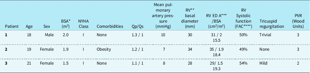
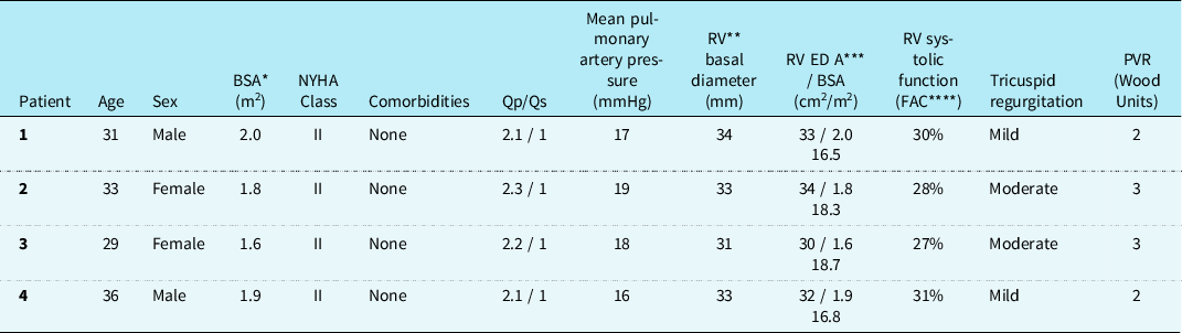
Cases were divided into two groups considering the amount of pulmonary lobe involved in the anomalous connection (Tables 1 and 2).
Group 1. Isolated partial anomalous pulmonary venous connection from the right inferior pulmonary lobe to inferior vena cava through suprahepatic veins. Patients’ characteristics

*BSA: body surface area.
**RV: right ventricle.
***RV ED A: right ventricular end diastolic area.
Group 2. Partial anomalous pulmonary venous connection of the entire right lung to inferior vena cava through suprahepatic veins. Patients’ characteristics

*BSA: body surface area.
**RV: right ventricle.
***RV ED A: right ventricular end diastolic area.
Group I: isolated partial anomalous pulmonary venous connection from one pulmonary lobe, in particular partial anomalous pulmonary venous connection from the right inferior pulmonary lobe to inferior vena cava through suprahepatic veins (Supplementary Figure S1).
This group includes three patients, one male and two females, with a median age of 19 years.
They were all asymptomatic and the diagnosis of partial anomalous pulmonary venous connection occurred as incidental findings.
Indeed there was a moderate volume overload of the right heart chambers at echocardiogram.
At right heart catheterisation, Qp/Qs were slightly elevated; all cases had Qp/Qs ≤ 1.5:1, with normal mean pulmonary artery pressure and pulmonary vascular resistance.
Group II: partial anomalous pulmonary venous connection of the entire right lung to inferior vena cava, all through suprahepatic veins (Supplementary Figures S2 and S3). They presented an “incomplete” form of scimitar syndrome, in fact they lacked a systemic arterial supply of the right lung, the right lung, and the right pulmonary artery were not hypoplastic and there was no pulmonary hypertension.
This group includes four patients, two females and two males, with a median age of 32 years.
They were all symptomatic for dyspnoea in NYHA class II. The CT imaging confirmed the diagnosis in all cases (Supplementary Figure S4).
The echocardiogram showed right heart volume overload with mild dilation and mild hypokinesis of the right ventricle, mild to moderate tricuspid regurgitation, paradoxical movement of interventricular septum.
At right heart catheterisation, there was a significant shunt with Qp/Qs > 2:1 in all cases.
The mean pulmonary artery pressure was <30 mmHg and the pulmonary vascular resistance was normal.
Results
Considering the low pathophysiological relevance of the shunt and the surgical difficulty due to the long distance between partial anomalous pulmonary venous connection and left atrium, patients in group I have been managed conservatively.
At 5 years follow up, all three patients were still asymptomatic with unchanged clinical conditions and echocardiographic results.
All patients in group II were symptomatic for dyspnoea in NYHA class II, with right heart volume overload, mild dilation and mild hypokinesis of the right ventricle, mild to moderate tricuspid regurgitation and with significant shunt.
Therefore, partial anomalous pulmonary venous connection of group II were surgically corrected using long right intra-atrial baffles (Gore Tex baffle in cases 1 and 2, and baffle of autologous pericardium in case 3 and 4). The long baffle was placed within the lumen of the IVC to channel the anomalous pulmonary vein effluent to the right atrium, then through an iatrogenic atrial septal defect into the left atrium (Figs 1 and 2).
Surgical repair of PAPVC to inferior vena cava using long right intra-atrial baffles.

CT image of long tunnel redirecting partial anomalous pulmonary venous connection in the left atrium.

Both patients 1 and 2 of group II experienced cough and hemoptysis 6 months after surgery. They were both diagnosed with complete baffle thrombosis and loss of right lung function.
Selective angiography showed complete exclusion of the right pulmonary artery with retrograde thrombotic occlusion of the pulmonary veins. However, there were neither signs of pulmonary hypertension nor elevation of total pulmonary vascular resistance. Resting cardiac output was at the lower limit of the normal range, balanced by the increase of heart rate.
Both two patients started medical therapy with Warfarin, indeed oral anticoagulation failed to recanalise the tunnel.
Patient 1 was lost at follow up, while patient 2 continued his follow up. He showed mild elevation of heart rate, but was persistently asymptomatic at rest at 10 years of follow up.
Considering thrombosis of the baffle as an early and serious complication, patients 3 and 4 of this group were both empirically and preventively treated with vitamin K antagonists immediately after surgery, with good outcome on long term follow-up.
The patency of the long intra-atrial baffles was routinely confirmed at CT scan.
Because of an ambiguous image at CT scan of patient 4, he even underwent catheterisation 7 months after cardiac surgery. Patency of his intra-atrial baffle was confirmed. Cardiac output was normal, as well as pulmonary pressure, and QP/QS was 1:1.
Therefore, oral anticoagulation was continued indefinitely.
Median follow up of the last two cases was 3 years. At their last visit, patients were asymptomatic with normal right ventricle size and function and improvement of functional capacity.
Discussion
Partial anomalous pulmonary venous connection is a rare congenital anomaly, often clinically silent and diagnosed in adulthood.
Partial anomalous pulmonary venous connection appears with a wide spectrum of clinical presentations, from the most common clinical silence, to dyspnoea with exertion.
Although the lack of symptoms, in some patients pulmonary overcirculation can potentially result in severe volume overload of the right heart, until development of secondary pulmonary artery hypertension.
Considering the wide spectrum of anatomical and physiopathological presentation, we analysed the subgroup of partial anomalous pulmonary venous connection to below the heart, or infradiaphragmatic partial anomalous pulmonary venous connection. All our cases were detected in adulthood.
Management of infradiaphragmatic partial anomalous pulmonary venous connection is apparently challenging, because of its complex anatomy with long descending vertical veins. Furthermore, facing its rarity and there is a lack of a standardised therapy.
It is therefore always necessary to consider a case-by-case approach to tailor the most suitable management.
In the asymptomatic patients with low shunt fraction and no evidence of right ventricle dysfunction, conservative management is recommended with close monitoring. Reference Stout, Daniels and Aboulhosn7
In our experience, isolated partial anomalous pulmonary venous connection from one pulmonary lobe (right lung to inferior vena cava) was not linked to a significant shunt fraction (Qp/Qs < 1.5 in all three cases), nor to pulmonary artery hypertension. All our cases were managed conservatively and maintained on regular follow up, until possible detection of significant shunt.
Finally, surgical treatment of partial anomalous pulmonary venous connection of the entire right lung in inferior vena cava is questionable and it largely depends on clinical presentation. Reference Alsoufi, Cai, Van Arsdell, Williams, Caldarone and Coles8
Partial anomalous pulmonary venous connection to below the heart is a very rare congenital anomaly, often silent until adulthood. We collected seven cases and tailored their management on the basis of clinical cases, anatomy and medical experience.
There is evidence for surgical treatment of partial anomalous pulmonary venous connection in case of significant shunt with Qp/Qs > 1.5:1, but the anatomical complexity of the defect should be related to repair feasibility and surgical result.
A conservative approach, may also be considered in case of high anatomical complexity, when redirecting the anomalous drainage in the left atrium is hardly feasible.
Treatment of partial anomalous pulmonary venous connection of the entire right lung to inferior vena cava with significant shunt is often a technical challenge.
When surgical treatment is clinically indicated, the main challenge consists in redirecting the anomalous infradiaphragmatic connection to the left atrium. Most common surgical technique consists of creating a long intra-atrial tunnel from the partial anomalous pulmonary venous connection to the left atrium. Slow blood flow inside the long baffles inclines to thrombosis and occlusion, as we observed in our first two cases.
To overcome this risk, we then prescribed oral anticoagulation as prevention.
Therefore, management decisions should also consider to balance the risk of chronic right heart volume overload and haemorrhagic risk secondary to potential long term anticoagulation.
To the best of our knowledge, there are a few cases of thrombosis of the intra-atrial baffle and its relative management in literature.
The necessity to use long-term oral anticoagulation should be considered in this setting. In our small experience, prophylactic oral anticoagulation showed good results at long-term follow-up in terms of patency of the long intra-atrial baffle.
Supplementary material
To view supplementary material for this article, please visit https://doi.org/10.1017/S1047951122001494
Acknowledgements
None.
Financial support
This research received no specific grant from any funding agency, commercial, or not-for-profit sectors.
Conflicts of interest
None.
Consent statement
The authors have obtained informed consent from the patients.