Introduction
The Mediterranean fruit fly (medfly), Ceratitis capitata (Wiedemann, 1824) (Diptera: Tephritidae), is one of the most important agricultural pests worldwide. Similarly to all other insects it is colonized by microorganisms, and the analyses of the bacterial community associated to its digestive tract, either by culture-dependent and independent methods, have shown that Enterobacteriaceae are the dominant populations with Klebsiella oxytoca as the most common species (Marchini et al., Reference Marchini, Rosetto, Dallai and Marri2002; Behar et al., Reference Behar, Jurkevitch and Yuval2008a).
It is generally accepted that the microbiota associated with the medfly's digestive tract plays a significant role in the development and physiology of the host (Behar et al., Reference Behar, Yuval and Jurkevitch2005, Reference Behar, Jurkevitch and Yuval2008b). More intriguing, it has also been reported that the sexual competitiveness of male fruit flies increased when diets were enriched with K. oxytoca in the laboratory and in field cages (Gavriel et al., Reference Gavriel, Jurkevitch, Gazit and Yuval2011).
Recently, it has been reported that the extracellular symbiotic bacterium Enterococcus mundtii associated with the generalist herbivore Spodoptera littoralis facilitates the normal development of host gut microbiota by secreting a stable bacteriocin active against invading bacteria (Shao et al., Reference Shao, Chen, Sun, Ishida, Hertweck and Boland2017). Following this path, we look back with a renewed interest at those bacterial strains identified as K. oxytoca isolated from the esophageal bulb (a foregut diverticulum of adult tephritids) of wild populations of C. capitata throughout the 1990s (Marchini et al., Reference Marchini, Rosetto, Dallai and Marri2002).
Materials and methods
Bacterial strains and growth conditions
The panel of bacteria used in this work includes 11 Klebsiella strains (KD1, KD2, KD3, KD5, KD6, KD7, KD12, KD16, KL22, KL25, and KL26) that were isolated from the esophageal bulb of wild populations of C. capitata collected in Italy from several fruit tree species in Florence, Tuscany (peach tree: KD1, KD2, KD3; apple tree: KD16, KL22, KL25, KL26; persimmon tree: KD12; orange tree: KD5), and in Catania, Sicily (prickly pear: KD6, KD7) during the first long-term study concerning the association of culturable bacteria with the medfly gut. Their presumptive identification was initially carried out by using the ATB 32E and/or ATB 32GN (API, Bio-Mérieux, France) microbial identification systems (Marri et al., Reference Marri, Valentini and Venditti1995; Marchini et al., Reference Marchini, Rosetto, Dallai and Marri2002). The 16S rRNA sequence of each strain was further established and all sequences showed similarity levels of 100% to their closely related species that was indeed K. oxytoca (Martinez-Sanudo et al., Reference Martinez-Sañudo, Savio, Mazzon, Girolami, Ciolfi and Marri2011). The 16S rRNA gene sequences and their GenBank accession numbers are: MK455094–101, MN161200–161202.
For the antagonistic assay, five Gram-positive and five Gram-negative American Type Culture Collection (ATCC) reference strains were initially used: Staphylococcus aureus 25923, Enterococcus faecalis 29212, Bacillus subtilis 6633, Bacillus cereus 9634, Micrococcus luteus 4698, Salmonella typhimurium 23853, Enterobacter cloacae 13047, Proteus vulgaris 6380, Pseudomonas aeruginosa 27853, and Escherichia coli 23739 (Marri et al., Reference Marri, Dallai and Marchini1996).
All strains were grown in LB broth, and maintained in glycerol (30%) stock solution at −80°C.
Antagonistic assay
Antibacterial activity of Klebsiella strains (producers) was determined by an Agar diffusion assay (Long and Azam, Reference Long and Azam2001) against the ten reference strains (testers). A lawn was prepared by mixing 5 ml soft-agar (0.7% agar) with 59 µl of the tester strain (OD600, 0.1) and poured over LB medium in 12 cm × 12 cm plates. Three microliter portions (OD600, 0.3) of eight potential producers were then spotted on the lawn. The plates were incubated for 4 days at room temperature (20–24°C) and examined daily for zones of inhibition. The zones of inhibition (measured from the edge of the bacterial growth to the edge of the clear zone) were then recorded. From this test, the strains KD1 and KD2 that had the same origin of KD3 (wild medflies collected from peach tree), and KD6 that shared the origin with KD7 (wild individuals collected from prickly pear) were excluded.
Subsequently, the Agar overlay method as essentially described by Anand et al. (Reference Anand, Bhat, Shouche, Roy, Siddharth and Sarma2006) was performed with all Klebsiella strains. Broth cultures of the producers (2–3 µl) were spotted on LB agar plates (diameter = 6 cm) and allowed to grow for 2 days. Then 2 ml of soft agar (0.7% agar) was added with 20 µl (OD600, 0.45) of each reference strain (tester), mixed thoroughly, and gently overlaid over the spotted strains. The overlaid plates were incubated at room temperature for 24–48 h. The width of the clear zone (R) was calculated as reported by Carasi et al. (Reference Carasi, Diaz, Racedo, Antoni, Urdaci and Serradell2014): R = (dInhib − dSpot)/2, where dInhib is the diameter of the zone without tester growth and dSpot is the diameter of the producer spot. Afterwards, inhibition scores were considered as essentially reported by the same authors: negative, R < 2 mm; low inhibition capacity, 2 mm < R < 5 mm; and high inhibition capacity, R > 6 mm (Carasi et al., Reference Carasi, Diaz, Racedo, Antoni, Urdaci and Serradell2014). Three independent experiments were performed. Statistical analysis of data was performed using GraphPad Prism version 8.00 for Windows, GraphPad Software, La Jolla California USA, http://www.graphpad.com. One-way ANOVA followed by Bonferroni test was used to determine significant differences of R values produced by all Klebsiella strains against the same tester. Differences were considered significant when P ≤ 0.05.
Detection of klebicin genes by PCR and DNA sequence analysis
Identification of individual klebicin types klebicins B, C, D, and CCL (Chavan et al., Reference Chavan, Rafi, Wertz, Goldstone and Riley2005) was performed by PCR using specific primer pairs: KlebB (Genbank: AF156893) forward primer: 5′-CAACACCAAGACTTTTTGTACAGG-3′; reverse primer: 5′-CCCATAACTCTCTCCTTTGTTGTT-3′; KlebC (GenBank: AY578794) forward primer: 5′-GGAACTTACACTCAATGACGGCAG-3′; reverse primer: 5′-CGGCTGCCAGTAGATGAAGTCCTG-3′; Kleb D (GenBank: AY578792) forward: 5′-CTTAACACTCAATGACGGCAGCGT-3′; reverse primer: same sequence of KlebC reverse primer; KlebCCL (GenBank: AF190857) forward primer: 5′-AAGCCGGTTTATGTGTCGGT-3′; reverse primer: 5′-ATCCAGCGTTTACGCCTACC-3′. All producer strains were tested with colony PCR. A bacterial colony was picked with a sterile inoculation loop and resuspended in 100 µl of autoclaved water and boiled for 10 min. For each individual PCR reaction, 1 µl of cell suspension was added to the reaction containing 1× PCR buffer, 1.5 mM MgCl2, 0.5 mM each dNTP and 2,5U Gotaq® DNA Polymerase (Promega, Madison, WI, USA) and 0.25 mM of each primer PCR were performed at the following conditions: 94°C for 5 min, followed by 40 cycles of 94°C for 1 min, 57°C for 1 min, and 72°C for 2 min and an extension step at 72°C for 10 min. All primer pairs were tested against all strains in order to reveal potential cross-reactivity of similar sequences. PCR products were run on a 1% agarose gel, then selected DNA bands were purified using Wizard® SV Gel and PCR Clean-Up System (Promega), cloned into pGEM®-T easy vector system (Promega), and sequenced on both strands at BMR Genomics (Padua, Italy).
DNA sequences were analyzed by the BLAST algorithm (Altschul et al., Reference Altschul, Gish, Miller, Myers and Lipman1990, Reference Altschul, Madden, Schäffer, Zhang, Zhang, Miller and Lipman1997), and the deduced amino acid sequence was aligned with the Clustal Omega program (Sievers et al., Reference Sievers, Wilm, Dineen, Gibson, Karplus, Li, Lopez, McWilliam, Remmert, Söding, Thompson and Higgins2011).
Results
Antagonistic assay
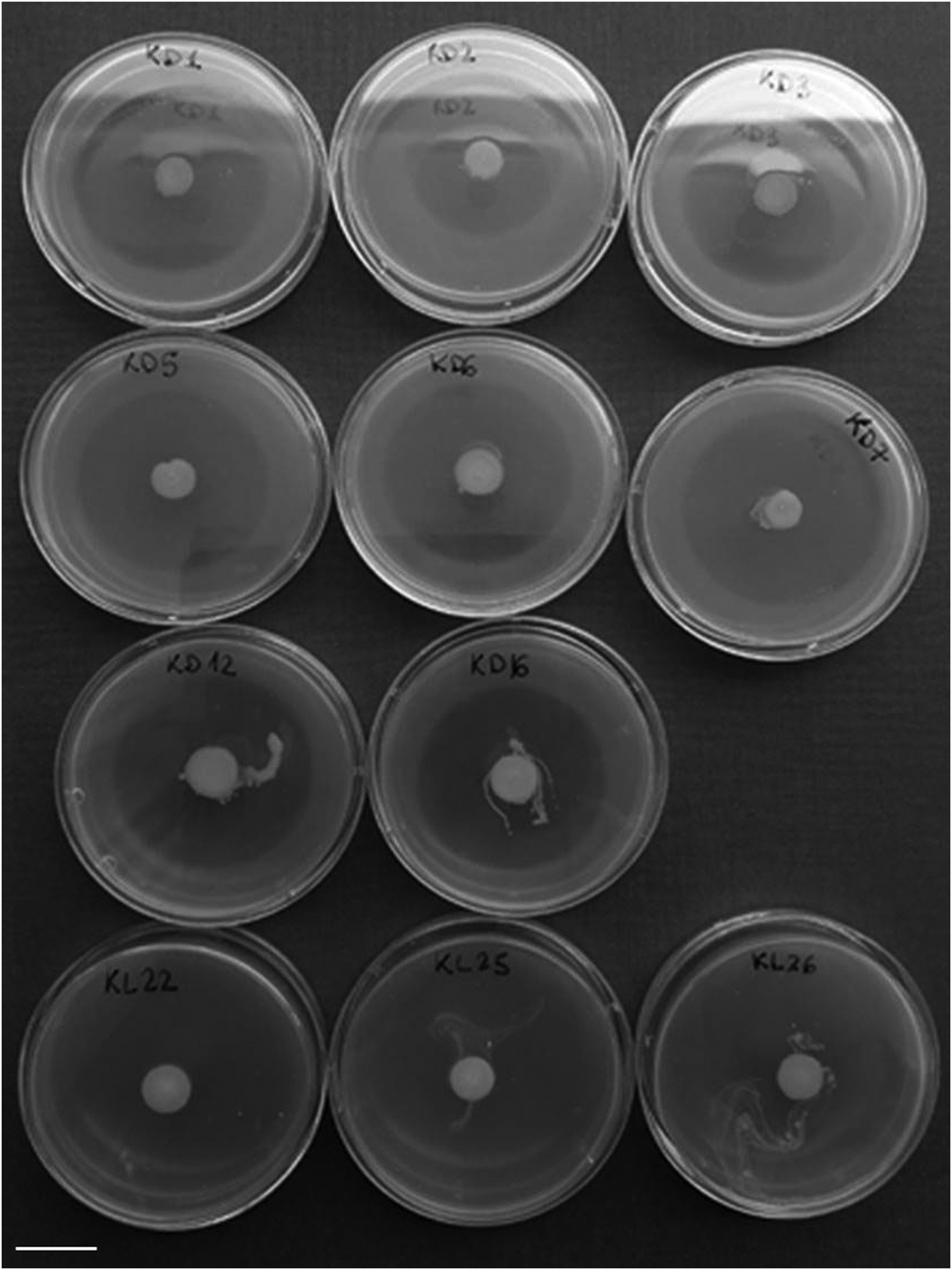
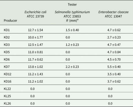
The antibacterial activity of eight Klebsiella strains was initially tested with the Agar diffusion assay (Long and Azam, Reference Long and Azam2001). Five strains showed activity against E. coli ATCC 23739. The zones of inhibition recorded were: 1 mm (KD12 and KD16), 2 mm (KD5), 3 mm (KD3), and 4 mm (KD7). Zones of inhibition <1 mm were detected against E. cloacae (KD3, KD5, KD7, KD12, and KD16), and against S. typhimurium (KD3 and KD7). Strains KL22, KL25, and KL26 did not display any activity against the tester strains at the experimental conditions applied. Assays were then repeated with the Agar overlay method (Anand et al., Reference Anand, Bhat, Shouche, Roy, Siddharth and Sarma2006) using all the 11 Klebsiella strains against the three enterobacterial species resulted sensitive in the diffusion test. The Agar diffusion method showed a stronger inhibition effect by the producer strains previously identified, but it also confirmed the range of activity for each producer. The key point remains although the difference among the inhibition zones (R) exhibited by all Klebsiella strains against E. coli was not statistically significant (Supplementary material, fig. S1). With regard to the strains that were not included in the diffusion assay, strain KD1 showed the same range of activity of KD3, while KD2 and KD6 results were correlable with KD5. Klebsiella KD7 exhibited again a stronger antibacterial activity, and a broader spectrum (fig. 1; table 1); afterwards the strain has been deposited in the Centre de Ressources Biologiques de l'Institut Pasteur CRBIP and in the Leibniz Institute DSMZ under accession number CIP 111713 and DSM 109964, respectively.
Example of antimicrobial activity of Klebsiella strains against Echerichia coli ATCC 23739 in the Agar overlay method. Scale bar, 1.5 cm.

Antimicrobial activity of Klebsiella strains in the Agar overlay method

All data are expressed in mean ± SD.
a R = (dInhib − dSpot)/2; dInhib: the diameter of the zone without tester growth, dSpot: the diameter of the producer spot (Carasi et al., Reference Carasi, Diaz, Racedo, Antoni, Urdaci and Serradell2014).
Detection of klebicin genes and DNA sequence analysis
It has been estimated that the vast majority of all bacteria and archaea produce at least one bacteriocin (Dobson et al., Reference Dobson, Cotter, Ross and Hill2012). Bacteriocins are a heterogeneous group of peptides that kill or inhibit members of the same or closely related species (Tagg et al., Reference Tagg, Dajani and Wannamaker1976). For Klebsiella spp., the first bacteriocin gene, klebicin B, was isolated from K. pneumoniae (Riley et al., Reference Riley, Pinou, Wertz, Tan and Valletta2001). Later, Chavan et al. (Reference Chavan, Rafi, Wertz, Goldstone and Riley2005) discovered two klebicin gene clusters: klebicin C both in K. pneumoniae and K. oxytoca isolates, and klebicin D just in K. oxytoca isolates. In the same paper, they also refer to klebicin CCL found in K. pneumoniae (Chavan et al., Reference Chavan, Rafi, Wertz, Goldstone and Riley2005). Klebicins are predicted to function as nucleases, and they are represented in all three highly divergent lineages of nuclease bacteriocins: the DNase family (klebicin B), the rRNase family (klebicin D), and the tRNase family (klebicins C and CCL) (Chavan et al., Reference Chavan, Rafi, Wertz, Goldstone and Riley2005).
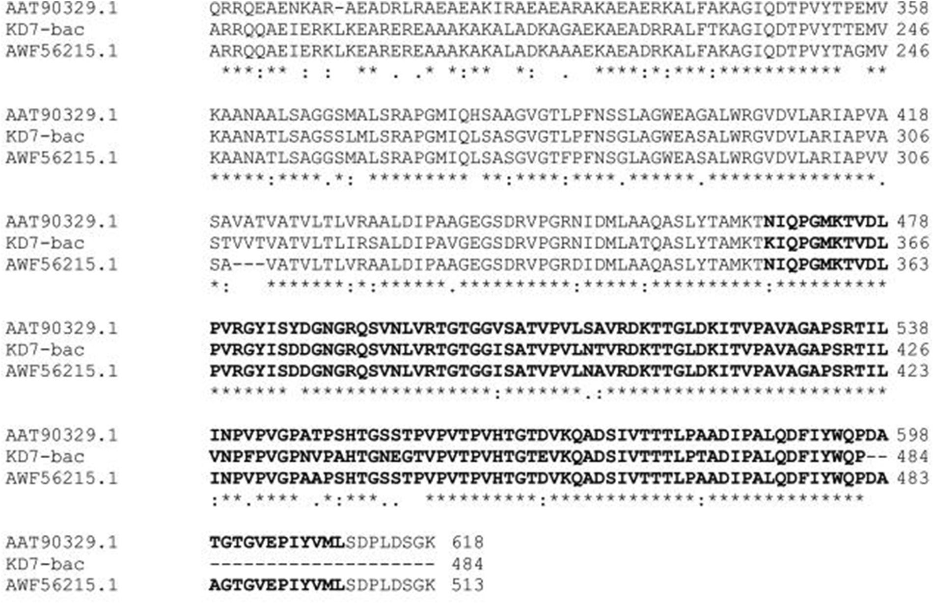
Primers, designed from each klebicin sequence, were used in order to detect the presence of their homologs in all Klebsiella strains object of the study. For all strains showing a killing phenotype in the antagonistic assay, a single amplification product of about 1650-bp was obtained using only KlebD primers. At first the PCR product from KD7, named KD7-bac, was sequenced, and the complete sequence, 1639-bp, was deposited in the GenBank/EMBL data library under accession number MN196477. Indeed, all the amplicons that have been sequenced next showed a full identity with KD7-bac DNA, except for two single-base substitutions in the KD12 derived sequence, and one single-base substitution in those derived from KD5 and KD6, respectively (Supplementary material, fig. S2). BLASTn analysis of KD7-bac DNA sequence showed 93% identity (e-value: 0.0) with a plasmid-carried colicin-D gene of Klebsiella michiganensis strain AR375 (plasmid unnamed3, GenBank: CP029143.1), and 86% identity with the sequence encoding for the klebicin D activity protein of K. oxytoca isolates (Chavan et al., Reference Chavan, Rafi, Wertz, Goldstone and Riley2005). Similarly, KD7-bac DNA sequence translation in all six reading frames by BLASTx aligned positions 188–1639 of Frame +2 with colicin-D (K. michiganensis-plasmid unnamed3: 93% identity; e-value: 0.0) [AWF56215.1], and positions 872–1639 with klebicin D activity protein (K. oxytoca: 88% identity; e-value: 5e-126) [AAT90329.1]. The ‘ORFfinder’ program at NCBI identified within the KD7-bac DNA sequence a putative gene encoding a potential protein of 483 aa that was then aligned with AWF56215.1 and AAT90329.1. Results, showed in fig. 2, indicate that all three sequences share the Pyocin S killer protein signature (Sano et al., Reference Sano, Matsui, Kobayashi and Kageyama1993). The ‘ORFfinder’ also identified on Frame +2 a second potential protein of 60 aa that shared 93% (e-value: 3 × 10⁻³²) deduced amino acid similarities with the C-terminal of the ‘tail fiber domain-containing protein’ of K. michiganensis-plasmid unnamed3 (GenBank: WP_075912839.1), and 73% (e-value: 3 × 10⁻¹⁷) amino acid similarities with the klebicin D phage-associated protein (GenBank: AAT90328.1). These results suggest for KD7-bac a bacteriophage/bacteriocin organization similar to that inferred by Chavan et al. (Reference Chavan, Rafi, Wertz, Goldstone and Riley2005) for klebicin C/D operons.
Partial alignment of KD7-bac putative protein (483 aa) with colicin-D (Klebsiella michiganensis-plasmid unnamed3, AWF56215.1), and klebicin D activity protein (K. oxytoca, AAT90329.1). Bold characters indicate the Pyocin_S superfamily domain signature (Sano et al., Reference Sano, Matsui, Kobayashi and Kageyama1993).

Discussion
In this study, we examined the antagonistic potential of Klebsiella strains isolated from the esophageal bulb of wild populations of C. capitata captured at various locations in Italy over a long time span. Eight strains out of 11 showed antagonistic activity against two enterobacterial reference strains, E. coli ATCC 23739 and E. cloacae ATCC 13047. Strain KD7 showed also an inhibition effect against S. typhimurium, even if low according to the definition of the antimicrobial activity adopted in this study (Carasi et al., Reference Carasi, Diaz, Racedo, Antoni, Urdaci and Serradell2014). The association of Enterobacteriaceae with Mediterranean fruit flies has been well documented over the years. Although the association has usually been considered in relation to symbiosis, the potential capacity of C. capitata in the transmission of pathogenic bacteria was also reported. Wild medflies collected in three geographic locations in central Israel carried coliforms that, in some cases, were presumptively identified as E. coli (Sela et al., Reference Sela, Nestel, Pinto, Nemny-Lavy and Bar-Joseph2005); in addition, GFP-tagged E. coli cells were readily transmitted to Mediterranean fruit flies that continued to harbor viable GFP-tagged bacteria for at least 7 days following contamination (Sela et al., Reference Sela, Nestel, Pinto, Nemny-Lavy and Bar-Joseph2005). On the other side, however, E. cloacae and other Enterobacter spp. have been commonly found as secondary facultative symbionts in the alimentary tract of the fly (Marri et al., Reference Marri, Valentini and Venditti1995; Marchini et al., Reference Marchini, Rosetto, Dallai and Marri2002; Behar et al., Reference Behar, Ben-Yosef, Lauzon, Yuval, Jurkevitch, Bourtzis and Miller2009). The paper from which the present study has taken its cue reports that while K. oxytoca is the most representative species in wild populations, Enterobacter spp. are in laboratory-reared medflies: it could not be only a matter of chance. In fact, the most part of those Klebsiella strains exhibited a killing phenotype in vitro and, at the same time, they resulted to carry DNA sequences highly similar to genes encoding bacteriocin, with the best hit, 93%, to a plasmid-carried colicin-D gene. The K. michiganensis-plasmid unnamed3 (134,879-bp) comprises, inter alia, either sequences encoding for conjugative transfer system and transposase family proteins. Both features are deeply involved in horizontal gene transfer, and they are consistent with a rapid spread of plasmid-associated genetic traits among bacterial populations (Hall et al., Reference Hall, Brockhurst and Harrison2017).
Although more studies are undoubtedly needed in order to establish a direct link between the identified klebicin-like sequences and the killing phenotype displayed in the antagonistic assay, the finding reported in this study remains quite attractive. A functional trait, like bacteriocin production, could indeed affect the fitness of bacteria in the wild with respect to those found in individuals maintained in the laboratory on artificial diets. It was reported that microcins (low molecular weight bacteriocins) enable the probiotic bacterium E. coli EcN to limit the expansion of competing Enterobacteriaceae (including pathogens and pathobionts) during inflammation in the human gut (Sassone-Corsi et al., Reference Sassone-Corsi, Nuccio, Liu, Hernandez, Vu, Takahashi, Edwards and Raffatellu2016). Accordingly, antagonism along with horizontal gene transfer could play a significant role in shaping bacterial communities in the insect gut and lead to the prevalence of the ‘best-equipped’ (bacteriocin producers) in free-living medflies.
We are aware that the setting hypothesized above might be a simplification of the actual situation and we cannot ignore that the diversity of microbial communities inhabiting the medfly gut might be related to the antimicrobial peptides produced by the insect females as well as to the infested fruit that serves as an activity hub for the amplification and distribution of bacterial strains (Marchini and Marri, Reference Marchini and Marri2010; Zaada et al., Reference Zaada, Ben-Yosef, Yuval and Jurkevitch2018). Nonetheless, the ‘cold-case’ investigation reported herein could help to generate testable hypothesis to better understand the actual in vivo relationships among bacterial communities within the insect host. It has been recently reported that bacteria associated with wild-caught Drosophila melanogaster fruit flies have a much higher colonization capability, and that specific bacterial species can proliferate in the insect gut and establish a stable mutualistic association (Pais et al., Reference Pais, Valente, Sporniak and Teixeira2018). On the other hand, further investigations may possibly lead to the use of selected probiotics with bacteriocin functionality for the quality improvement of mass-rearing system in the context of eco-friendly approaches for pest control (Noman et al., Reference Noman, Liu, Bai and Li2019).
Supplementary material
The supplementary material for this article can be found at https://doi.org/10.1017/S0007485319000816.
Acknowledgements
This work was supported by funding from the University of Siena (project numbers 2264-2018-BC-PSR2018_001, 2264-2019-BC-PSR2019_001) to SC and LM.