Kaposiform lymphangiomatosis is a rare anomaly characterised by the proliferation of abnormal lymphatic vessels that exhibits features of both malformation and neoplasia. Reference Croteau, Kozakewich and Perez-Atayde1 Kaposiform lymphagiomatosis shares overlapping patterns of clinical symptoms, anatomical location and imaging features with other lymphatic anomalies. Reference Goyal, Alomari and Kozakewich2 KLA is distinct in histopathology, clinical course, response to therapy and prognosis. Reference Croteau, Kozakewich and Perez-Atayde1,Reference Goyal, Alomari and Kozakewich2 Due to its uncommon and varied clinical presentation, kaposiform lymphagiomatosis is often diagnosed late. Reference Goyal, Alomari and Kozakewich2
Kaposiform lymphagiomatosis is commonly complicated by pleural and pericardial effusions. Reference Croteau, Kozakewich and Perez-Atayde1,Reference Perez-Atayde, Debelenko and Al-Ibraheemi3 Presently, treatment is multimodal with both medical and procedural therapies providing temporary symptomatic relief. Reference Croteau, Kozakewich and Perez-Atayde1,Reference Safi, Gupta, Adams, Anandan, McCormack and Assaly4 Procedural interventions can provide transient improvement and serve as a bridge to medical therapy. Reference Croteau, Kozakewich and Perez-Atayde1 Novel procedural techniques are necessary for the management of prolonged chylous effusions to reduce associated morbidity, including long hospital stay and repeated procedures.
We present a case of a 4-year-old boy with recurrent pericardial effusion with novel use of the Indigo™ aspiration system (Penumbra) where conventional drainage was minimally effective.
Case report
A 4-year-old Caucasian boy presented to the emergency department with 1 week of easy bruising. Initial investigations noted pancytopenia, consumptive coagulopathy, marked hepatosplenomegaly, and cardiomegaly. Echocardiography confirmed a large pericardial effusion with tamponade physiology.
Pericardiocentesis with insertion of an 8.5Fr Pigtail drain resulted in an initial drainage of 410 ml of serosanguinous fluid with elevated triglyceride and lymphocyte content consistent with chyle. A broad infectious and rheumatological work-up was undertaken to narrow the differential diagnosis which included macrophage activation syndrome, atypical HUS/TTP, and systemic lupus erythematosus. A bone marrow biopsy did not demonstrate malignancy. A CT scan demonstrated mediastinal oedema, extensive cervical, axillary and mesentery lymphadenopathy, massive splenomegaly, patchy bilateral pulmonary opacities, and diffuse airway thickening. PET and MRI scans demonstrated areas of marrow replacement most prominent in his right proximal femur. Percutaneous fluoroscopic-guided bone marrow aspiration yielded no significant results and lymphatic stains were negative.
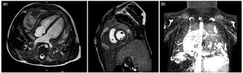
He continued to have high output drain losses (400–500 ml/day). He was commenced on methylprednisolone but had no improvement in his drainage. Following commencement of sirolimus due to presumptive diagnosis of kaposiform lymphagiomatosis, he had significant improvement in drainage (38 ml/day) and the drain was removed 8 days later. A pericardial drain was re-inserted urgently 4 days later due to re-accumulation and clinical and echocardiographic evidence of tamponade. There was minimal drainage due to loculation of the pericardial effusion confirmed on MRI (Fig 1).
(a) T2 weighted MRI imaging demonstrating a large, circumferential, heterogenous pericardial effusion with extensive loculations. (b) On T2 weighted lymphatic imaging, there is increased signal within the pericardium, consistent with collections of lymphatic fluid. The abdominal lymphatic system is dilated and tortuous beginning at the level of the kidneys with continued tortuosity and dilation through the cisterna chyli through the thoracic duct. There is also hyperintensity in the mediastinum and neck surrounding the great vessels without evidence of compression.

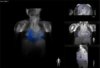
The multidisciplinary team decided to return the patient to the catheterisation laboratory for pericardial washout and biopsy. The pericardial drain was transected, and 4 mg tissue plasminogen activator (tPA) was instilled. Reconstructed 3D MRI images were used as an overlay with our VesselNavigator system to determine the location of the loculations (Fig 2). Under echocardiographic and fluoroscopic guidance with the overlay technology, the existing drain was accessed with an 0.035” Whooley wire and the tract was dilated to allow a 12Fr sheath to be placed. An 8Fr 50 cm Penumbra CAT3 catheter attached to the Indigo™ aspiration system was advanced through the 12Fr sheath over the wire. Dilute tPA was infused through the side port of the 12Fr sheath whilst the system was active. The catheter was manipulated into the pericardial space and 400 ml of fluid was removed along with semi-solid thrombus. Finally, a 6Fr Bioptome was used to sample the pericardium anteriorly from three separate areas for histopathology. The 12Fr sheath was then removed, and a 14Fr Pigtail drain was placed in the pericardial space.
VesselNavigator allowed use of 3D anatomical information from patient’s existing MRA dataset as a 3D roadmap overlay on a live X-ray image allowing visualization of loculations in the pericardial space during the procedure.

Following the procedure, drain output remained low, and the drain was subsequently removed after 10 days. Echocardiography demonstrated a loculated pericardial effusion posterior to the left ventricle, which remained stable until the day of discharge. Four months following discharge, the pericardial effusion had completely resolved.
Discussion
This patient presented with a recurrent pericardial effusion requiring multiple procedural interventions. Based on the clinical scenario, radiological features and modest improvement with sirolimus therapy, kaposiform lymphagiomatosis was diagnosed. Despite medical therapy, he had ongoing re-accumulation of pericardial fluid which became loculated and difficult to evacuate through conventional drainage. The use of the Indigo™ aspiration system (Penumbra) and overlay imaging with our VesselNavigator system enabled the safe and effective removal of thrombus in the pericardial space in this patient.
Kaposiform lymphagiomatosis is an aggressive lymphatic anomaly that, due to the varied presentation, is often diagnosed late leading to poor outcomes. Reference Croteau, Kozakewich and Perez-Atayde1 The 5-year survival for kaposiform lymphagiomatosis is 51% and overall survival is 34% with the most common cause of death being cardio-respiratory failure. Reference Croteau, Kozakewich and Perez-Atayde1,Reference Fernandes, Fargo and Saini5 Thoracic involvement is more common and extensive in kaposiform lymphagiomatosis compared to other lymphatic anomalies. Reference Goyal, Alomari and Kozakewich2 Croteau and colleagues reported 17 of 20 patients diagnosed with kaposiform lymphagiomatosis over a 16-year period developed a pleural or pericardial effusion or both. High volume drain output (>1 L/day) and the need for multiple procedures due to rapid accumulation of fluid are common. Reference Croteau, Kozakewich and Perez-Atayde1
The diffuse, infiltrative nature of kaposiform lymphagiomatosis make attempt at surgical intervention, including resection of lymphatic mediastinal masses, pleurodesis, and thoracic duct ligation, only temporising with marginal success in the medium-to-long term. Reference Ji, Chen, Peng, Xia and Li6 Thoracic duct embolisation with sclerotherapy has proven to be one valuable, safe treatment alternative for persistent chylous effusions. Reference Chen and Itkin7 However, the likelihood of recurrence and need for repeated interventions remains high. Reference Bundy, Ootaki, McLean, Hays, Miller and Downing8 Medical therapies, including sirolimus, vincristine and interferon, used in conjunction with procedural therapies have shown promising results. Sirolimus, particularly, has been found to reduce lymphatic tissue volume and improve clinical symptoms. Reference Wang, Li, Yao, Dong, Xiao and Zheng9
Catheter-directed thrombolysis and percutaneous mechanical aspiration have been safely and effectively used in the management of pulmonary embolism and peripheral vessel thrombus. Reference Sista, Horowitz and Tapson10 The Indigo™ aspiration system (Penumbra) is an endovascular mechanical thrombectomy device comprised of a continuous aspiration source attached to robust, trackable, and atraumatic CAT family of catheters. Its benefit in the management of pulmonary embolism has been demonstrated Reference Sista, Horowitz and Tapson10 but its use in the removal of thrombus in the pericardial space has not previously been described. In our patient, with the assistance of Vessel-Nav system to identify loculations, this was an easy-to-use, safe, and effective tool in removing thrombus in the pericardial space.
Conclusions
We encountered a rare case of kaposiform lymphagiomatosis in a patient presenting with recurrent pericardial effusion. The effusion was successfully managed in the short-to-medium term with the use of the Indigo™ aspiration System (Penumbra).
Acknowledgments
The authors would like to thank Dr. Jenny Zablah who was instrumental in the performance of the procedure and additionally providing pre-procedural advanced imaging planning and guidance.
Financial support
This research received no specific grant from any funding agency, commercial, or not-for-profit sectors.
Conflicts of interest
None.