Introduction
Haemangiomas are benign vascular tumours that commonly manifest on cutaneous or mucosal surfaces in childhood. Less than 1 per cent of them present in muscles.Reference Wolf, Daniel, Krause and Kaufman1 Most intramuscular haemangiomas occur in the extremities, with only 13.5–21 per cent presenting in the head and neck region.Reference Scott2–Reference Rossiter, Hendrix, Tom and Potsic4 Only five cases of haemangiomas occurring in the digastric muscle have been previously reported.
Accurate pre-operative diagnosis of intramuscular haemangiomas is difficult due to a low index of clinical suspicion, as a result of their low incidence and more importantly, due to their non-specific manifestation. Intramuscular haemangiomas of the anterior belly of the digastric are often mistaken for submandibular gland tumours, and those located in the posterior belly may be thought to be originating from the parotid gland.
This report describes a case of an intramuscular haemangioma of the posterior belly of the digastric muscle. We discuss the reasons for the common failure of pre-operative investigations to establish the correct diagnosis. We also emphasise the role of meticulous review of magnetic resonance imaging (MRI), and the safety of core-needle biopsy.
Case report
A 28-year-old woman presented with a four-month history of a slowly growing mass in the right parotid region. She was otherwise asymptomatic, and had no significant past medical history.
The patient was referred for an ultrasound scan, which described a 20 × 23 × 34 mm, very well-defined, hypoechoic, heterogeneous mass of the deep lobe of the parotid, consistent with a benign tumour. The ultrasound scan did not demonstrate any large, high-flow vessels. Therefore, a core-needle biopsy was considered safe, and was performed using a Temno 18-gauge needle.
Histology was suggestive of an intramuscular haemangioma. As in all suspected vascular lesions, the patient was scheduled for MRI and angiography.
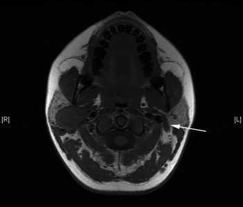
Magnetic resonance imaging revealed a well-defined mass medial to the parotid gland and displacing it laterally and anteriorly. A slightly increased signal was seen on T1-weighted images. Both T2-weighted images and gadolinium-enhanced T1-weighted images demonstrated a markedly increased signal (Figure 1). These findings were consistent with the previously suggested diagnosis of an intramuscular haemangioma.
Axial, T1-weighted magnetic resonance image showing a tumour of the posterior belly of the digastric muscle. An arrow indicates the contralateral digastric muscle. R = right; L = left

Angiography showed an abnormal vascular ‘blush’ deep to the parotid gland, supplied by a branch of the occipital artery.
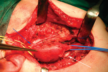
Following a standard parotidectomy incision and identification of the main trunk of the facial nerve, it was apparent that the tumour was arising from the posterior belly of the digastric muscle (Figure 2). The mass was well defined, and was removed, with the whole posterior belly of the digastric muscle, without any significant bleeding.
Intra-operative photograph showing a tumour arising within the posterior belly of the digastric muscle. Arrows point to the facial and hypoglossal nerves.

The surgery and the post-operative course were uneventful.
The final histology report described striated muscle together with a lesion composed of mainly thin-walled, vascular spaces of varying calibre, i.e. an intramuscular haemangioma.
At the time of writing, the patient had remained free of recurrence, with normal facial nerve function, for more than eight months after excision.
Discussion
Intramuscular haemangiomas of the digastric muscle are very rare lesions, which may mimic submandibular or parotid gland masses. Unlike paediatric haemangiomas, intramuscular haemangiomas do not appear to regress spontaneously.Reference Harar, Kalan, Brown, Path and Kenyon5
The origin of intramuscular haemangiomas remains controversial. The majority of authors propose a congenital origin because of these tumours' high incidence in early and late childhood. Others suggest that intramuscular haemangiomas are formed after repeated mechanical injury in malformed tissue.Reference Giudice, Piazza, Bolzoni and Peretti6 Wolf et al. speculate about a possible hormonal role.Reference Wolf, Daniel, Krause and Kaufman1
Intramuscular haemangiomas are usually found in large muscles. Therefore, the most common muscle involved in the head and neck region is the masseter, followed by the trapezius, orbital, sternocleidomastoid and temporalis muscles. Intramuscular haemangiomas usually present in patients under 30 years of age,Reference Slack, Milroy and Parker7 unlike cutaneous and mucosal haemangiomas of infancy.Reference Lee and Lim8 Chaudhary et al. reported a slight male preponderance.Reference Chaudhary, Jain, Gudwani, Kapoor and Motwani9 In the five previously reported cases (reviewed here) and our case there were a total of two male and four female patients.
Histopathological findings and prognosis vary between the three subtypes of skeletal muscle haemangioma – capillary, cavernous and mixed – as classified by Allen and Enzinger.Reference Allen and Enzinger10 In intramuscular haemangiomas, the capillary variant accounts for 68 per cent of lesions, and has a recurrence rate of approximately 20 per cent following excision. The cavernous subtype, comprising vessels with a larger diameter, accounts for 26 per cent, with a recurrence rate of less than 9 per cent. Lastly, the mixed type, seen in 6 per cent of patients, contains a combination of capillaries and large vessels, and has a recurrence rate of 28 per cent.Reference Giudice, Piazza, Bolzoni and Peretti6
Difficulty in establishing an accurate pre-operative diagnosis of intramuscular haemangioma is well described. Such difficulty is due to the non-specific clinical manifestation and rarity of intramuscular haemangiomas in the head and neck region. They are pre-operatively diagnosed in only 8 per cent of cases.Reference Giudice, Piazza, Bolzoni and Peretti6 Despite their vascular origin, intramuscular haemangiomas lack typical symptoms such as pulsation, fluctuation and audible bruits, because they are typically encapsulated by a fibrotic sheet of muscle.
In our institution, patients with a neck lump are first seen in the ‘lump clinic’ and are referred for an ultrasound scan. Provided the ultrasound does not demonstrate a vascular lesion, the radiologist performs either fine-needle aspiration or core-needle biopsy. Ultrasound-guided core-needle biopsy is an established technique used in numerous anatomical sites. The development of non-advancing needles has recently resulted in an increased use in head and neck masses.
There is a widespread perception that core-needle biopsy is contraindicated in haemangiomas due to a high risk of bleeding.Reference Colagoclu, Tafiran, Yazici, Buyrac and Ünsal11 In contrast to other haemangiomas, intramuscular haemangiomas contain variable amounts of non-vascular tissues such as fat, smooth muscle and fibrous tissue.Reference Clement, Graham, Rawlings, Ablett and Dempster12 These non-vascular elements, together with compression by surrounding structures preventing stagnation of blood within the tumour, may mask the tumour's vascular nature.
Fine-needle aspiration biopsy seems to be inconclusive in intramuscular haemangiomas, as it reveals blood only. Multiple fine-needle aspiration biopsies have not been shown to be useful to obtain representative cellular material.Reference Slack, Milroy and Parker7, Reference Sichel, Rosenmann, Eliashar and Chisin13 In the presented patient, the correct pre-operative diagnosis of an intramuscular haemangioma was established with core-needle biopsy. This technique is our primary method of tissue retrieval from head and neck pathologies (with the exception of vascular and thyroid tumours), and has been used in over 2000 procedures.
All patients with a suspected vascular lesion should be referred for MRI and angiography. Magnetic resonance imaging is usually diagnostic for haemangiomas, with a high signal being seen on both T1- and T2-weighted images and on gadolinium-enhanced images (Figure 1).
In view of the infiltrative nature of intramuscular haemangiomas, the optimal management is wide surgical excision including a cuff of surrounding muscle.Reference Lee and Lim8
Out of all six reported cases of intramuscular haemangioma of the digastric muscle, only two cases were successfully identified pre-operatively (i.e. our case and that of Sayan et al. Reference Sayan, Kogo, Koizumi, Watatani, Saka and Matsuya14) (Table I). The case report by Sayan et al. was published long before the wide availability of MRI scanners.Reference Sayan, Kogo, Koizumi, Watatani, Saka and Matsuya14 The lesion in these authors' paper was diagnosed with computed tomography (identifying the vascularity of the lesion) and plain X-ray (showing phleboliths).Reference Sayan, Kogo, Koizumi, Watatani, Saka and Matsuya14 In our patient, the definitive diagnosis was established by core-needle biopsy and MRI scan.
Reports of intramuscular haemangioma of digastric muscle

Y = years; F = female; M = male; post = posterior; ant = anterior; FNA = fine-needle aspiration; CT = computed tomography; US = ultrasound; MRI = magnetic resonance imaging; pre-op = pre-operative
Clement et al. have commented that more careful interpretation of MRI scans may lead to a precise pre-operative diagnosis of intramuscular haemangioma.Reference Clement, Graham, Rawlings, Ablett and Dempster12 This opinion was relevant to our case, as the initial report of the MRI scan failed to place the lesion accurately within the posterior belly of the digastric muscle. However, closer review clarified its true position.
• Head and neck intramuscular haemangiomas are rarely accurately diagnosed pre-operatively
• Their vascularity is often not evident on ultrasonography
• Core-needle biopsy is a safe diagnostic procedure for such tumours
• Magnetic resonance imaging is helpful for anatomical localisation
• Treatment involves a wide excision, following identification of the facial nerve main trunk
Accurate pre-operative diagnosis allows better pre-operative planning. However, we disagree with Clement and colleagues' statement that knowing the correct diagnosis pre-operatively could enable excision of the lesion without the need to expose the facial nerve.Reference Clement, Graham, Rawlings, Ablett and Dempster12 We believe that only a superficial parotidectomy approach with wide exposure of the facial nerve can enable safe excision of a lesion within the posterior belly of the digastric muscle.
Conclusion
This report demonstrates the usefulness of the combined investigative modes of ultrasound-guided core-needle biopsy and meticulous MRI review in the accurate pre-operative diagnosis of intramuscular haemangioma of the posterior belly of the digastric muscle.
Acknowledgement
We gratefully acknowledge the critical and thorough review of the manuscript given by Mr S Toynton.