

Introduction
Adipose tissue, once thought to be ‘just fat’, is biologically active tissue that can secrete hormones which play a role in the control of body mass and energy. Leptin is a peptide hormone produced by adipocytes that acts on the central nervous system to inhibit food intakeReference Havel1, Reference Hadley and Levine2 and increase metabolic rate or thermogenesisReference Jequier3. Leptin is positively correlated with fat mass in horsesReference Kearns, McKeever and Malinowski4, Reference Kearns, McKeever, Roegner, Brady and Malinowski5 and humansReference Jequier3, Reference Castracane, Kraemer, Franken, Kraemer and Gimpel6, Reference Moller, O'Brien and Sreekumaran-Nair7. Functionally, leptin inhibits food intake and interacts with a variety of metabolic pathways to facilitate increases in energy expenditure. As such, leptin plays a major role in the defence of overall energy homeostasis, which in turn affects body composition and body mass via adjustments in total fat massReference Havel1, Reference Hadley and Levine2. Disruption in the control of leptin production, secretion, receptor number or receptor sensitivity has been linked to a spectrum of health-related disorders associated with energy homeostasis, including decreases in food intake with exercise training and excessive food intake and obesityReference Havel1, Reference Hadley and Levine2.
The role of leptin in the control of appetite is complex, and it serves as a signalling protein in a series of interactive endocrine, paracrine and cytokine pathways communicating the magnitude of the total energy stored in the adipose tissue of the bodyReference Myers, Cowley and Munzberg8. Leptin receptors are present in the nuclei of the hypothalamus and control appetite by stimulating the synthesis of pro-opiomelanocortin, a precursor to α-melanocyte-stimulating hormone, which is a signal for anorexiaReference Myers, Cowley and Munzberg8. Leptin also inhibits the synthesis of neuropeptide Y that is known to be orexigenicReference Myers, Cowley and Munzberg8. A failure of this mechanism is known as leptin resistance. Research has shown deficiencies in leptin receptors in cases of obesity, where food consumption may not decrease despite elevated leptin concentrationReference Myers, Cowley and Munzberg8.
There appears to be an age-related alteration in the control of leptin production, secretion and receptor sensitivity as well as its influence on metabolism in humans and rodentsReference Myers, Cowley and Munzberg8, Reference Fatouros, Tournis, Leontsini, Jamurtas, Sxina and Thomakos9. Disruption in the control of the feedback mechanisms affecting leptin may contribute to the increase in obesity seen with age. In a study of 122 elderly women with high–normal body mass index, leptin was negatively correlated with ageReference Neuhauser-Berthold, Herbert, Lührmann, Sültemeier, Blum and Frey10. This was not observed in menReference Neuhauser-Berthold, Herbert, Lührmann, Sültemeier, Blum and Frey10. However, in the study by Moller et al. Reference Moller, O'Brien and Sreekumaran-Nair7, it was found that relative fat mass was positively correlated with leptin in young humans [~25 with a decline in the statistical association between relative fat mass and plasma leptin concentration as age progressed in both men and women (~70 years)]. Castracane et al. Reference Castracane, Kraemer, Franken, Kraemer and Gimpel6 reported substantially higher plasma concentrations of leptin in older women, both with and without hormone replacement therapy, versus younger women not on oral contraceptives. The influence of gender has been noted in several studies where it has been documented that males have a lower leptin concentration than femalesReference Castracane, Kraemer, Franken, Kraemer and Gimpel6, Reference Moller, O'Brien and Sreekumaran-Nair7, Reference Kedzierski and Kapica11. This may be related to gender differences in body fat deposition. However, it also may be related to endocrine factors, as noted by JequierReference Jequier3 and othersReference Havel1, Reference Hadley and Levine2 who have reviewed the relationship between leptin and sex hormones in men and women. The latter would certainly have an impact on whole body energy balance and the endocrine factors related to maintenance of overall body adiposity.
Acute exercise does not appear to have a profound effect on circulating leptin concentrations in humans or horsesReference Gordon, McKeever, Betros and Manso Filho12–Reference Gordon and McKeever16. For example, in a study of humans, no changes in plasma leptin were shown in fit male rowers exercising at maximum sustained effort for 20 min, demonstrating that there is no effect of intense acute exerciseReference Jurimae, Purge and Jurimae17. Similarly, no changes were seen in the production or concentration of leptin in untrained males who performed 60 min of moderate exercise on a cycle ergometerReference Racette, Coppack, Landt and Klein13. Similarly, Hickey et al. Reference Hickey, Considine, Israel, Mahar, McCammon and Tyndall18 suggested that acute exhaustive exercise has no immediate effects on leptin concentrations in trained individuals. However, those studiesReference Racette, Coppack, Landt and Klein13–Reference Hickey, Considine, Israel, Mahar, McCammon and Tyndall18 only measured the immediate effects of one exercise session on plasma leptin concentrations. As in humans, Gordon et al. Reference Gordon, McKeever, Betros and Manso Filho12 demonstrated that acute exercise and exercise training do not affect leptin during exercise or immediately after, but a decrease in leptin was shown after a 24 h recovery period. A study of trained jumping horses showed no change in leptin immediately after and 24 h after an exercise sessionReference Piccione, Grasso, Costa, Fazio and Caolo19. The former study was in Standardbred mares on an equine treadmill, with the latter study being conducted in an arena setting. Methodology and equipment may have played roles in the intensity and variation of the exercise and the discrepancy of resultsReference Piccione, Grasso, Costa, Fazio and Caolo19. In contrast, Kedzierski and KapicaReference Kedzierski and Kapica11 found that plasma leptin concentration increased over resting concentrations in samples taken at the end of exercise and at 30 min post-exercise in 1.5-year-old Standardbreds in training for 6 months. This effect was observed only in winter and not in summer. Furthermore, the authors did not observe an effect of acute exercise in horses older than 1.5 yearsReference Kedzierski and Kapica11. Together, this suggests that plasma leptin concentration is influenced by both by seasonal and age-related effects. Higher plasma leptin concentrations were also observed in fillies, and the authors speculated that this increase in plasma leptin concentration in the youngest group of horses was due to a strong physiological challenge, where the role of glucocorticoids would alter metabolismReference Kedzierski and Kapica11.
However, repeated exercise that challenges energy stores has been shown to influence plasma leptin concentrations and alter body compositionReference Gordon, McKeever, Betros and Manso Filho12, Reference Racette, Coppack, Landt and Klein13. Gordon et al. Reference Gordon, McKeever, Betros and Manso Filho12 demonstrated that fit Standardbreds have substantially lower fat mass and consequentially lower leptin concentrations than unfit Standardbreds, where fit Standardbreds had been in training for over 1 year. Another study by Gordon et al. Reference Gordon, McKeever, Bokman, Betros, Manso and Filho15 observed that trained horses had lower plasma leptin concentrations than untrained horses, where the training period was 10 weeks. In a study of unfit older men, Fatouros et al. Reference Fatouros, Tournis, Leontsini, Jamurtas, Sxina and Thomakos9 examined the effect of three different training intensities and found that plasma leptin concentration decreased with training and then increased with detraining.
There have been a number of published papers that have demonstrated that various physiological systems, including those related to the endocrine response to acute exercise, are affected by ageing and training (or the lack of exercise training analogous to a sedentary lifestyle in humans and horses)Reference McKeever and Bertone20. It follows logically that, since leptin is a key signalling hormone involved in whole body energy balanceReference Havel1, then the disruption of the normal leptin signalling response in the horse could be linked to the onset of obesity and the resultant negative effects of excessive body adiposity. This would make leptin a hormone that is potentially important in the ageing process. Modulating or reversing the negative effects of ageing on the endocrine control of metabolic processes would be a beneficial effect of moderate exercise training. Therefore, the objective of this study was to test the hypothesis that ageing and training would alter whole body adiposity as well as plasma concentrations of the peptide hormone leptin.
Materials and methods
All tests and laboratory procedures were approved by the Rutgers University Institutional Review Board for the Care and Use of Animals. Twelve unfit and mature Standardbred mares, ‘young’ (7.3 ± 0.6 years; n = 6) and ‘old’ (22 ± 0.7 years; n = 6), were used in the study. Prior to the study, all of the mares were examined by a veterinarian and confirmed to be healthy. Each horse received routine deworming treatments, vaccinations, hoof trims and other care as per the standard operating procedures of the Rutgers Animal Care staff. Mares were unfit and had not been trained for several years. Mares were maintained in groups at the equine research facility on 24 h turnout in which they were all acclimated to the treadmill, the Equi-ciser and all procedures used in the laboratory. Water, grass hay and a salt block were offered ad libitum. One kilogram of commercially available pelleted feed was fed twice daily as maintenance. The calories provided exceeded the minimum requirements stipulated by the National Research Council21 for mature horses weighing 500 kg doing moderate work (~23.3 Mcal day− 1).
Data and sample collection presented in the present paper were part of a multiple-investigator study designed to address several hypotheses related to the effects of ageing and training. As such, the general experimental design featured measurement periods centred on the performance of three exercise tests, as outlined below.
Graded exercise tests (GXTs) and plasma collection
Each mare performed an incremental GXT in June, August and October of 2009. The mares were fed only grass hay (concentrates withheld) on the morning of the testReference McKeever and Malinowski22, Reference Kearns and McKeever23. Each test was performed between 08.00 and 11.00 hours in a climate-controlled treadmill laboratory. Heart rate (HR) was measured using an external equine heart rate monitor (Polar Equine, Lake Success, NY, USA), and gas exchange was measured using an open flow calorimeter (Oxymax-XL; Columbus Instruments, Columbus, OH, USA). The HR versus speed relationship was used to calculate the velocity used in the training sessions, as detailed belowReference Kearns and McKeever23. The maximal rate of oxygen consumption was determined as per prior methodsReference Kearns and McKeever23 and used to document an exercise training effect between weeks 0 and 8 and the maintenance of that state of training between weeks 8 and 16. During the GXT, the treadmill was set at a 6% incline. The tests started at an initial speed of 4 m s− 1 for 60 s, after which the speed was increased to 6 m s− 1 for 60 s and then increased by 1 m s− 1 every 60 s until fatigue was reached. Fatigue was defined as the point where the mare could not keep up with the treadmill despite humane encouragement. Blood samples (30 ml) were collected via an intravenous catheter placed in the left jugular vein 30 min before each test. Blood was collected prior to each of the three GXTs when the mares were standing quietly in stalls. Aliquots were placed into 10 ml tubes containing the anticoagulant EDTA (Vacutainer; Becton Dickinson, Parsippany, NJ, USA) and placed on ice until being centrifuged for 10 min at 4°C. Plasma was then aliquoted into two separate 2 ml cryovials and placed in a freezer at − 80°C until analysis.
Exercise training protocol
Exercise training was performed at the Rutgers Equine Exercise Physiology Laboratory using a free-stall motorized circular equine exercise machine (Equi-Ciser; Equi-Master International, Sudre, AB, Canada). Mares were divided into two evenly matched groups of six based on the HR data collected during their first GXT. Training was conducted between 06.00 and 10.00 hours from June to October, and lasted for 40 min on Mondays, Wednesdays and Fridays. Mares were weighed weekly prior to training. The 40 min session on the Equi-Ciser included a 5 min warm-up period at a brisk walk, 30 min of trotting and 5 min of cool-down at a walk. The speed of the Equi-Ciser was set based on the HR data from the pre-training GXT of one randomly selected horse for each training session, in each group of six, wearing the HR monitor. The goal was to keep the individual with the monitor working at a submaximal work intensity of 60% HRmax as determined from GXT data, for the trotting period (20 min) of the session. The Equi-Ciser was programmed automatically to change direction every 5 min.
Body composition
Body weight was measured weekly with an electronic livestock scale prior to each GXT session. Rump fat thickness, the directly measured variable used in indirect calculations of body fat percentageReference Westervelt, Stouffer, Hintz and Schryver24, Reference Kearns, McKeever and Abe25, was determined using ultrasound and the location on the rump was as described by Westervelt et al. Reference Westervelt, Stouffer, Hintz and Schryver24, 5–10 cm below the dorsal midline between the point of the hip and sacral bone on each side and used previously to assess rump fat thickness in Standardbred maresReference Kearns, McKeever and Malinowski4, Reference Kearns, McKeever, Roegner, Brady and Malinowski5.
Leptin radioimmunoassay
A multi-species radioimmunoassay kit (Cat no. XL-85K; Millipore, Stillwater MN, USA) validated for horsesReference McManus and Fitzgerald26 was used to measure plasma leptin concentration. Samples were stored at − 80°C from the time of collection until analysis. Samples were run in duplicate in a single assay run with a within-assay coefficient of variation of 3.8%. A sample volume of 200 μl was used in this assay. Plasma leptin concentrations are expressed as ng ml− 1 human equivalents.
Statistical analysis
Data were analysed for main effects and interactions using two-way ANOVA with repeated measures (Quattro Pro 8.0; Corel, Mountain View, CA, USA; Sigmastat 3.12; SPSS, Chicago, IL, USA). Where appropriate, the Student–Newman–Keuls test was used to identify post hoc differencesReference Little and Hills27. To protect against a type II error, the null hypothesis was rejected when P < 0.10Reference Little and Hills27.
Results
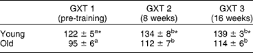
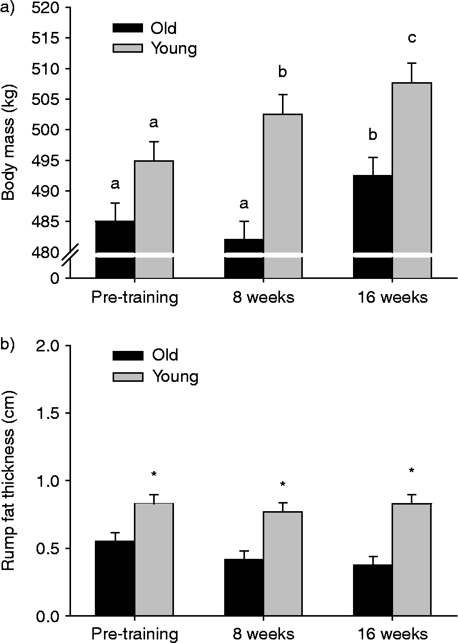
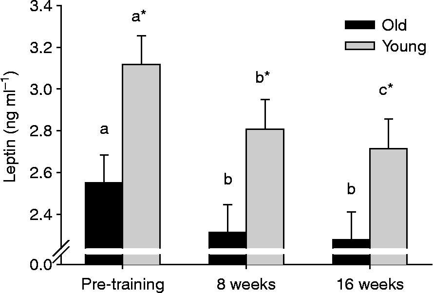
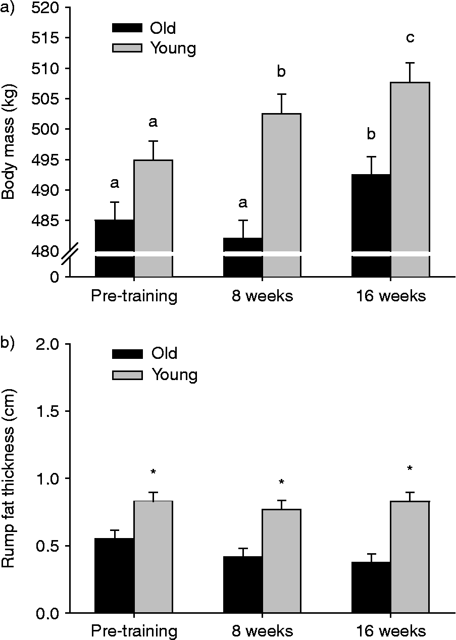
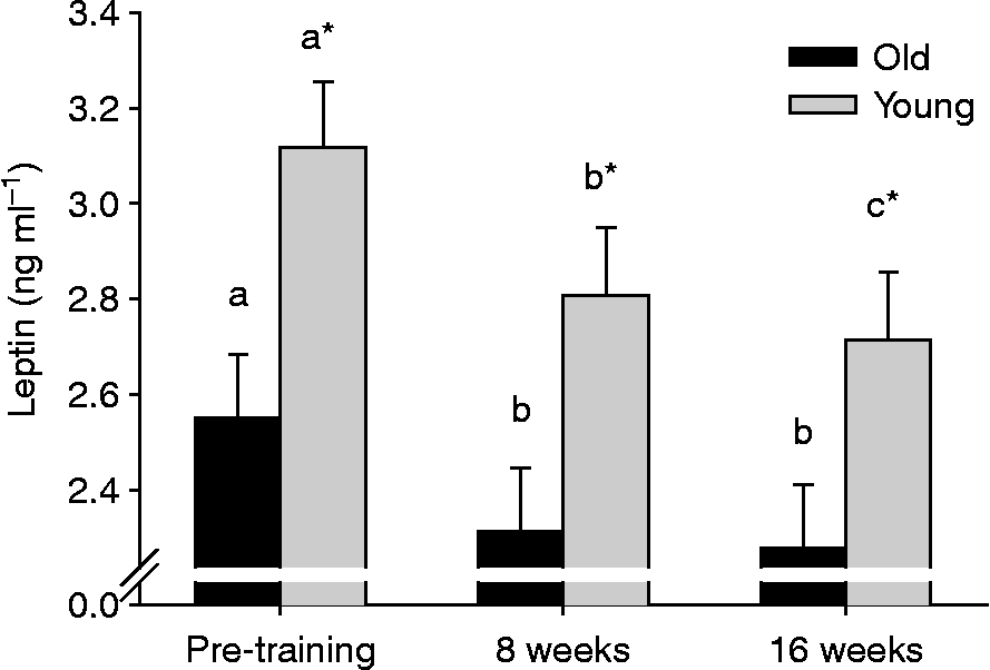
There was a significant difference in maximal aerobic capacity (VO2max) due to age (Table 1). Training increased VO2max in both groups by 8 weeks. Between 8 and 16 weeks of training, there were no decreases or further increases in the intensity or duration of exercise, and thus there was a maintenance of aerobic capacity and no change in VO2max. There were no differences in body mass (Fig. 1a) due to age (P>0.10); however, training caused an increase (P < 0.10) in body mass in both groups. This increase was significant in the young horses by 8 weeks and in the old horses by 16 weeks of training. Interestingly, when one examines rump fat thickness (Fig. 1b), the directly measured parameter that reflects fat mass, one observes that there were age-related differences in rump fat thickness before training and at 8 and 15 weeks of training in both groups. However, there was no effect of training on rump fat thickness. An increase in body mass without a concurrent increase in rump fat thickness indicates an increase in fat-free mass, the latter reflecting primarily muscle mass. Finally, there were significant differences in plasma leptin concentration due to ageing and training, with lower concentration in the old horses at all time points. Training resulted in a reduction (P < 0.10) in plasma leptin concentration in both groups of horses (Fig. 2).
Maximal oxygen uptake (ml kg−1 min−1)i
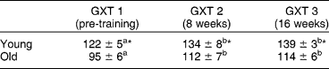
a,b Within an age group, the symbol denotes that the mean was significantly different from pre-training, and the same symbol indicates that there was no difference between 8 and 16 weeks.
* A sample time denotes a difference due to age.
i Mean ± SE values for maximal oxygen uptake (VO2max) measured in young and old horses before and after 8 and 16 weeks of submaximal exercise training.
(a) Mean ± SE values for body mass and (b) rump fat thickness (VO2max) measured in young and old horses before and after 8 and 16 weeks of submaximal exercise training. There were no differences in body mass (a) due to age; however, within an age group, means with a different letter were significantly different. For rump fat thickness (b), an asterisk (*) within a sample time point denotes a difference due to age
Mean ± SE values for plasma leptin concentration measured in young and old horses before and after 8 and 16 weeks of submaximal exercise training. An asterisk (*) within a sample time denotes a difference due to age. Within an age group, means with different letters are significantly different
Discussion
The major finding of the present study was an effect of ageing and training on fat-free mass and plasma leptin concentration. The lower pre-training plasma leptin concentration measured in the old horses before training was consistent with their lower fat mass, as reflected in their lower rump fat thickness. A lower pre-training plasma leptin concentration would be expected with a lower fat mass as established and consistent with previous published studies of horsesReference Kearns, McKeever, Roegner, Brady and Malinowski5, Reference Gordon, Betros, Manso and McKeever14.
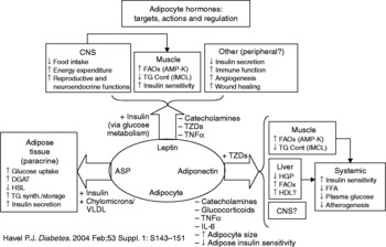
Interestingly, while training resulted in a lower plasma leptin concentration, the decrease did not appear to be related to a decrease in fat mass. Fat mass remained relatively constant for each age group throughout the study. However, overall body mass increased, which logically must have come about via an increase in fat-free mass that is primarily muscle. Most studies focus on the relationship between fat mass and plasma leptin concentration; however, the metabolic demand of a training-induced increase in muscle mass would also be expected to have a substantial effect on whole body energy demand and overall energy stores. This would appear to be contrary to what has been previously observed in horses, where the relationship between total body adiposity and plasma leptin concentration has been a major focus. However, there is information in the literature to suggest that leptin is involved in signalling pathways associated with all metabolically active tissues affected by whole body energy balance (Fig. 3)Reference Havel1. The lower leptin concentrations observed in the present experiment are consistent with the reported action of leptin in pathways affecting metabolically active tissues like muscle in addition to adipose tissue. The results are also consistent with vertical studies of fit versus unfit Standardbred horses. For example, Gordon et al. Reference Gordon, Betros, Manso and McKeever14 demonstrated that fit horses have significantly lower plasma leptin than unfit horses and that leptin is related to fat mass. The energy required to increase body mass must come from the diet. A decline in plasma leptin therefore must allow for the caloric intake necessary to alter body composition according to training, or plasma leptin could be further suppressed in an attempt to maintain pre-training body compositionReference Havel1. The decline in plasma leptin and fat mass may be related to the challenge to maintain overall energy homeostasisReference Havel1.
Schematic diagram of suggested pathways for the regulatory actions and interactions of leptin and other adipokines in metabolically active tissues (used with permissionReference Havel1)

In conclusion, age and training appear to alter the relationship between body composition and leptin, where leptin is not related to a change in body fat mass but has a more complex signal integration and role in overall energy homeostasis. It is clear from past studies of humans and horses that endocrine, immune and other systemic disruptions of the integrative physiological response to exercise occur with age and exercise trainingReference McKeever and Bertone20, Reference Little and Hills27–Reference Malinowski, Betros, Flora, Kearns and McKeever30. Those alterations in the integration of multiple systems to meet the challenge of exercise may have implications for how the ageing horse's ability to perform exercise is assessedReference McKeever and Bertone20.
Acknowledgements
The authors would like to thank the graduate and undergraduate students from Rutgers University and the Rutgers Equine Science Center for their contributions to this project. Special thanks to Clint Burgher, Joanne Powell and Anthony Sachetti of the Rutgers University Animal Care Staff for their invaluable assistance. Support for the study was provided by the New Jersey State Equine Initiative, the New Jersey Agricultural Experimentation Station and the George H. Cook Scholars Program. None of the authors of this paper has a financial or personal relationship with other people or organizations that could inappropriately influence or bias the content of the paper.