Acute myocardial infarction is rarely reported in children. Most of the cases are secondary to congenital anomalies or Kawasaki disease.Reference Cui 1 – Reference Thomas, Ramachandran, Jefferies, Beekman, Hor and Lorts 4 Coronary artery total occlusion caused by fibromuscular dysplasia has never been reported in young children.Reference Hill and Sheppard 5 Here we report a case of a 12-year-old boy with fibromuscular dysplasia, who underwent successful coronary intervention for acute myocardial infarction, as well as his long-term outcome.
Case report
A 12-year-old boy presented with chest pain, pallor, and vomiting that occurred suddenly in the early morning. The boy denied any cold sweating, numbness, dizziness, weakness, or shortness of breath. He had a history of fibromuscular dysplasia with multiple renal artery stenoses, and an episode of hypertensive encephalopathy. His hypertension was under good control after successful percutaneous renal artery angioplasty and regular intake of amlodipine.Reference Chen, Lee, Chang, Lin, Fu and Jan 6 His weight was 37 kg, height 145 cm, and blood pressure 100/70 mmHg. Physical examination revealed bradycardia of 55 beats/minute with no heart murmurs. He had clear breath sounds, no hepatosplenomegaly, and no pitting oedema of the lower limbs.
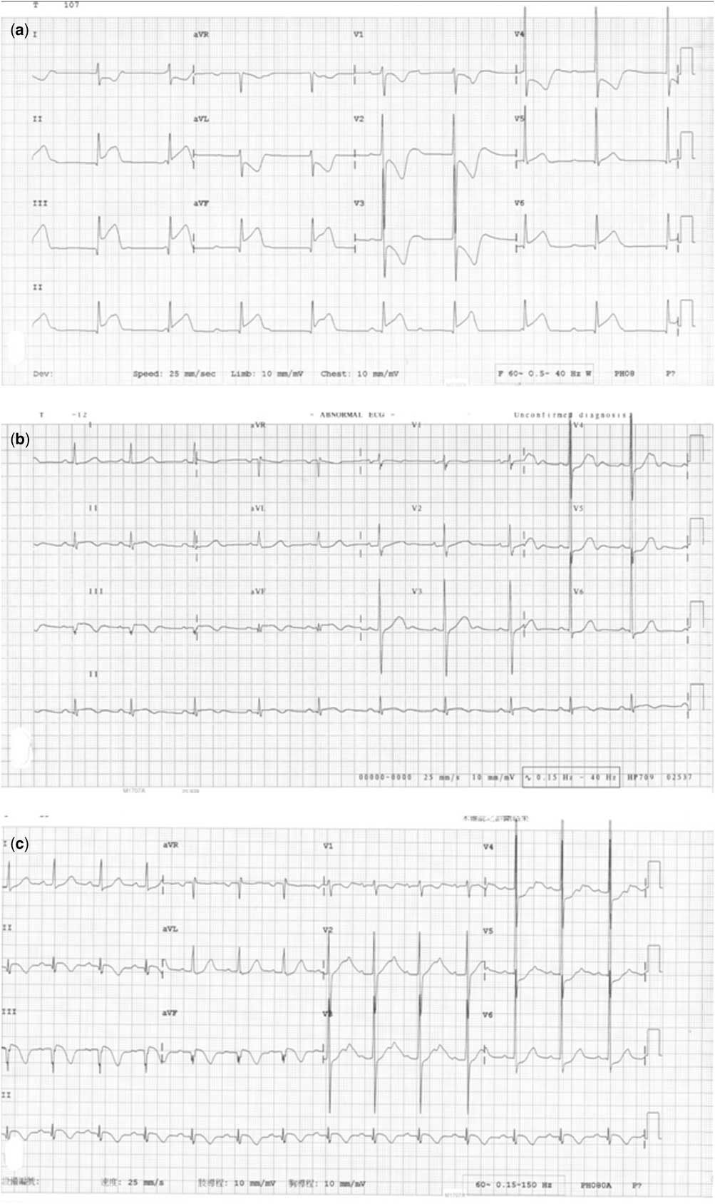
Electrocardiogram at the emergency room revealed complete atrioventricular block, with a ventricular rate of 55 bpm (Fig 1a). The serum creatine kinase-MB isoenzyme level was 9 U/L (reference range, <16 U/L), with creatine kinase 146 U/L (reference range, 10~160 U/L). Troponin I level was 0.042 ng/ml (reference range, <0.034 ng/ml), while C-reactive protein was 0.19 mg/dl. Acute myocarditis was suspected and the patient was admitted to the paediatric intensive care unit for observation.
( a ) Initial electrocardiography revealed complete atrioventricular block with a ventricular rate of 55 beats/minute and ischaemic changes in leads II-III-aVF. ( b ) The heart rate returned to sinus rhythm with Q wave in lead III after admission. ( c ) The electrocardiography later showed significant ST elevation at the inferior, reciprocal changes on the left precordial leads, and Q wave in lead III and aVF.

After admission, his heart rate returned to sinus rhythm (Fig 1b). Echocardiography showed an ejection fraction of 67%, with past noted problems of a mildly dilated aortic root (2.82 cm) and a bicuspid aortic valve. As he was being observed at the intensive care unit, his chest pain suddenly worsened. Repeat electrocardiogram showed significant ST elevation in the inferior with reciprocal changes in the left precordial leads (Fig 1c). The troponin I level also elevated to 30.7 ng/ml. Emergency coronary angiography and percutaneous transluminal coronary angioplasty were performed.
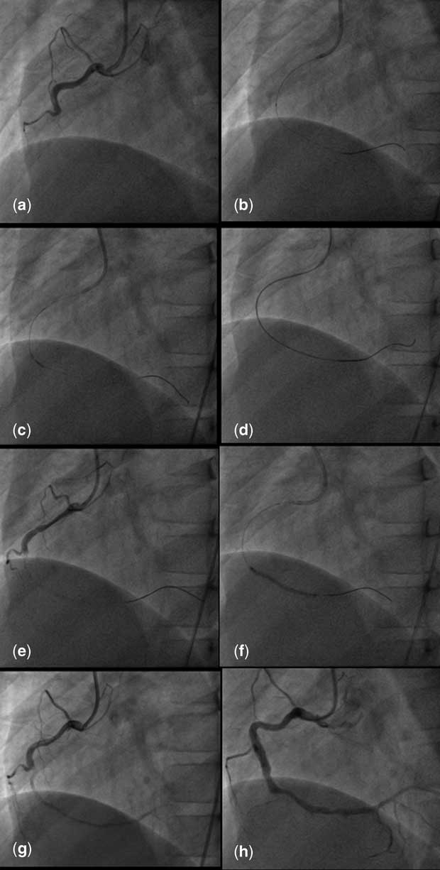
After establishing vascular access (6 Fr sheath) via the right femoral artery, coronary angiography showed total occlusion at the mid-portion of the right coronary artery (Fig 2a; Movie 1). On angiography, the left coronary artery was seen to be intact. A 5 Fr JR4 guide catheter was used to enter the right coronary artery and the long completely occluded segment was transverse with a Fielder FC coronary guide wire (Asahi Intecc Co., Aichi, Japan) supported by a 1.8 Fr FineCross MG microcatheter (Terumo Co., Tokyo, Japan) (Fig 2b).
( a ) Angiography of the right coronary artery showed total occlusion at the mid-portion. ( b ) The long, totally occluded segment was wired by a Fielder FC coronary guide wire and ( c ) dilated with a 1.25 mm balloon catheter. ( d ) The tightly occluded distal segment was crossed with a Tornus microcatheter and Grandslam wire. ( e ) The test shot showed no antegrade flow after 1.5 mm balloon catheter. ( f ) The right coronary artery mid-portion was further dilated with 2 and 2.5 mm balloon catheters and ( g ) antegrade flow was re-established. ( h ) Coronary angiography taken 6 months later revealed a patent right coronary artery without re-stenosis.

The lesion at the mid-portion of the right coronary artery was first dilated with a 1.25×15 mm balloon catheter up to 10 atmospheric pressures (Fig 2c). However, the distal portion could not be crossed with either a 1.25 mm or a 1.5 mm balloon catheter. The tightly occluded distal segment was then crossed with a Tornus microcatheter (Asahi Intecc Co.) and the wire was exchanged for a 0.014-inch Grandslam guide wire (Asahi Intecc Co.) (Fig 2d). The mid- to distal portion was dilated using a 1.5×20 mm balloon catheter up to 10 atmospheric pressures, but there was no antegrade flow (Fig 2e).
The FineCross micro-catheter was advanced again to the distal portion of the right coronary artery and intraluminal placement of the wire was verified by injection of contrast. Then the right coronary artery mid-portion was further dilated with 2×20 mm and 2.5×20 mm balloon catheters up to 10 atmospheric pressures (Fig 2f) and the posterior lateral branch by a 1.5×20 mm balloon catheter up to 10 atmospheric pressures. Antegrade flow was finally re-established despite type E dissection at the mid-portion of the right coronary artery (Fig 2g).
We did not stent because of possible complications with growth in this still young patient. He tolerated the procedure well and had an uneventful hospital course. He was then put on dual anti-platelets of aspirin and clopidogrel. Coronary angiography 6 months later revealed a patent right coronary artery without restenosis (Fig 2h; Movie 2). His medication was then changed into regular aspirin therapy.
Discussion
Although fibromuscular dysplasia is a common cause of renal-vascular hypertension in children,Reference Bayazit, Yalcinkaya and Cakar 7 – Reference Tullus, Brennan and Hamilton 9 the involvement of coronary arteries is very rare.Reference Hill and Sheppard 5 For children as young as 12 years, acute myocardial infarction caused by fibromuscular dysplasia has never been described.
Acute myocardial infarction is rarely reported in children. Most of the cases are secondary to congenital anomalies or Kawasaki disease.Reference Cui 1 – Reference Thomas, Ramachandran, Jefferies, Beekman, Hor and Lorts 4 Coronary intervention by percutaneous transluminal coronary angioplasty in young children for acute myocardial infarction is quite uncommon.Reference Cui 1 – Reference Thomas, Ramachandran, Jefferies, Beekman, Hor and Lorts 4 , Reference Akagi 10 Although some authors describe good immediate outcome, long-term outcome is lacking in those reports.
The real cause of fibromuscular dysplasia is unknown, although some hormonal and mechanical factors have been proposed. Familial cases account for 10% of cases. The renal artery is the most commonly involved site. It plays an important role in the pathogenesis of paediatric secondary hypertension.Reference Bayazit, Yalcinkaya and Cakar 7 – Reference Tullus, Brennan and Hamilton 9 However, involvement of the coronary artery is rarely seen. A large series from the United Kingdom between 1994 and 2008 had only two cases of fibromuscular dysplasia of the coronary arteries in 50 non-atherosclerotic coronary cardiac cases, accounting for 0.1% of all-cause sudden cardiac death. The ages of the two patients were 23 and 18 years.Reference Hill and Sheppard 5 Coronary events due to fibromuscular dysplasia among school-aged children have never been reported.
The occlusion of the right coronary artery is caused by fibromuscular dysplasia rather than by acute thrombosis. Two-and-a-half years after the percutaneous transluminal coronary angioplasty, coronary angiography revealed critical stenosis in the same site.
In summary, although the renal and carotid arteries are the most commonly affected vessels by fibromuscular dysplasia,Reference Bonvini, Rastan, Sixt, Righini, Hofstetter and Zeller 11 the coronary arteries should also be considered. Percutaneous transluminal coronary angioplasty is an effective therapeutic option for children as young as 12 years.
Acknowledgement
This research received no specific grant from any funding agency, commercial or not-for-profit sectors. We also thank Dr Sheng-Ling Jan for sharing his experience on the critical care for this boy.
Conflicts of Interest
None.