Introduction
Rhinella arenarum females, which are toads widely found in Argentina, present an eminently seasonal reproductive cycle as do other amphibian species (Díaz-Paniagua, Reference Díaz-Paniagua1990). In fact, the reproductive ability is limited potentially to the months of the year in which not only the physiological conditions but also the environmental conditions (temperature, photoperiods, food availability and rainfall) are adequate for the development of the processes of gametogenesis, ovulation, fertilization and embryogenesis (Fernández & Ramos, Reference Fernández, Ramos and Jamieson2003).
In the reproductive cycle of this species we can identify a reproductive and a postreproductive stage. During the former stage, the ovary reaches its maximum degree of structural and functional development and the female is prepared physiologically for ovulation. The fully grown ovarian oocytes that have already completed their cytoplasmic and nuclear maturation are released into the coelomic cavity to enter the oviduct, pass through the whole duct and finally emerge to be fertilized (Winik et al., Reference Winik, Alcaide, Crespo, Medina, Ramos and Fernández1999; Fernández & Ramos, Reference Fernández, Ramos and Jamieson2003; Medina et al., Reference Medina, Ramos, Crespo, González-Calvar and Fernández2004).
During the postreproductive period, characterized by the recovery of the reproductive system, oviductal development and differentiation as well as follicle growth occur and the final stage of oogenesis is reached (Valdez Toledo & Pisanó, Reference Valdez Toledo and Pisanó1980). During this process, and under the coordinated action of the gonadotrophic and steroid hormones (Ramos et al., Reference Ramos, Cisint, Crespo, Medina and Fernández2001), the gamete starts an orderly and defined sequence of metabolic and cytological changes that determine cytoplasmic and nuclear maturation.
In numerous amphibian species it has been demonstrated that only those oocytes that pass through the oviduct and contact the products synthetised and released into the lumen of this duct acquire the capacity to be fertilized (Elinson, Reference Elinson1971; Barbieri & Budeguer de Atenor, Reference Barbieri and Budeguer de Atenor1973; Miceli et al., Reference Miceli, Fernández, Raisman and Barbieri1978a,Reference Miceli, Fernández and del Pinob; Fernández et al., Reference Fernández, Mansilla and Miceli1984; Katagiri, Reference Katagiri1987; Omata, Reference Omata1993; Olson & Chandler, Reference Olson and Chandler1999).
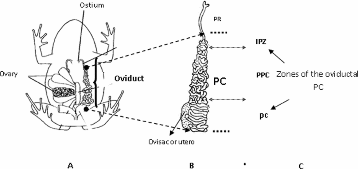
In these vertebrates, the oviducts appear as a pair of elongated ducts arranged on both sides of the body axis (Fig. 1A). From a structural and functional point of view, we can differentiate three main zones: pars recta (PR), pars convoluta (PC) and ovisac, also called pars uterina or uterus by some authors (Moreno, Reference Moreno1972; Fernández et al., Reference Fernández, Mansilla and Miceli1989a; Winik et al., Reference Winik, Alcaide, Crespo, Medina, Ramos and Fernández1999; Medina et al., Reference Medina, Crespo, Ramos, Cisint and Fernández2007; Fig. 1B). The PR, the most cephalic portion, communicates freely with the pleuroperitoneal cavity of the animal through an opening called the ostium (Fig. 1A). In Rhinella arenarum, the structural organization of this portion of the oviduct was analyzed by optical microscopy (OM) (Moreno, Reference Moreno1972) and transmission electron microscopy (TEM) (Fernández et al., Reference Fernández, Mansilla and Miceli1989a,Reference Fernández, Mansilla and Micelib; Fernández & Ramos, Reference Fernández, Ramos and Jamieson2003).
(A) Anatomical and functional organization of the reproductive system of the Rhinella arenarum female. In the diagram the position of the oviduct was plotted on one side of the body axis and the ovary on the other. (B) Two large regions of the oviduct: pars recta (PR) and pars convoluta (PC). (C) Zones of the PC involved in the secretion of jelly coat eggs: intermediate proximal zone (IPZ), pars preconvoluta (PPC) and pars convoluta (pc) itself.

From the physiological point of view, oviductal secretion contains an enzyme with proteolytic activity called oviductin (Miceli & Fernández, Reference Miceli and Fernández1982; Miceli et al., Reference Miceli, Fernández, Mansilla and Cabada1987; Hardy & Hedrick, Reference Hardy and Hedrick1992) that induces structural and functional modifications in the vitelline envelope (VE) of the oocytes, making it sensitive to sperm lysins and penetrable by sperm (Miceli & Fernández, Reference Miceli and Fernández1982; Katagiri et al., Reference Katagiri, Iwao and Yoshizaki1982; Bakos et al., Reference Bakos, Korosky and Hedrick1990; Llanos et al., Reference Llanos, Barrera, Valz-Gianinet and Miceli2006).
After the PR we find the main body of the oviduct, the PC, constituted by three regions: the intermediate proximal zone (IPZ), the pars preconvoluta (PPC) and the pars convoluta (pc) itself, which is the most distal zone of the duct (Fig. 1C). Functionally, in anuran amphibians, this region is in charge of secreting the products that form the jelly coats that surround the oocytes at the time of deposition and that represent an extracellular matrix indispensable for fertilization (Crespo et al., Reference Crespo, Ramos, Medina and Fernández2009).
At present, data concerning the histological organization of the PC in relation to the process of secretion of the jelly components are scarce. It is known that the IPZ, only described and partially studied in Rhinella arenarum (Moreno, Reference Moreno1972; Winik et al., Reference Winik, Alcaide, Crespo, Medina, Ramos and Fernández1999; Medina et al., Reference Medina, Crespo, Ramos, Cisint and Fernández2007) and Rhinella paracnemis (Alcaide & Cruz López, Reference Alcaide and Cruz López2002), represents a zone of morphological, structural and functional transition. Morphologically, in this region, the oviduct is no longer straight and the first convolutions appear, represented by the first two handles of the PPC (Moreno, Reference Moreno1972). At the ultrastructural level, from the terminal portion of the PR, incipient acinous glands appear and begin to form the glandular layer that acquires a progressive development throughout the whole PC (Winik et al., Reference Winik, Alcaide, Crespo, Medina, Ramos and Fernández1999). Finally, from the functional viewpoint, it is from this zone that the synthesis and secretion of the components of the jelly coats that surround the oocytes at the moment of fertilization begin (Crespo et al., Reference Crespo, Ramos, Medina and Fernández2009).
The PPC represents the main body of the oviduct, occupying almost two-thirds of it, and is constituted by numerous short tight handles (Fig. 1B). The rest of the duct, which corresponds to the pc, presents long wide horizontal handles with prominent curves in their tips that are arranged parallel to each other and also perpendicular to the oviduct axis. The last region of the pc and the beginning of the ovisac constitute another transition zone called intermediate distal zone.
Studies carried out in different anuran species have demonstrated that, even after traversing the PR, only those oocytes that pass through the PC and contact the products of its secretion are fertilizable. In agreement with these data, there is experimental evidence that the fertilizability of the oocytes increases during the passage of the gametes through the PC and varies as a function of the number of jelly coats that surround them (Barbieri & Budeguer de Atenor, Reference Barbieri and Budeguer de Atenor1973). In the particular case of Rhinella arenarum, the layers are four: two concentric inner layers that surround each oocyte called J1 in apposition to the VE followed by J2 and two outer tubular layers (J3 and J4), an arrangement that permits the spatial organization of the gametes resembling beads on a necklace (Barbieri & Budeguer de Atenor, Reference Barbieri and Budeguer de Atenor1973).
The aim of the present work was to analyze and determine the histological characteristics of the mucosa of the PC, with special attention to the degree of development of the secretory cells (SC) present at the epithelial and glandular level. This study was carried out in all sectors of the duct and during each period of the reproductive cycle. We also determined for the first time in anuran amphibians the sequential morphological organization of the jelly during the passage of the oocytes through the different sectors of the PC.
Materials and methods
Animals
Sexually mature Rhinella arenarum females were collected during the 2007–2011 year period in the neighbourhood of San Miguel de Tucumán, Argentina. The animals were used within 24 h after capture or stored for brief periods under appropriate humidity and temperature conditions.
Animal maintenance and experimental procedures were in accordance with the Guide for the Care and Use of Laboratory Animals (European Communities Council Directive, 1986).
Ovulation procedure
This study was performed with specimens captured during the following periods of the reproductive cycle: preovulatory period (n = 4), immediately before ovulation; ovulatory period (n = 4), just after the transit of the oocytes through the duct; early postovulatory period (n = 4), 7 days after oviposition; and late postovulatory period (n = 4), during hibernation. Specimens from the preovulatory and ovulatory periods were selected after careful control of ovarian state and ovulation signs with a Stemi DV4/DR stereomicroscope (Zeiss).
Ovulation was achieved either spontaneously or induced by intraperitoneal injection of a suspension of homologous hypophysis (Houssay et al., Reference Houssay, Giusti and Lascano González1929) preserved according to Pisanó (Reference Pisanó1956).
Histology
The oviducts obtained from the females collected during the different periods of the reproductive cycle were dissected carefully and prefixed for 5 h at 4°C in 10% formol solution–0.1 M disodium phosphate, pH 7.4. Then the PC (Fig. 1B) was sliced into the different segments to be analyzed: IPZ, PPC and pc (Fig. 1C), which were postfixed for 19 h at 4°C with the above solution. After this time, the samples were processed according to the technique of Moreno (Reference Moreno1972), embedded in Paraplast® and cut into 5-μm thick sections. In order to eliminate the Paraplast® the sections were subjected to two consecutive baths of xylol of 2 min each. Then the samples were rehydrated by passage through a series of graded alcohol baths of decreasing concentration.
For staining we used either Erlich's haematoxylin (haematoxylin 0.67 g, absolute alcohol 33 ml, distilled water 33 ml, glycerine 33 ml, glacial acetic acid 3.3 ml, aluminum potassium sulfate 0.825 g) or Mayer's haematoxylin (0.1% haematoxylin, 0.02% sodium iodide and 5% aluminum potassium sulfate in distilled water). After 2 min the sections were washed with tap water for 10 min and stained for 2 min with 1% eosin in 96° ethyl alcohol.
After this time, the slices were dehydrated by passage through a series of graded alcohol baths of increasing concentration, subjected to two baths in xylol and finally mounted with Canada balsam.
The preparations were observed with an optical microscope Axiolab (Zeiss) and photographed with a Canon Powershot A620 camera connected to the optical equipment.
All reagents were purchased from Sigma Chemical Co., St. Louis, Missouri, USA.
Results
Histological organization of the oviductal PC
The observations carried out show that the whole PC wall is organized on the basis of three layers or tunics arranged concentrically around the duct lumen, and that, considered from the inside out, constitute the mucosal, the muscular and the serosal layers. The mucosa presents an epithelial lining of pseudostratified type arranged as simple folds with no lateral branching, oriented towards the oviductal lumen, and whose height decreases from the IPZ to the pc. In this layer we can identify SC with microvilli and ciliated cells (CC), arranged in a regular alternating pattern (Fig. 2B). We can also see the presence of small cells called basal cells (BC) located in the depths of the epithelial layer that show a different aspect from those of the other epithelial cells because they lack membrane specializations (Fig. 2B).
Oviductal pars convoluta (PC) of Rhinella arenarum during the preovulatory period. (A) intermediate proximal zone (IPZ) (×630); (B) pars preconvoluta (PPC) (×630); (C) pars convoluta (pc) (×630); and (D) pc (×400). Notice in the microphotographs the presence of an epithelium containing ciliated cells (CC), epithelial secretory cells (ESC) and basal cells (★) and of a glandular layer (Gl) with glandular secretory cells (GSC). L, oviductal lumen. In (C) we can see ESC, some of them in the process of secretion (arrows). In (D), notice the detail of cytoplasmic granulations (*) in the GSC.
Under the epithelium we find the glandular layer, formed by GSC that delimit the lumen towards which they release their content, forming tubular cylindrical and/or simple tubuloacinar glands (Figs. 2A and 3B). Characteristically, these cells have numerous microvilli oriented toward the gland lumen (Fig. 3B). This layer, of incipient development in the ulterior portion of the PR, increases progressively its thickness and structural complexity in a cephalocaudal direction along the whole PC (Fig. 1B) until it occupies two-thirds of the duct wall between the epithelium and the muscle layer.
Oviductal pars convoluta (PC) of Rhinella arenarum during the ovulatory period. (A, B) Intermediate proximal zone (IPZ) (×630), (C) pars preconvoluta (PPC) (×400) and (D–F) pars convoluta (pc) (×630). The microphotographs show the epithelium (Ep) and the glands (Gl) with dilated acinus (arrows). Notice in (C) the apices of the folds (arrows) in intimate contact with the jelly (G) surrounding the oocyte (Ov). (*) Secreted material in the gland lumen. In (D) we can see secretion from the epithelial secretory cells (ESC) (arrow with dotted line). In (E) notice the longitudial cross-section at the glandular layer showing secreted fibrillar material (*) accumulated in the lumen. In (F) we can see the ESC in secretion process into oviductal lumen (arrows with dotted line) and glandular secretory cells (GSC) releasing their content into the duct delimited by the epithelial folds (arrow). G, jelly. Ov, oocyte. L, lumen.
Our observations allowed us to determine that, although the structure described is basically maintained along the whole organ, there are variations in the tissue characteristics according to the period of the reproductive cycle analyzed.
Reproductive cycle
Preovulatory period
During the preovulatory period, the PC shows, in all sectors analyzed, the maximum degree of development of the mucosa at both the epithelial and the glandular level (Fig. 2A–D). The epithelium reaches a considerable thickness and exhibits a high number of SC of great volume whose cytoplasm is loaded with contents, so that most of them occupy practically the whole layer (Fig. 2B–D). The stored products are released directly into the lumen (Fig. 2C), bathing the free surface of the membranes (Fig. 2C, D). In this way the SC, especially the ones located in the apex of the folds, frequently exhibit the microvilli bathed by the secreted material (Fig. 2C).
The CC that predominate in the upper portion of the lateral faces and in the apex of the folds exhibit during this period the shape of a triangular wedge due to the pressure exerted on them by the SC (Fig. 2B, C).
The images of the preparations stained with haematoxylin–eosin show the presence of cilia in the apical surface of the CC (Fig. 2B, C), which also usually appear bathed with the secretion (Fig. 2C).
With respect to the glandular layer, the SC present a polyedric, usually hexagonal shape (Fig. 2A, C, D) due to the pressure they exert on each other because of the great amount of material stored in their cytoplasm, a characteristic that also determines a marked decrease in the lumen of the glandular acini in the PPC and pc (Fig. 2B–D).
In the IPZ we can also see these types of glands, which characteristically in this zone are not stained with eosin. They co-exist with glands that show dilated lumens and are extremely reactive to the stain (Fig. 2A).
When the observations are carried out with low luminic intensity we can see the presence of cytoplasmic granulations that correspond to the secretory granules (SG), that almost fill the cytoplasm (Fig. 2D).
In the basal zone of the epithelium and almost in contact with the basal lamina we find BC (Fig. 2B).
Ovulatory period
In the ovulatory period the mucosa, throughout its extension, exhibits secretory activity at both the epithelial and the glandular levels. In Fig. 3A we can see in the IPZ the presence of voluminous ESC, some of these in the secretion process.
The glands show the acinous lumens dilated by accumulated material (Fig. 3B) and ready to be secreted into the lumen, a process that takes place at the bottom of the epithelial folds.
The images obtained at the level of the PPC (Fig. 3C) show that the oocytes, during their transit through this zone, are surrounded by fibrillar material with a partially structured appearance and show that at the level of the pc (Fig. 3D) the classic jelly coats are already forming. These images also show that ESC and GSC participate in jelly coat formation.
As shown in Fig. 3C, D during the passage of the oocytes through the duct lumen, the external layer of their coats contacts only the apex of the epithelial folds, at which level the SC release abundant secretion that becomes deposited on the surface of the jelly coats.
Conversely, although there is no evidence of contact with the lateral edges and the bottom of the folds, there is an important contribution of material from the SC that release their content in those areas. Thus, in the ducts delimited by the lateral faces of the epithelial folds that are in direct communication with the oviductal lumen, there is a large amount of secretion provided by the SC located in these zones of the epithelium (Fig. 3C, F).
With respect to the participation of the GSC, these too show secretory activity that is evidenced in the material accumulated in the acinar lumen (Fig. 3E) and in the observation of the glands in the process of secretion from the bottom of the epithelial folds towards the lumen of the oviduct. It is frequent to observe the rupture of the folds due to the abundant secretion (Fig. 3C, F).
Postreproductive period
During the early postovulatory period (7 days after ovulation) we can see a remarkable decrease in the thickness of the epithelial and glandular layers in all zones of the PC (Fig. 4A, B). Thus, at the bottom fold level, it is easy to observe the connective tissue that is not apparent during the reproductive phase. Unlike during the preovulatory period, the CC now predominate in the epithelial folds and show recovery of their shape by decompression of the adjacent SC due to the decrease in their number and volume. The glands also are of a smaller size because of the reduction in content of the GSC. However, a characteristic of note is that during this period the secretory activity continues, as shown in the inset of Fig. 4A, where we can see at higher magnification an ESC during the process of secretion.
Oviductal pars convoluta (PC) of Rhinella arenarum during the early (A, B) and late (C, D) postovulatory period. (A) Pars preconvoluta (PPC) (×630); inset, ESC during the process of secretion (arrowhead). Notice in the epithelium the ciliated cells (CC) and the epithelial secretory cells (ESC). (B) Pars convoluta (pc) (×630), (C) pc (×400) and (D) pc (×630). In (C, D) we can see the recovery of both oviductal mucosal layers and in (D) the detail of cytoplasmic granulations in the glandular secretory cells (GSC); Ep, epithelium; L, oviductal lumen.
Late postovulatory period
During the late postovulatory period and concurrently with the hibernation stage of the species, we can see the recovery of the morphological characteristics of the oviductal mucosa with respect to the period described above (Fig. 4C, D). The development and thickness of both layers increase because of the increase in the number and volume of the SC, which again exhibit a large number of SG that appear as cytoplasmic granulations (Fig. 4D).
Discussion
It has been demonstrated in numerous amphibian species that the oviduct, through its secretions, plays a fundamental role in the fertilization process (Barbieri & Budeguer de Atenor, Reference Barbieri and Budeguer de Atenor1973; Miceli et al., Reference Miceli, Fernández, Raisman and Barbieri1978a,Reference Miceli, Fernández and del Pinob; Fernández et al., Reference Fernández, Mansilla and Miceli1984; Takamune et al., Reference Takamune, Yoshizaki and Katagiri1986; Katagiri, Reference Katagiri1987; Hedrick & Nishihara, Reference Hedrick and Nishihara1991; Hardy & Hedrick, Reference Hardy and Hedrick1992; Omata, Reference Omata1993; Lindsay et al., Reference Lindsay, Wieduwilt and Hedrick1999; Olson & Chandler, Reference Olson and Chandler1999; Fernández & Ramos, Reference Fernández, Ramos and Jamieson2003; Peavy et al., Reference Peavy, Hernandez and Carroll2003; Llanos et al., Reference Llanos, Barrera, Valz-Gianinet and Miceli2006; Hedrick, Reference Hedrick2007).
Investigations carried out by our work group showed that in Rhinella arenarum the SC of the PC located both at the epithelial and at the glandular levels are involved in the secretion of Ca2+ (Medina et al., Reference Medina, Ramos, Crespo, González-Calvar and Fernández2004) and of proteins (Crespo et al., Reference Crespo, Ramos, Medina and Fernández2009), biomolecules that, by integrating the jelly coats, constitute the medium surrounding the oocyte at the time of its encounter with the spermatozoon (Medina et al., Reference Medina, Crespo, Ramos and Fernández2009). Both components would participate through mechanisms at present under study in the induction of the acrosome reaction. From this point of view, these coats present an excellent analogy with the mammalian oviduct, an organ that through its secretions creates the optimum microenvironment to secure fertilization (Killian, Reference Killian2004).
The results obtained by means of the present histological study show that in the preovulatory period the mucosa of the PC reaches its maximum degree of morphological and functional development, presenting a high number of ESC and GSC full of contents that can be seen as cytoplasmic granulations. Some of these cells exhibit processes of secretion of the stored material that, on being released into the lumen, have a disorganized aspect with granular and flocculent components.
Confirming our results, previous data obtained by TEM (Winik et al., Reference Winik, Alcaide, Crespo, Medina, Ramos and Fernández1999) demonstrated that the secretion contains abundant material of heterogeneous aspect constituted by SG released from the GSC and flocculent material from the ESC and that, although exocytosis still prevails, we can often observe processes of apocrine and holocrine secretion (Winik et al., Reference Winik, Alcaide, Crespo, Medina, Ramos and Fernández1999; Medina et al., Reference Medina, Winik, Crespo, Ramos and Fernández2000; Reference Medina, Crespo, Ramos, Cisint and Fernández2007). That is to say that in this species the release of the contents that will form the structure of the jelly coats includes several mechanisms that have not been demonstrated in other amphibians.
The above results, associated with the present morphological analysis, would indicate that there is evidence of secretion of the jelly components before the passage of the gametes and parallel to the process of their storage in SG.
It is known that in this species and during the preovulatory period the serum levels of testosterone (T), dihydrotestosterone (DHT) and progesterone (P), unlike those of estradiol (E2), reach the highest levels in the cycle (Fernández et al., Reference Fernández, Mansilla and Miceli1984; Medina et al., Reference Medina, Ramos, Crespo, González-Calvar and Fernández2004). From the functional point of view it has been demonstrated that DHT stimulates the development of the biosynthetic machinery of the SC and that, as with P, acts as a secretagogue (Medina et al., Reference Medina, Crespo, Ramos, Cisint and Fernández2007). Associated with these data and given that the present results indicate that under this endocrine profile the oviduct exhibits a highly developed mucosa together with secretory activity, it is possible to suggest that both events would be regulated by these hormones.
During the transit of the oocytes through the PC, the histological analysis allowed us to verify that the epithelial and glandular layers have the greatest secretory activity. In fact, the products released into the oviductal lumen, already abundant during the preovulatory period, increase considerably during the ovulatory period with the evident participation in this process of all the SC. Our observations showed that while the ESC located in the apex of the folds release their content almost in contact with the oocytes, the GSC secrete their material from the bottom of the epithelial folds by exocytosis and possibly by merocrinia. In fact, the rupture of the bottoms through which the secretory pathway of a large number of products is established is evident.
During their transit toward the lumen, these products become mixed with the secretion of the ESC of the lateral faces of the folds, causing the formation of a structured fibrillar product. Due to this last feature, it is possible that this secretion pattern, which is repeated in every zone of the PC and involves a great variety of components, will allow us to determine a chemical composition and a structural and spatial arrangement characteristic of each jelly coat.
During this period we also observe in parallel with the secretion that the passage of the oocyte through the PC is absolutely necessary for the arrangement of the jelly coats around the gamete. In this work we demonstrate, and as far as we know for the first time, that in a sequential way and along the different regions of the PC, each zone is specialized in the formation of the different jelly coat layers. In effect, the first handles of the PC (IPZ and initial PPC) release the components that form the concentric jelly coats (J1 and J2). In these zones we can already see SC in the process of secretion and secreted material both in the lumen of the tubulo-acinous glands and in the ductal lumen. The rest of PC, PPC and pc participate in the secretion of J3 and J4, tubular external layers that allow the characteristic spatial arrangement of the gametes during oviposition.
The participation in this process of a large number of ESC and GSC would be the origin of the abundant additional secretion in which there is a large amount of Ca2+ (Medina et al., Reference Medina, Winik, Crespo, Ramos and Fernández2000) and of proteins that, as reported by Crespo et al. (Reference Crespo, Ramos, Medina and Fernández2009), are characteristic only of the ovulatory period. This differential secretion, which occurs under the same endocrine profile as the one determined in the previous period, suggests the possibility that the ESC and the GSC release their content probably induced not only by hormonal but also by mechanical stimulus generated by the pressure and distension that the gamete exerts against the walls of the conduct as it traverses it. In this process the innervation of the oviduct would acquire particular relevance because of its relationship with distension-sensitive mechanoreceptors, an aspect that is currently under study in our laboratory.
Our results allow us to confirm that oviductal secretion is a seasonally continuous process that begins before ovulation with the release of disorganized material into the oviductal lumen, and that this process is completed with the passage of the oocyte. During this stage the molecules secreted become arranged around the gametes to form the different jelly coats. This process, in which the SC from all the zones of the oviduct participate, occurs progressively and along the whole PC, from the IPZ where the coats are simple to the pc in which the jelly coats form complex structures. In agreement with the above statement, the images obtained show that even in the last portions of the oviduct (pc) the contribution of the SC allows the aggregation of material to the outer surface of these coats.
It is important to remark that from the observations performed it is evident that the coats would not become organized without the presence of the oocyte in the oviduct. In our laboratory, we are now studying the physicochemical interactions between the gamete and the mucosa of the PC that would be involved in this process and that undoubtedly determine the spatial and structural arrangement of the components of each layer.
Once ovulation has occurred, the secretion pattern of the PC shows modifications associated with the passage of the oocytes. Thus, in the early postovulatory stage, 7 days after ovulation, the histological study shows a decrease in the thickness of the epithelial and glandular layers as a consequence of the reduction in the number of the ESC and GSC and of the decrease in the volume of the remaining SC by the release of their contents. At the same time we can see an obvious decrease in the amount of secretion toward the duct lumen. However, we should highlight the fact that although the images show a mucosa in regression, the presence of some SC releasing material would reaffirm the results of Crespo et al. (Reference Crespo, Ramos, Medina and Fernández2009) concerning the fact that oviductal secretion is a continuous process in the species. At this stage it would probably be regulated by E2, whose circulating levels begin to increase after ovulation (Fernández et al., Reference Fernández, Mansilla and Miceli1984; Medina et al., Reference Medina, Ramos, Crespo, González-Calvar and Fernández2004).
With respect to the late postovulatory period, although the morphological characteristics of the oviductal mucosa start to recover and we can see an increase in the number and volume of the SC by the increase in their contents, the results obtained show scarce secretion into the lumen.
On the whole, the low functional activity registered during this phase of the reproductive cycle, evidenced by the limited spectrum of secreted proteins (Crespo et al., Reference Crespo, Ramos, Medina and Fernández2009), would indicate that important metabolic processes must take place in the oviductal PC of Rhinella arenarum. In a short time these processes allow the change in an oviduct from a state of morphological and functional regression characteristic of the late postovulatory period to a state of maximum development and differentiation characteristic of the preovulatory period. This latter period is characterized by the presence of a large amounts of material stored in the SC and ready to be secreted (Medina et al., Reference Medina, Crespo, Ramos, Cisint and Fernández2007) and by an abundant protein secretion (Crespo et al., Reference Crespo, Ramos, Medina and Fernández2009).
In our laboratory the study of neuroendocrine system participation in secretion of the components of the jelly coat is under way, as well as the study of the biomolecules that induce the acrosome reaction.
Acknowledgements
This work was supported by a grant (026/D425) from the Consejo de Investigaciones de la Universidad Nacional de Tucumán (CIUNT) and by a grant (PICT 2006 No. 2006) from Agencia Nacional de Promoción Científica y Tecnológica (ANPCYT), Argentina.