Introduction
The whale shark (Rhincodon typus) is the largest fish in the ocean and represents, along with the basking (Cetorhinus maximus) and megamouth (Megachasma pelagios) sharks, the only filter-feeding sharks. Similar to the filter-feeding baleen whales, whale sharks feed primarily on planktonic organisms, including copepods and other small marine invertebrates, as well as coral spawn, fish eggs, and small squid and fish (Motta et al., Reference Motta, Maslanka, Hueter, Davis, De La Parra, Mulvany, Habegger, Strother, Mara and Gardiner2010). Three methods of filter feeding have been observed in whale sharks: surface or underwater ram filter feeding; stationary suction feeding (horizontal or vertical); and subsurface passive feeding, with all three feeding methods observed within the first 2 m below the surface (Clark & Nelson, Reference Clark and Nelson1997; Heyman et al., Reference Heyman, Graham, Kjerfve and Johannes2001; Nelson & Eckert, Reference Nelson and Eckert2007; Taylor, Reference Taylor2007) where broadband light is available for visual processes. Whale sharks are capable of dives into the mesopelagic (200–1000 m) and bathypelagic (1000–4000 m) zones (Brunnschweiler et al., Reference Brunnschweiler, Baensch, Pierce and Sims2009) where narrowband blue light dominates; however, there is no empirical evidence that foraging occurs at these depths. Although it has been suggested that these dives represent a form of search behavior for foraging opportunities (Brunnschweiler et al., Reference Brunnschweiler, Baensch, Pierce and Sims2009; Meekan et al., Reference Meekan, Fuiman, Davis, Berger and Thums2015), the relative brevity of these deep-dives compared with the longer surface foraging periods suggests a behavioral response other than foraging.
This project was initiated to characterize the spectral sensitivity and function of the whale shark retinal pigments to determine if these pigments have evolved and adapted to the broadband spectrum of light available for surface foraging or to the narrowband blue-shifted spectrum of light available for foraging at depth. Whether at the surface or at depth, the spectral tuning of the rod and cone pigments has evolved to maximize photon capture in the light environment where the animal lives and forages. We have addressed this question by modeling both the whale shark rod (rhodopsin or Rh1) and long-wavelength-sensitive (LWS) cone opsin proteins and identifying amino acid substitutions at critical positions that influence the spectral tuning of these visual pigments. Because the whale shark has been well documented as a surface forager, we hypothesized that both the whale shark rod and cone visual pigments were spectrally tuned to optimize photon capture of the broadband light available at or near the surface. This hypothesis was based on the observation that both marine and freshwater fish that forage at or near the surface of the water typically possess rod visual pigments that absorb maximally around 500 nm, whereas those species that forage at depth tend to have rod and cone pigments that are shifted toward shorter wavelengths (Denton & Warren, Reference Denton and Warren1956, Reference Denton and Warren1957; Munz, Reference Munz1957; Denton & Shaw, Reference Denton and Shaw1963; Loew & Lythgoe, Reference Loew and Lythgoe1978; Bowmaker et al., Reference Bowmaker, Govardovskii, Shukolyukov, Zueva, Hunt, Sideleva and Smirnova1994; Lythgoe et al., Reference Lythgoe, Muntz, Partridge, Shand and Williams1994). We have also examined the spectral tuning properties and deactivation kinetics of whale shark melanopsin (Opn4). Whale shark Opn4 was modeled to identify amino acids that may influence spectral tuning by a comparison with other vertebrate Opn4 amino acid sequences whose absorbance spectra have been previously described either by using direct expression of the pigment or by using electrophysiological methods (Koyanagi et al., Reference Koyanagi, Kubokawa, Tsukamoto, Shichida and Terakita2005; Panda et al., Reference Panda, Nayak, Campo, Walker, Hogenesch and Jegla2005; Qiu et al., Reference Qiu, Kumbalasiri, Carlson, Wong, Krishna, Provencio and Berson2005; Torii et al., Reference Torii, Ojima, Okano, Nakamura, Terakita, Shichida, Wada and Fukada2007; Bailes & Lucas, Reference Bailes and Lucas2013). With regard to whale shark Opn4 deactivation kinetics, a calcium imaging assay was performed to better understand the dynamics of the pupillary light reflex (PLR) in this species. Previous unpublished data have demonstrated that the deactivation rates of Opn4 from rod monochromats are significantly slower than the deactivation rates from vertebrates possessing a duplex retina containing both rods and cones (unpublished data are from the lab of coauthor PRR). This translates into significantly slower pupil dilation, or mydriasis, in the rod monochromat than animals possessing a duplex retina, with the former maintaining a tightly constricted, or myotic, pupil after light activation (Somasundaram et al., Reference Somasundaram, Wyrick, Fernandez, Ghahari, Pinhal, Richardson, Rupp, Cui, Wu and Brown2017). This adaptation most likely evolved in rod monochromats to prevent photobleaching of rod photoreceptors as the animals move rapidly back and forth between photopic and scotopic environments, as is the case with the mysticete whales during traveling and foraging behaviors (Panigada et al., Reference Panigada, Zanardelli, Canese and Jahoda1999; Croll et al., Reference Croll, Acevedo-Gutierrez, Tershy and Urban-Ramirez2001; Goldbogen et al., Reference Goldbogen, Calambokidis, Shadwich, Oleson, Mcdonald and Hildebrand2006; Heide & Nielsen, Reference Heide and Nielsen2007; Parks et al., Reference Parks, Warren, Stamieszkin, Mayo and Wiley2011; Wiley et al., Reference Wiley, Ware, Bocconcelli, Cholewiak, Friedlaender, Thompson and Weinrich2011; Goldbogen et al., Reference Goldbogen, Calambokidis, Friedlaender, Francis, Deruiter, Stimpert, Falcone and Southall2013). Based on documented observations of surface foraging in whale sharks, we hypothesized that the whale shark Opn4 deactivation rate should be relatively fast and that the resulting rapid mydriasis should be consistent with the pupil response from an animal possessing a duplex retina. Alternatively, if the whale shark possesses nonfunctional cone photoreceptors, a relatively slower Opn4 deactivation rate would be expected along with prolonged myosis consistent with the pupil response from an animal possessing a rod-only retina.
Materials and methods
Identification of whale shark retinal opsins from genome assemblies, sequence analysis, and homology modeling
Currently, there are two published whale shark (Rhincodon typus) genomes: the first consisting of 1,213,000 contigs and 997,976 scaffolds, with approximately 15% completeness in coverage (Read et al., Reference Read, Petit, Joseph, Alam, Weil, Ahmad, Bhimani, Vuong, Haase and Webb2017) and the second being a reassembly of the Read et al. (Reference Read, Petit, Joseph, Alam, Weil, Ahmad, Bhimani, Vuong, Haase and Webb2017) genome and consisting of 15,923,929 contigs and 155,537 scaffolds, with approximately 79% completeness in coverage (Hara et al., Reference Hara, Yamaguchi, Onimaru, Kadota, Koyanagi, Keeley, Tatsumi, Tanaka, Motone and Kageyama2018). The original whale shark genome (National Center for Biotechnology Information (NCBI) taxid: 259,920, Read et al., Reference Read, Petit, Joseph, Alam, Weil, Ahmad, Bhimani, Vuong, Haase and Webb2017) was investigated with BLASTN at the National Center for Biotechnology Information (NCBI, https://www.ncbi.nlm.nih.gov/) using elephant shark (Callorhinchus milii) rhodopsin (Rh1; GenBank accession number: EF565167.1), rhodopsin-like 2 (Rh2; accession number: EF565168.1), long-wavelength-sensitive 1 cone (LWS-1; accession number: EF565165.1 and LWS-2; accession number: EF565166.1), melanopsin (Opn4; accession numbers: opn4m1, JQ172797; opn4m2, JQ172798; opn4xlong, JQ172799; and opn4xshort, JQ172800), zebrafish (Danio rerio) (Rh2; accession number: NM131253, Rh2.1; NM182891, Rh2.2; NM182892, Rh2.3; NM131254, and Rh2.4), short-wavelength-sensitive 1 cone (SWS-1; accession number: NM_131319.1 and SWS-2; accession number: NM_131192.1, LWS; KT008400, LWS1; KT008401, and LWS2), Nile tilapia (Oreochromis niloticus) (SWS-1; accession number: XM_003448756, SWS-2; accession number AF247116, SWS2A; AF247120, SWS2B, and LWS; accession number: XM_003442629), and the following LWS sequences: mouse (Mus musculus; accession number: AF190672); pouched lamprey (Geotria australis; accession number: AY366491); spotted wobbegong (Orectolobus maculatus; accession number: JX534165); and ornate wobbegong (Orectolobus ornatus, accession number: JX534166) as query sequences optimizing for either highly similar or somewhat similar sequences using default settings including word size (11), match/mismatch scores (2/3), and gap costs (existence: 5; extension: 2). Correspondingly, the second whale shark genome (Rtypus_kobe_v1.0; https://figshare.com/projects/sharkgenome1-phyloinfokobe/28863) was uploaded into Geneious Prime 2019 (https://www.geneious.com) and was analyzed by BLASTN, MEGABLAST, discontiguous MEGABLAST, and TBLASTX using the NCBI accession numbers given previously as queries with the same search parameters described earlier to examine the raw sequence reads.
Phylogenetic trees were generated after sequence alignments using a Bayesian inference method with a Metropolis Markov chain Monte Carlo method in MrBayes (Geneious Prime 2019) with the following settings: HKY85 substitution model; a gamma-distributed rate of variation across all sites with a gamma category value of four; chain length of 1,100,000 generations with subsampling frequency of 200, burn-in length of 100,000, and four heated chains with 14,283 random seeds. The resulting trees were visualized by FigTree v1.4.4 (Rambaut et al., Reference Rambaut, Suchard, Xie and Drummond2014). The amino acid sequences for whale shark Rh1, LWS, and Opn4 were used for homology-based three-dimensional structural modeling using LOMETS software (Wu & Zhang, Reference Wu and Zhang2007). Top ranked LOMETS structures were imported into PyMOL software (The PyMOL Molecular Graphics System, Version 2.0 Schrödinger, LLC) and aligned with either bovine Rh1 [Protein Database (PDB) ID 1U19; Okada et al., Reference Okada, Sugihara, Bondar, Elstner, Entel and Buss2004] or squid Rh1 (PDB ID 2Z73; Murakami & Kouyama, Reference Murakami and Kouyama2008). The molecular basis of spectral tuning for the analysis of Rh1 and LWS visual pigments is based on previously described mutagenic studies (Asenjo et al., Reference Asenjo, Rim and Oprian1994; Sun et al., Reference Sun, Macke and Nathans1997; Fasick & Robinson, Reference Fasick and Robinson1998; Hunt et al., Reference Hunt, Dulai, Partridge, Cottrill and Bowmaker2001; Yokoyama, Reference Yokoyama2008; Yokoyama et al., Reference Yokoyama, Tada, Zhang and Britt2008; Musilova et al., Reference Musilova, Cortesi, Matschiner, Davies, Patel, Stieb, De Busserolles, Malmstrøm, Tørresen, Brown, Mountford, Hanel, Stenkamp, Jakobsen, Carleton, Jentoft, Marshall and Salzburger2019).
Polymerase chain reaction amplification of whale shark LWS exon 5
The coding region of the whale shark LWS opsin gene was isolated with polymerase chain reaction (PCR) using oligonucleotide primers designed to amplify exon 5 between coding nucleotide positions 822 and 984. Gene-specific sense and antisense primers annealing within exon 5 were designed based on the published LWS gene sequence (Hara et al., Reference Hara, Yamaguchi, Onimaru, Kadota, Koyanagi, Keeley, Tatsumi, Tanaka, Motone and Kageyama2018). The upstream sense oligonucleotide primer was 5′-AAAGCCGAGAGGGAAGTTTCCC-3′; the downstream antisense oligonucleotide primer was 5′-CTGTCTGTTCATGAAGACGTAGAT-3′. Each amplification was carried out in a 50-μL mixture containing 0.2 μM of each primer, 1 ng of genomic DNA, and AmpliTaq Gold Master Mix (Invitrogen, Carlsbad, CA). The thermal cycle parameters began with 94°C for 9 min, followed by 45 cycles consisting of 94°C for 1 min, 54°C for 1 min, and 72°C for 1 min, with a final 10-min incubation at 72°C. PCR products were sequenced directly, with the final opsin cDNA sequence being based on consensus double-stranded sequences from at least two independent PCR amplifications. Sequences were confirmed using NCBI BLAST and homology alignments with whale shark and other vertebrate LWS opsin sequences.
Opn4 constructs
The whale shark Opn4 carboxyl-terminal domain (CTD) was identified by alignment with other vertebrate Opn4 coding sequences. The synthetic whale shark CTD was designed with 5′ KpnI and 3′ NotI restriction sites and consisted of the terminal 484 bp of the full-length whale shark Opn4 sequence. The coding sequence of the 1D4 epitope (corresponding to the terminal eight amino acids of bovine rhodopsin) was added to the whale shark Opn4 CTD sequence immediately 5′ to, and in-frame with, the stop codon. The entire construct was then synthesized as a gene fragment (Eurofins Genomics, Louisville, KY). The synthesized whale shark Opn4 CTD gene fragment was resuspended at 10 ng/µL in nuclease-free water and PCR amplified with 1 μM of each forward and reverse primers consisting of 5′-ACTCGGTACCCATCCGAAATACA-3′ and 5′-TTTTCCTTTTGCGGCCGCTT-3′, respectively, using AmpliTaq Gold 360 Master Mix (Thermo Fisher Scientific, Waltham, MA) with the following cycle parameters: 95°C, 1 min; 55.5°C, 1 min; 72°C, 1 min for 45 cycles with a final extension at 72°C for 10 min. The resulting amplicon was gel purified in 2% UltraPure agarose (Invitrogen, Carlsbad, CA) and subjected to restriction digests with KpnI and NotI (Invitrogen, Carlsbad, CA). The double-digested PCR product was gel purified and ligated with T4 DNA ligase (Invitrogen) into pMT3-mouse-Opn4m previously digested with KpnI and NotI to generate the pMT3-mouse-whale shark-Opn4 chimeric construct (pMT3; Franke et al., Reference Franke, Sakmar, Oprian and Khorana1988). DH5α competent cells (Invitrogen, Carlsbad, CA) were heat-shock transformed with the pMT3-mouse-whale shark-Opn4 chimeric construct and 10 colonies were used to confirm construct sequence.
Coding sequence for bowhead whale (Balaena mysticetus) Opn4 was obtained from the Bowhead Whale Genome Resource (Identifier: bmy_16888; Keane et al., Reference Keane, Semeiks, Webb, Li, Quesada, Craig, Madsen, Van Dam, BraWand and Marques2015). The synthetic bowhead whale Opn4 gene was designed with 5′ EcoRI and 3′ NotI restriction sites flanking 1,434 bp of the full-length Opn4 coding region as well as the coding sequence of the 1D4 epitope appended to the 3′ terminus. The bowhead Opn4 gene fragment was optimized for human cell line expression and synthesized (Integrated DNA Technologies, Coralville, IA). The synthesized bowhead whale Opn4 gene fragment was resuspended at 10 ng/μL in nuclease-free water and PCR amplified with 10 μM of each forward and reverse primers consisting of 5′-CCGGAATTCATGAACTCACCTTTGGGGACCGGA-3′ and 5′-TTTTCCTTTTGCGGCCGCTTAAGCTGGTGCAAC-3′, respectively, using the following cycling parameters: 95°C, 15 s; 61.8°C, 1 min; and 68°C, 90 s for 21 cycles. The amplicon was digested with EcoRI and NotI (Invitrogen) and cloned into the pMT3 mammalian expression vector in a similar fashion as described previously and sequenced (GENEWIZ, South Plainfield, NJ). Opn4 phosphonull was previously generated by mutating the serine and threonine residues to alanine residues in the mouse Opn4 CTD (see Blasic et al., Reference Blasic, Matos-Cruz, Ujla, Cameron, Hattar, Halpern and Robinson2014 for details).
Heterologous expression of Opn4 constructs in HEK 293 cells
Opn4 activity from wild type and mutant murine Opn4 in a calcium imaging assay was previously described (Blasic et al., Reference Blasic, Brown and Robinson2012; Blasic et al., Reference Blasic, Matos-Cruz, Ujla, Cameron, Hattar, Halpern and Robinson2014). In brief, HEK293 cells (ATCC, Manassas, VA) were seeded in six-well plates (Corning, Corning, NY) containing Dulbecco’s modified Eagle medium (DMEM; Thermo Fisher Scientific, Waltham, MA) supplemented with 10% (v/v) fetal bovine serum (Thermo Fisher Scientific) and 1% (v/v) antibiotic antimitotic solution (Thermo Fisher Scientific), and incubated in 5% CO2 at 37°C. Cells were transiently transfected at 70–80% confluency with 3 μg of OPN4 construct DNA, 1 µg of Gαq DNA (cognate G-protein; Panda et al, Reference Panda, Nayak, Campo, Walker, Hogenesch and Jegla2005), and polyethylenimine (Sigma, St. Louis, MO) as per the manufacturer’s protocol in preparation for in vitro calcium imaging assay.
Signaling kinetics of Opn4 constructs using fluorescent calcium imaging
Transfected cells were harvested 24 h post-transfection and reseeded into a 96-well plate (Corning, Corning, NY) at a density of 1 × 10⁵ cells per well and dark adapted for 18–24 h in 5% CO2 at 37°C. Forty-eight h post-transfection, cells were incubated with 20 μM 9-cis-retinal (Sigma) in the presence of fluorescent-sensitive dye (Fluo-4 AM; Invitrogen, Carlsbad, CA) containing 5 mM probenecid (Invitrogen) for 1 h in the dark. Fluorescence measurements were taken at an excitation of 485 nm and emission of 525 nm every second for 300 s after a flash exposure (40 Hz) on a Tecan Infinite M200 microplate reader (Tecan, Männedorf, CH).
Statistical analysis of Opn4 deactivation rates
Data from three transfections (8 replicates per transfection) were normalized and averaged, and the deactivation rates were calculated. The deactivation rate of signaling kinetics corresponds to data measurements taken after the peak of fluorescence from calcium imaging assay data. The deactivation rate was fitted to an exponential decay function (y = y(0) × e (k×x)) where y(0) is the fluorescence value at t = 0, e is the decay constant (2.71828), k is the rate constant, and x is time in seconds. Statistical significance was calculated by an unpaired t-test of Opn4 constructs with respect to the mouse–whale shark opn4 chimera. Error bars represent standard error of the mean. All data were analyzed and plotted using GraphPad Prism software (GraphPad Software Inc., Sand Diego, CA).
Results
Mining the whale shark genome for retinal opsin genes
Blast analysis of the published whale shark (Rhincodon typus) genome (Read et al., Reference Read, Petit, Joseph, Alam, Weil, Ahmad, Bhimani, Vuong, Haase and Webb2017) using the elephant shark (Callorhinchus milii) rhodopsin (Rh1) nucleotide sequence as the query sequence resulted in a single whale shark annotated ortholog (Rh1; accession number XM_020522486.1), with 78% identity to elephant shark Rh1 and located on an unplaced genomic scaffold (accession number NW_018044092.1). The predicted whale shark Rh1 gene (Gene ID: 109922438) was reported to be 20,915 bp and consisted of five exons, resulting in a deduced polypeptide consisting of 329 amino acids. When aligned with a variety of full-length mammalian, avian, and teleost Rh1 amino acid sequences (not shown), it was evident that the published whale shark Rh1 sequence from Read et al. (Reference Read, Petit, Joseph, Alam, Weil, Ahmad, Bhimani, Vuong, Haase and Webb2017) was missing 25 amino acids from the highly conserved N-terminal domain including the start codon, most likely the result of either incomplete sequence coverage of the genome or incorrect identification of the start codon in the annotated sequence. To determine the sequence of these missing residues, the more recent reassembled whale shark genome from Hara et al. (Reference Hara, Yamaguchi, Onimaru, Kadota, Koyanagi, Keeley, Tatsumi, Tanaka, Motone and Kageyama2018) was examined. Here, the elephant shark Rh1 nucleotide sequence was used as query against the unassembled nucleotide sequence files found in Rtypus_kobe_v1.0 (Hara et al., Reference Hara, Yamaguchi, Onimaru, Kadota, Koyanagi, Keeley, Tatsumi, Tanaka, Motone and Kageyama2018). BLASTN analysis resulted in a nucleotide sequence (ID name: scf_rhity00001532) that was 77.4% homologous to the first 363 bases from elephant shark Rh1. The first 75 bases of this homologous whale shark sequence from the Hara et al. (Reference Hara, Yamaguchi, Onimaru, Kadota, Koyanagi, Keeley, Tatsumi, Tanaka, Motone and Kageyama2018) genome assembly were used to replace the missing 75 bases from the Read et al (Reference Read, Petit, Joseph, Alam, Weil, Ahmad, Bhimani, Vuong, Haase and Webb2017) whale shark Rh1 sequence. This complete Rh1 construct (1,062 bp) was then blasted again against the Hara et al (Reference Hara, Yamaguchi, Onimaru, Kadota, Koyanagi, Keeley, Tatsumi, Tanaka, Motone and Kageyama2018) unassembled nucleotide sequence files, resulting in five sequences that were 100% identical to query sequence including the 987 bases identified from the Read et al. (Reference Read, Petit, Joseph, Alam, Weil, Ahmad, Bhimani, Vuong, Haase and Webb2017) genome.
The presence of whale shark cone opsins was initiated by BLAST analysis of the whale shark genome (Read et al., Reference Read, Petit, Joseph, Alam, Weil, Ahmad, Bhimani, Vuong, Haase and Webb2017) with rhodopsin-like 2 (Rh2), short-wavelength sensitive (SWS) and LWS cone opsins from elephant shark, and zebrafish (Danio rerio) as query sequences, but failed to identify orthologs for these genes. These initial results strongly suggested that the whale shark genome from Read et al. (Reference Read, Petit, Joseph, Alam, Weil, Ahmad, Bhimani, Vuong, Haase and Webb2017) lacked cone opsin genes and led to an early conclusion that the whale shark was a rod monochromat possessing a rod-only retina. However, a full-length LWS cone opsin gene was identified by Hara et al. (Reference Hara, Yamaguchi, Onimaru, Kadota, Koyanagi, Keeley, Tatsumi, Tanaka, Motone and Kageyama2018) using the reassembled short sequence reads previously generated by Read et al. (Reference Read, Petit, Joseph, Alam, Weil, Ahmad, Bhimani, Vuong, Haase and Webb2017). No Rh2 genes have previously been identified in elasmobranchs and was not identified in the reassembled whale shark genome (Hara et al., Reference Hara, Yamaguchi, Onimaru, Kadota, Koyanagi, Keeley, Tatsumi, Tanaka, Motone and Kageyama2018). To confirm the absence of Rh2 sequences in the whale shark, zebrafish Rh2.1-4 nucleotide sequences were used as query against the unassembled nucleotide sequence files found in Rtypus_kobe_v1.0 (Hara et al., Reference Hara, Yamaguchi, Onimaru, Kadota, Koyanagi, Keeley, Tatsumi, Tanaka, Motone and Kageyama2018). Discontinuous megablast analysis for all four zebrafish Rh2 queries resulted in poor homology with whale shark sequences (Score = 190; E value = 3.85 × 10−45 with zebrafish Rh2.3 as query as an example of the most homologous blast result), with whale shark Rh1 (ID name: scf_rhity00001532) as the top hit in each query. This result confirms the analysis by Hara et al. (Reference Hara, Yamaguchi, Onimaru, Kadota, Koyanagi, Keeley, Tatsumi, Tanaka, Motone and Kageyama2018) that whale shark most likely lacks an Rh2 opsin. BLAST analyses of the whale shark genome from Hara et al (Reference Hara, Yamaguchi, Onimaru, Kadota, Koyanagi, Keeley, Tatsumi, Tanaka, Motone and Kageyama2018) using the SWS-1 and SWS-2 sequences from zebrafish and tilapia had similar results to the RH2 queries, confirming the analyses of Hara et al. (Reference Hara, Yamaguchi, Onimaru, Kadota, Koyanagi, Keeley, Tatsumi, Tanaka, Motone and Kageyama2018) that the whale shark genome most likely lacks cone opsins other than the LWS class.
To determine if the whale shark genome possesses additional LWS genes, the whale shark genome from Hara et al. (Reference Hara, Yamaguchi, Onimaru, Kadota, Koyanagi, Keeley, Tatsumi, Tanaka, Motone and Kageyama2018) was analyzed by BLASTN, TBLASTX, MEGABLAST, and discontiguous MEGABLAST using the whale shark, elephant shark, zebrafish, tilapia, lamprey, wobbegong, and mouse LWS sequences as queries. Each BLASTanalysis resulted in the same combination of raw sequence reads which make up the LWS opsin gene published by Hara et al. (Reference Hara, Yamaguchi, Onimaru, Kadota, Koyanagi, Keeley, Tatsumi, Tanaka, Motone and Kageyama2018), confirming the presence of a single LWS class in the whale shark genome. This analysis, however, revealed a region of the whale shark LWS gene that was consistently missing or lacking high sequence homology in the BLAST analyses, and consisted of approximately 160 bp corresponding to exon 5. Hara et al. (Reference Hara, Yamaguchi, Onimaru, Kadota, Koyanagi, Keeley, Tatsumi, Tanaka, Motone and Kageyama2018) assembled the whale shark LWS gene with the following gene IDs: scf_rhity20000003, scf_rhity00023674, scf_rhity00145207, scf_rhity00143710, scf_rhity20000004, and scf_rhity00134969; however, gene IDs scf_rhity20000003 and scf_rhity20000004 could not be found in the Rtypus_kobe_v1.0 genome of Hara et al (Reference Hara, Yamaguchi, Onimaru, Kadota, Koyanagi, Keeley, Tatsumi, Tanaka, Motone and Kageyama2018). To confirm the sequence of exon 5 of the whale shark LWS gene, we PCR amplified whale shark genomic DNA corresponding to exon 5 using gene specific primers. This resulted in a 163–base pair amplicon that was 100% identical to exon 5 from the whale shark LWS gene reported by Hara et al. (Reference Hara, Yamaguchi, Onimaru, Kadota, Koyanagi, Keeley, Tatsumi, Tanaka, Motone and Kageyama2018) and supports the hypothesis that whale shark possesses a single, intact LWS opsin gene.
To identify the whale shark melanopsin (Opn4) gene, the whale shark genome (Read et al., Reference Read, Petit, Joseph, Alam, Weil, Ahmad, Bhimani, Vuong, Haase and Webb2017) was blasted using the elephant shark Opn4m and Opn4x nucleotide sequences as query sequences. This resulted in the identification of two whale shark annotated orthologs (accession numbers XM_020523467 and XM_020511520), with approximately 80% and 74% identity to both the elephant shark Opn4m1 and Opn4m2 sequences, respectively; and 67% and 69% identity to the elephant shark Opn4x-long sequence, respectively. Both whale shark Opn4 sequences were located on unplaced genomic scaffolds (Reference sequence: NW_018045904 and NW_018031206). These two sequences were not the result of two different Opn4 genes in whale shark but rather a single gene located on separate genomic scaffolds. When the two predicted whale shark Opn4 orthologous sequences were combined, the resulting open reading frame resulted in a 1644-bp fragment that encoded a deduced polypeptide of 547 amino acids and contained no frameshifts or premature stop codons. BLAST results of the published predicted whale shark Opn4-like sequences resulted in only two fish Opn4 subject sequences with 80% or greater sequence identity: melanopsin photopigment Opn4m1 and Opn4m2 from elephant shark (accession number NM_001292428 and NM_001292116), respectively. These results suggested that the whale shark Opn4 is a member of the mammalian-like melanopsin (Opn4m) subtype. Interestingly, the whale shark Opn4 identified in the Read et al. (Reference Read, Petit, Joseph, Alam, Weil, Ahmad, Bhimani, Vuong, Haase and Webb2017) genome possessed a deduced intracellular loop-3 (ICL-3) domain containing an additional 41 amino acids not found in other vertebrate Opn4 sequences. When these 41 residues were blasted for similar sequences in the NCBI database, the single resulting subject sequence was whale shark Opn4. The more recent whale shark genome (Hara et al., Reference Hara, Yamaguchi, Onimaru, Kadota, Koyanagi, Keeley, Tatsumi, Tanaka, Motone and Kageyama2018) also identified Opn4; however, these additional residues in ICL-3 were absent in the published results. To confirm the ICL-3 sequence, the Opn4 sequence from Read et al. (Reference Read, Petit, Joseph, Alam, Weil, Ahmad, Bhimani, Vuong, Haase and Webb2017) was used as the query sequence in a BLASTN analysis of the Hara et al (Reference Hara, Yamaguchi, Onimaru, Kadota, Koyanagi, Keeley, Tatsumi, Tanaka, Motone and Kageyama2018) whale shark genome. The results showed that almost the entire whale shark Opn4 gene, including nucleotide positions 123–1644 of the coding sequence, is placed on a single sequence read (scf_rhity00007234) with 100% homology to the Read et al (Reference Read, Petit, Joseph, Alam, Weil, Ahmad, Bhimani, Vuong, Haase and Webb2017) Opn4 sequence, including the extended ICL-3 loop spanning nucleotide positions 782–907. Exon 1 was found on a separate sequence read (scf_rhity00046926) and contained a single nonsynonymous transversion at nucleotide position 125 (adenine to thymine; Glu42Val) between the Read et al (Reference Read, Petit, Joseph, Alam, Weil, Ahmad, Bhimani, Vuong, Haase and Webb2017) and Hara et al. (Reference Hara, Yamaguchi, Onimaru, Kadota, Koyanagi, Keeley, Tatsumi, Tanaka, Motone and Kageyama2018) sequences, respectively.
Alignment and phylogeny of whale shark retinal pigments
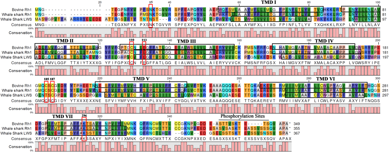
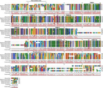
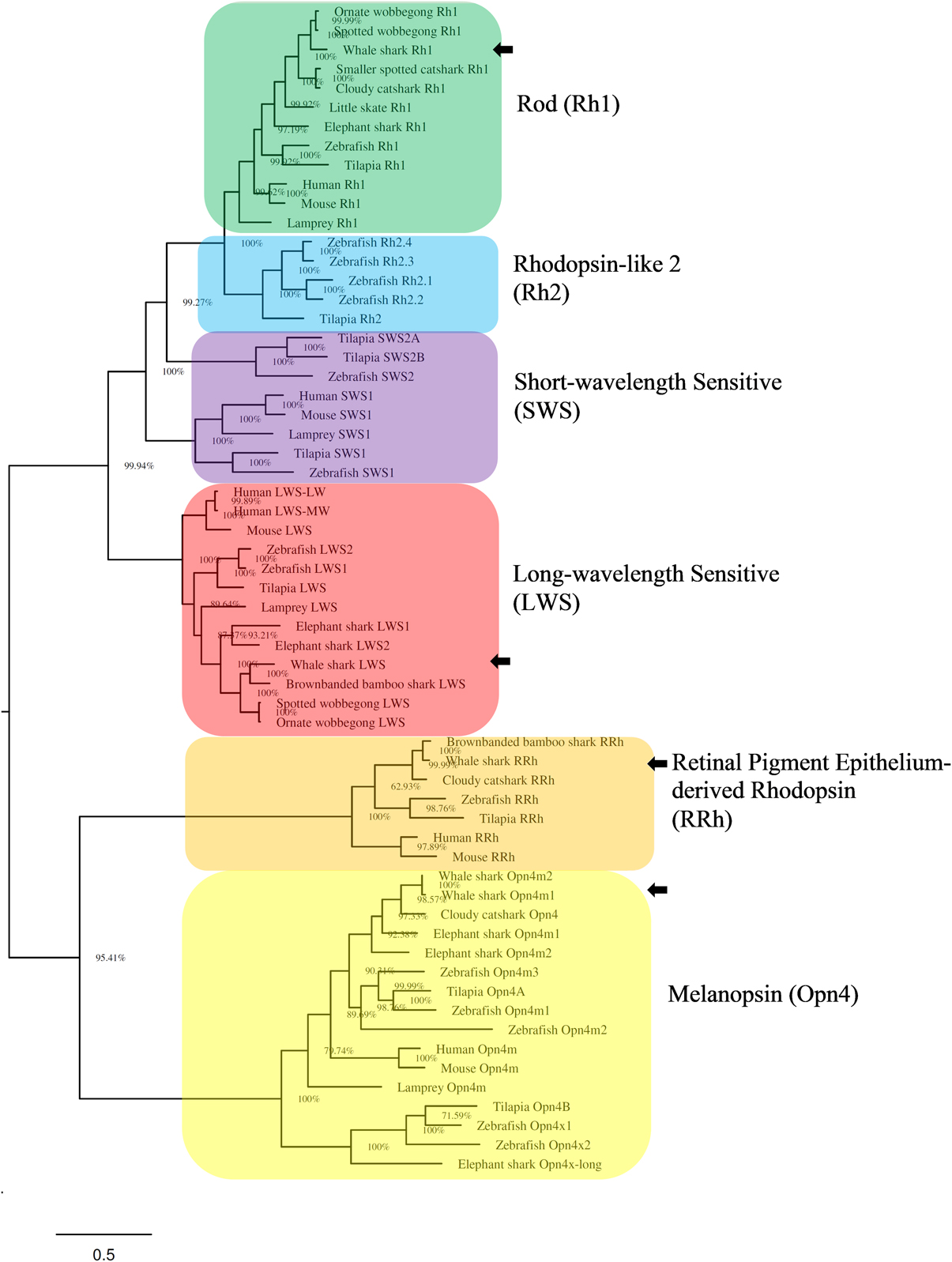
Alignment and analysis of the whale shark Rh1, LWS, and Opn4 opsin amino acid sequences are summarized in Figs. 1 and 2, showing conserved features to other opsin proteins including seven transmembrane domains (TMDs), conserved residues at the chromophore attachment site, and the Schiff base counterion. To determine the evolutionary relationships of the whale shark retinal opsins, we inferred the phylogeny of shark and other vertebrate opsin genes using Bayesian analysis to generate the consensus tree shown in Fig. 3. The whale shark Rh1 and LWS nucleotide sequences were placed in clades with other shark Rh1 and LWS sequences and were nearer to species from the order Orectolobiformes (wobbegong and bamboo sharks) than to species from the orders Chimaeriformes and Carcharhiniformes (elephant and cat sharks, respectively), which is in agreement with current elasmobranch phylogenies (Vélez-Zuazo & Agnarsson, Reference Vélez-zuazo and Agnarsson2011; Naylor et al., Reference Naylor, Caira, Jensen, Rosana, Straube, Lakner, Carrier, Musick and Heithaus2012).
Alignment of whale shark rod and cone opsin sequences. Whale shark rod and LWS cone opsin sequences were aligned with the sequence for bovine rhodopsin (Rh1) which was used for comparison and amino acid numbering. Transmembrane domains (TMDs; boxed in black) for bovine Rh1 are based on the crystal structure (Palczewski et al., Reference Palczewski, Kumasaka, Hori, Behnke, Motoshima, Fox, Le Trong, Teller, Okada and Stenkamp2000). TMDs for whale shark Rh1 and LWS cone were predicted using TMHMM Server version 2.0 (http://www.cbs.dtu.dk/services/ TMHMM). Conserved residues are boxed in red and include: (1) asparagine (N) at position 15 in the amino terminal domain [predicted using NetNGlyc Server 1.0 (http://www.cbs.dtu.dk/services/ NetNGlyc)]; (2) the counterion glutamate (E) at position 113; (3) cysteine (C) residues at positions 110, 185, and 187 involved in disulfide bond formation; (4) lysine (K) at position 296 that links to the chromophore; and (5) serine (S) and threonine (T) phosphorylation sites spanning positions 334–343 in the CTD.

Alignment of whale shark melanopsin (Opn4). Whale shark Opn4 sequences were aligned with elephant shark and cloudy catshark Opn4m sequences. Mouse Opn4 was used for comparison and amino acid numbering. Transmembrane domains (TMDs; boxed in black) for mouse Opn4 are from Provencio et al. (Reference Provencio, Rodriguez, Jiang, Hayes, Moreira and Rollag2000). TMDs for elephant shark, cloudy catshark, and whale shark Opn4 were predicted using TMHMM Server version 2.0 (http://www.cbs.dtu.dk/services/TMHMM). Conserved residues are boxed or circled in red and include (1) glycosylation sites (asparagine-N) in the amino terminal domain [predicted using NetGlyc Server 1.0 (http://www.cbs.dtu.dk/services/NetNGlyc)], (2) conserved cysteines (C) at positions 95 and 220 involved in disulfide bond formation, (3) potential counterion at position 145 (tyrosine-Y), (4) lysine (K) at position 337 that links to the chromophore, (5) potential serine (S), and threonine (T) phosphorylation sites spanning positions 372–395 in the CTD. Elephant shark Opn4 was previously referred to as Opn4m-1 (Davies et al., Reference Davies, Tay, Zheng, Danks, Brenner, Foster, Collin, Hankins, Venkatesh and Hunt2012).

Phylogeny of opsins in whale shark (Rhincodon typus) and other vertebrates. A Bayesian inference method performed with Markov chain Monte Carlo method to estimate the posterior distribution of model parameters (represented as percentages) at the base of each node. The tree shows the relative position of whale shark rod (Rh1), LWS cone, melanopsin (Opn4), and retinal pigment epithelium-derived rhodopsin (RRh), as well as rhodopsin-like 2 (Rh2) and SWS 1 and 2 cone from other vertebrates. Each opsin class is colored by clade: Rh1 (green), Rh2 (blue), SWS (violet), LWS (red), RRh (orange), and Opn4 (yellow). The degree of internal branching is expressed as a percentage. Scale bar indicates the number of nucleotide substitutions per site. Locations of whale shark Rh1, LWS, RRh, and Opn4m are indicated with black arrows. The opsin nucleotide sequences and GenBank accession numbers used to generate the tree are as follows: Rod opsin (Rh1): zebrafish (Danio rerio), NM131084; mouse (Mus musculus), NM_145,383; human (Homo sapiens), NM_000539; Nile tilapia (Oreochromis niloticus), AY775108; Arctic lamprey (Lethenteron japonicum), M63632; elephant shark (Callorhinchus milii), NM_001292252; little skate (Raja erinacea), U81514; smaller spotted catshark (Scyliorhinus canicula), Y17585; whale shark (Rhincodon typus), XM_020522486; spotted wobbegong (Orectolobus maculatus), JX534163; ornate wobbegong (Orectolobus ornatus), JX534164; Rod opsin-like 2 (Rh2): zebrafish, NM131253 (Rh2.1), NM182891 (Rh2.2), NM182892 (Rh2.3), NM131254 (Rh2.4); Nile tilapia, AF247124; Short-wavelength sensitive-1 (SWS1): human, NM_001708; mouse, NM_007538; zebrafish, NM_131319; Nile tilapia, XM_003448756; pouched lamprey (Geotria australis), AY366495; Short-wavelength sensitive-2 (SWS2): zebrafish, NM_131192; Nile tilapia, AF247116 (SWS2A), AF247120 (SWS2B); Long-wavelength sensitive (LWS): Nile tilapia, XM_003442629; zebrafish, KT008400 (LWS1), KT008401 (LWS2); mouse, AF190672; human, NM_000513 (LWS-MW), NM_020061 (LWS-LW); pouched lamprey, AY366491; spotted wobbegong, JX534165; ornate wobbegong, JX534166; elephant shark, NM_001317730 (LWS1), NM_001292806 (LWS2); Retinal pigment epithelium-derived rhodopsin (RRh): human, NM_006583; mouse, NM_009102; Nile tilapia, XM_003458164; zebrafish, NM_001004654; Melanopsin (Opn4): elephant shark, NM_001292428 (Opn4m1), NM_001292116 (Opn4m2), NM_001292471 (Opn4x-long); zebrafish, GQ925715 (Opn4m1), GQ925716 (Opn4m2), GQ925717 (Opn4m3), GQ925718 (Opn4x1), GQ925719 (Opn4x2); Nile tilapia, XM_019355631 (Opn4A), XM_003453905 (Opn4B); whale shark, XM_020523467 & XM_020511520; mouse, NM_013887; human, NM_033282; and Arctic lamprey, AB932626. The following curated gene IDs are from Hara et al. (Reference Hara, Yamaguchi, Onimaru, Kadota, Koyanagi, Keeley, Tatsumi, Tanaka, Motone and Kageyama2018): cloudy catshark (Scyliorhinus torazame) Rh1, Scyto2000054; brownbanded bamboo shark (Chiloscyllium punctatum) LWS, Chipu2000048; whale shark LWS, Rhity2000048; cloudy catshark RRh, Scyto2000062; whale shark RRh, Rhity2000054; brownbanded bamboo shark RRh, Chipu2000056; cloudy catshark Opn4, Scyto2000059; whale shark, Rhity2000050 (Opn4m2); and Human GPR21 and GPR52 (GenBank accession numbers NM005294 and NM005684, respectively) were used as outgroups.

Based on the whale shark genomes from both Read et al (Reference Read, Petit, Joseph, Alam, Weil, Ahmad, Bhimani, Vuong, Haase and Webb2017) and Hara et al (Reference Hara, Yamaguchi, Onimaru, Kadota, Koyanagi, Keeley, Tatsumi, Tanaka, Motone and Kageyama2018), the whale shark appears to possess a single Opn4 gene, unlike elephant shark which possesses three, including two Opn4m isoforms and a single Opn4x isoform (Davies et al., Reference Davies, Tay, Zheng, Danks, Brenner, Foster, Collin, Hankins, Venkatesh and Hunt2012). As shown in Fig. 2, the whale shark Opn4 amino acid sequences generated by the two published whale shark genomes are identical between the initial methionine and amino acid position 261 where the sequence from Read et al. (Reference Read, Petit, Joseph, Alam, Weil, Ahmad, Bhimani, Vuong, Haase and Webb2017) (named here whale shark Opn4-1) containing an additional 41 amino acids, that are absent in the whale shark Opn4 sequence from Hara et al. (Reference Hara, Yamaguchi, Onimaru, Kadota, Koyanagi, Keeley, Tatsumi, Tanaka, Motone and Kageyama2018) (named here whale shark Opn4-2). As described previously, this 41-amino acid region of the Opn4-1 sequence is unique and is not found in any other vertebrate Opn4 sequence in the NCBI database. The whale shark Opn4-1 and Opn4-2 sequences are identical after this 41-amino acid insertion but deviate again at the CTD where an additional 42 amino acids are found in the Opn4-2 sequence that are absent in the Opn4-1 sequence. The alignment of the related Opn4 sequences from elephant shark and cloudy catshark, along with whale shark Opn4-1 and Opn4-2 sequences reveal seven conserved TMDs and other conserved amino acids, each of which is described in Fig. 2. The phylogenetic tree shown in Fig. 3 places the whale shark Opn4-1 and Opn4-2 sequences with the Opn4 sequences from elephant shark and cloudy catshark, with a closer relationship to the latter rather than the Opn4m and Opn4x sequences from the former. This phylogenetic relationship suggests that the whale shark Opn4 sequences are more similar to the mammalian-like (m) Opn4 class rather than to the xenopus-like (x) Opn4 class and are labeled accordingly as whale shark Opn4m-1 and Opn4m-2 (Fig. 3).
Spectral tuning properties of whale shark retinal pigments
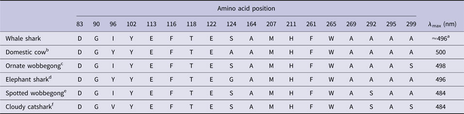
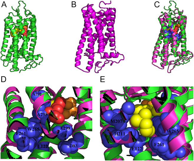
To better understand the spectral tuning properties of the whale shark Rh1, LWS, and Opn4 pigments, the amino acid sequences were deduced and key spectral tuning positions were examined. Eighteen amino acid positions that have been previously identified to contribute to the spectral tuning of Rh1 pigments (Hunt et al., Reference Hunt, Dulai, Partridge, Cottrill and Bowmaker2001; Yokoyama, Reference Yokoyama2008; Musilova et al., Reference Musilova, Cortesi, Matschiner, Davies, Patel, Stieb, De Busserolles, Malmstrøm, Tørresen, Brown, Mountford, Hanel, Stenkamp, Jakobsen, Carleton, Jentoft, Marshall and Salzburger2019) were examined in whale shark Rh1 as well as domestic cow and four other shark species with known Rh1 absorbance maxima. As shown in Table 1, whale shark Rh1 possesses the same residues at these 18 positions as the ornate wobbegong Rh1 [absorbance maximum (λ max) = 498 nm] except for position 299. Serine to alanine substitutions at this position result in a slight blue-shift of 2 nm which would provide the whale shark Rh1 with a predicted λ max = 496 nm. Comparing the spectral tuning sites of whale shark Rh1 with those of bovine Rh1 (λ max = 500 nm) and the elephant shark (λ max = 496 nm), two substitutions at potential tuning sites are observed: Iso96Tyr and Ser124Ala/Gly. The spectral shift associated with position 96 relies on the loss of tyrosine at position 102 (Yokoyama et al., Reference Yokoyama, Tada, Zhang and Britt2008); however, Tyr102 is conserved in all species examined here and negates any spectral tuning properties associated with position 96. Previous mutagenesis experiments at position 124 in bovine Rh1 resulted in a slight blue-shift of 3 nm (Ala124Thr λ max = 497 nm; Lin et al., Reference Lin, Kochendoerfer, Carroll, Wang, Mathies and Sakmar1998). Whale shark Rh1 possesses serine at position 124 which would have a similar influence on spectral tuning as in the bovine mutant, resulting in a whale shark Rh1 pigment with a predicted λ max ≈ 497 nm. When the predicted whale shark Rh1 structure was modeled and aligned with the structure of bovine rhodopsin, the helical alignments were nearly identical (Fig. 4A–4C). Alignment of nine spectral tuning residues (Fig. 4D and 4E) also appear to be identical between the predicted whale shark and bovine Rh1 structures. These results suggest that the whale shark Rh1 pigment possesses a λ max around 496 nm, a value shared by the elephant shark Rh1 (λ max = 496 nm; Davies et al., Reference Davies, Carvalho, Tay, Brenner, Hunt and Venkatesh2009) and near the ornate wobbegong Rh1 (λ max = 498 nm).
Amino acid substitutions critical to spectral tuning

a Absorbance maximum is predicted based on the Rh1 λ max values from domestic cow (500 nm), ornate wobbegong (498 nm), and elephant shark (496 nm). The influence of individual spectral tuning positions is described in Results.
b Accesion number NM_001014890.2; Rh1 λ max value from Oprian et al., Reference Oprian, Molday, Kaufman and Khorana1987.
c Accesion number JX534164.1; Rh1 λ max value from Hart et al., Reference Hart, Theiss, Harahush and Collin2011.
d Accesion number NM_001292252.1; Rh1 λ max value from Davies et al., Reference Davies, Carvalho, Tay, Brenner, Hunt and Venkatesh2009.
e Accesion number JX534163.1; Rh1 λ max value from Hart et al., Reference Hart, Theiss, Harahush and Collin2011.
f Curated gene ID Scyto2000054; Rh1 λ max value from Hara et al., Reference Hara, Yamaguchi, Onimaru, Kadota, Koyanagi, Keeley, Tatsumi, Tanaka, Motone and Kageyama2018.
Structural modeling of whale shark Rh1 spectral tuning amino acid positions. Bovine rhodopsin Rh1 (A; PDB ID 1U19; Okada et al., Reference Okada, Sugihara, Bondar, Elstner, Entel and Buss2004) and the predicted structure of whale shark Rh1 (B) were aligned (C) to determine relative positions of helices and residues. Spectral tuning residues proximal to the region surrounding the PSB (D) and the β-ionone ring of the chromophore (E) are highlighted in blue in the faded structural images. Chromophore (yellow), lysine 296 (red), and aspartate 113 (orange) are labeled in bovine rhodopsin for topological reference.

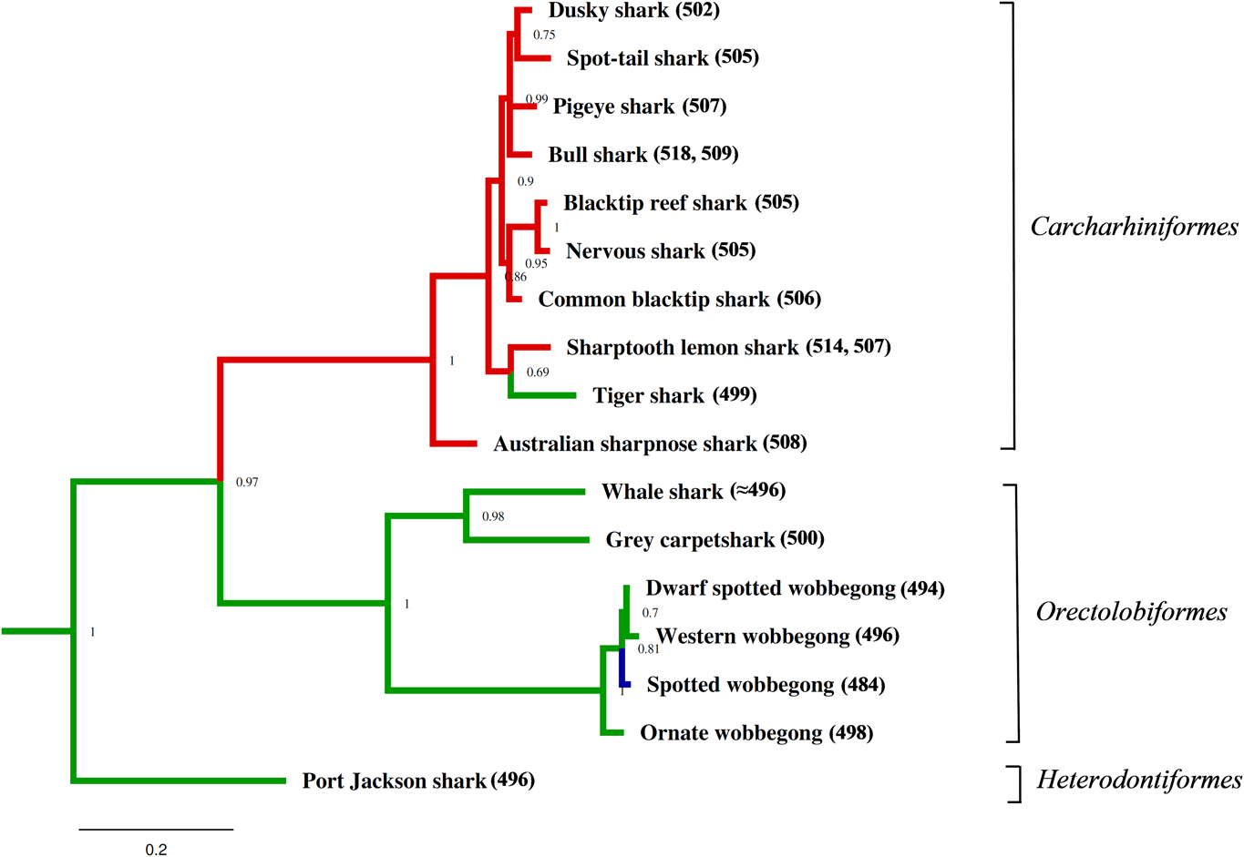
The evolution of elasmobranch Rh1 spectral tuning, in the context of a trait characteristic, was investigated by first inferring the phylogeny of the cytochrome oxidase 1 (CO1) gene previously reported from 17 different shark species using Bayesian analysis. The resulting consensus tree (Fig. 5) was appended by adding the Rh1 λ max values from species belonging to the superorder Galeomorphii, including the three orders Carcharhiniformes, Orectolobiformes, and Heterodontiformes. No Rh1 λ max values have been reported for species from the order Lamniformes. It appears that the ancestral phenotype with regard to Rh1 spectral tuning resides within Heterodontiformes as seen with the Port Jackson shark (Heterodontus portusjacksoni; λ max = 496 nm) which possesses a Rh1 pigment with similar spectral tuning properties as those predicted in whale shark Rh1 (λ max ≈ 496 nm). This phenotype is conserved within the members of Orectolobiformes (λ max = 494–500 nm), whereas the spotted wobbegong possesses a significantly blue-shifted Rh1 pigment (λ max = 484 nm) when compared with the other species. Alternatively, all members of the order Carcharhiniformes possess red-shifted Rh1 pigments (λ max = 502–508 nm) relative to the Heterodontiformes and Orectolobiformes except for tiger shark (λ max = 499 nm).
Evolution of rod (Rh1) spectral sensitivity from three orders of galean sharks with regard to absorbance maxima. The inferred phylogeny is based on Bayesian analysis of cytochrome oxidase subunit 1 (CO1) sequences from 17 shark species from which the absorbance maxima (λ max) for the Rh1 pigments has been experimentally determined (Hart et. al., Reference Hart, Theiss, Harahush and Collin2011). Branches are colored according to their λ max values relative to Port Jackson shark (496 nm): blue shift (<490 nm, blue); no shift (494–500 nm, green); and red shift (>500 nm, red). Bull shark (Carcharhinus leucas) and sharptooth lemon shark (Negaprion acutidens) λ max values are from direct photoreceptor (A2 chromophore) measurements and modeling with an A1 chromophore, respectively (Hart et al., Reference Hart, Theiss, Harahush and Collin2011). CO1 GenBank accession numbers: pigeye shark (Carcharhinus amboinensis), TZMSC247; nervous shark (Carcharhinus cautus), FOAF123; bull shark (Carcharhinus leucas), EWTGS097; common blacktip shark (Carcharhinus limbatus), FOAE316; blacktip reef shark (Carcharhinus melanopterus), FOAE146; dusky shark (Carcharhinus obscurus), TZMSC449; spot-tail shark (Carcharhinus sorrah), FOA067; tiger shark (Galeocerdo cuvier), EWTGS048; sharptooth lemon shark (Negaprion acutidens), FOA072; Australian sharpnose shark (Rhizoprionodon taylori), FOAE164; Port Jackson shark (Heterodontus portusjacksoni), FOAMP003; grey carpet shark (Chiloscyllium punctatum), FOAD569; whale shark (Rhincodon typus), ESHKD028; Western wobbegong (Orectolobus hutchinsi), FOAE411; spotted wobbegong (Orectolobus maculatus) FOA016; ornate wobbegong (Orectolobus ornatus), FOA013; and dwarf spotted wobbegong (Orectolobus parvimaculatus), FOAD510. The degree of internal branching is expressed as a decimal. Scale bar indicates the number of nucleotide substitutions per site. Goldfish (GenBank accession number JQ667509) and zebrafish (GenBank accession number MK572160) CO1 sequences were used as outgroups.

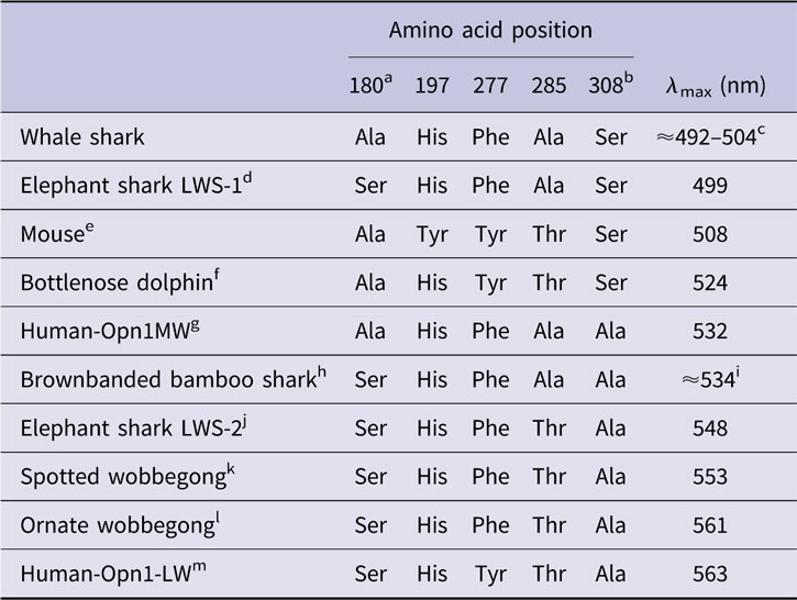
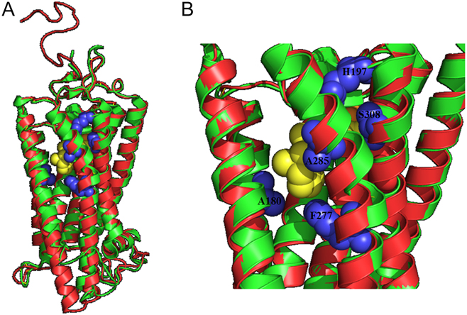
The whale shark LWS cone opsin amino acid sequence was deduced, and five key spectral tuning positions were examined. As shown in Table 2, the whale shark LWS opsin possesses a unique set of amino acid substitutions at these five positions not shared by previously examined vertebrates. The whale shark LWS opsin is most similar to the elephant shark LWS1 (Davies et al., Reference Davies, Carvalho, Tay, Brenner, Hunt and Venkatesh2009) and human Opn1MW (green cone opsin or LWS-MW), differing by only a single amino acid substitution when compared with these opsins, respectively. The whale shark LWS opsin possesses Ala180 and Ser308, whereas elephant shark possesses Ser180, and human Opn1MW possesses Ala308; however, both share the same amino acid substitutions with the whale shark LWS opsin at the remaining four spectral tuning positions. When modeled (Fig. 6A and 6B), the whale shark LWS opsin lacks the OH-side group found in elephant shark LWS-1 at position 180, proximal to the β-ionone ring of the chromophore. Unlike human Opn1MW, whale shark LWS possesses an OH-side group at position 308, proximal to the protonated Schiff base (PSB) linkage of the chromophore. Site-directed mutagenesis of the human red/green cone opsins have been previously examined, including the single-site mutation at position 180 (Asenjo et al., Reference Asenjo, Rim and Oprian1994), as well as studies examining the role of position 308 in the spectral tuning of the LWS cones (Sun et al., Reference Sun, Macke and Nathans1997; Fasick & Robinson, Reference Fasick and Robinson1998). As shown in Table 2, it is predicted that the whale shark LWS pigment should possess a λ max of either 492 nm, based on the 7-nm blue-shift expected from the serine to alanine substitution at position 180 using the elephant shark LWS1 absorbance spectrum (λ max = 499 nm), or a λ max of 504 nm, based on the 28 nm blue-shift expected from the alanine to serine substitution at position 308 using the human Opn1MW absorbance spectrum (λ max = 532 nm). Interestingly, when these two predicted λ max values for the whale shark LWS cone visual pigment are averaged, the result is 498 nm, which is near the predicted absorbance maximum of whale shark Rh1.
Opn1LW/MW amino acid substitutions critical to spectral tuning
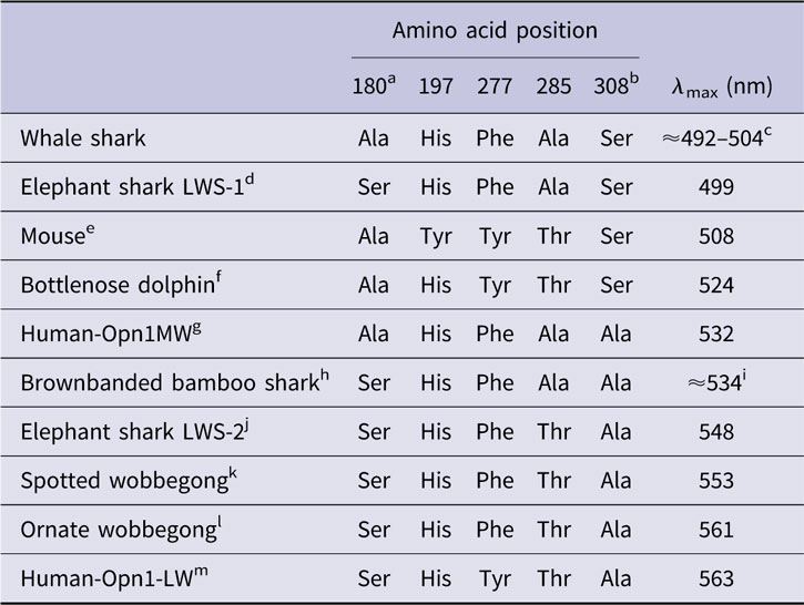
a Ser180 to Ala180 results in 7-nm blue shift; Ala180 to Ser 180 results in 2-nm red shift (Asenjo et al., Reference Asenjo, Rim and Oprian1994).
b Ala308 to Ser308 results in 28-nm blue shift (Sun et al., Reference Sun, Macke and Nathans1997; Fasick & Robsinson, Reference Fasick and Robinson1998).
c Estimated absorbance maximum (λ max) of 492 nm for whale shark LWS is based on the elephant shark LWS-1 (λ max = 499 nm) minus 7 nm from the whale shark Ala180 substitution. The estimated λ max of 504 nm is based on the human Opn1MW (λ max = 532 nm) minus 28 nm from the whale shark Ser308 substitution. Whale shark LWS sequence (Hara et al., Reference Hara, Yamaguchi, Onimaru, Kadota, Koyanagi, Keeley, Tatsumi, Tanaka, Motone and Kageyama2018).
d Accession number EF565165.1; LWS λ max value from Davies et al., Reference Davies, Carvalho, Tay, Brenner, Hunt and Venkatesh2009.
e Accession number AF190672; LWS λ max value from Sun et al., Reference Sun, Macke and Nathans1997.
f Accession number AF055457.1; LWS λ max value from Fasick et al., Reference Franke, Sakmar, Oprian and Khorana1998.
g Accession number NM_000513.2; LWS λ max value from Asenjo et al., Reference Asenjo, Rim and Oprian1994.
h Curated gene ID Chipu2000048; LWS λ max value from Hara et al., Reference Hara, Yamaguchi, Onimaru, Kadota, Koyanagi, Keeley, Tatsumi, Tanaka, Motone and Kageyama2018.
i The estimated λ max of 534 nm for brownbanded bamboo shark LWS is based on the human Opn1MW (λ max = 532 nm) plus 2 nm from the bamboo shark Ser180 substitution.
j Accession number EF565166.1; LWS λ max value from Davies et al., Reference Davies, Carvalho, Tay, Brenner, Hunt and Venkatesh2009.
k Accession number JX534165.1; LWS λ max value from Hart et al., Reference Hart, Theiss, Harahush and Collin2011.
l Accession number JX534166.1; LWS λ max value from Hart et al., Reference Hart, Theiss, Harahush and Collin2011.
m Accession number NM_020061.5; LWS λ max value from Asenjo et al., Reference Asenjo, Rim and Oprian1994.
Structural modeling of whale shark LWS cone spectral tuning amino acid positions. Bovine rhodopsin (green) and the predicted structure of whale shark LWS cone opsin (red) were aligned (A) to determine relative positions of helices and residues. Spectral tuning residues proximal to the β-ionone ring of the chromophore (A180) and protonate Schiff base (S308) are highlighted in blue (B). Chromophore (yellow), lysine 296/312 (red), and aspartate 113/129 (orange) are labeled in bovine rhodopsin for topological reference (cone numbering is equal to rod numbering plus 16 amino acids).

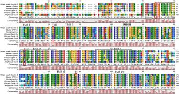
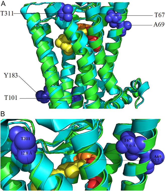
To examine the spectral tuning properties of whale shark Opn4, the whale shark Opn4 amino acid sequence was aligned with five divergent vertebrate Opn4 amino acid sequences whose absorbance maxima of ≈480 nm have been previously described by either direct expression of the pigment or by electrophysiological methods (Koyanagi et al., Reference Koyanagi, Kubokawa, Tsukamoto, Shichida and Terakita2005; Panda et al., Reference Panda, Nayak, Campo, Walker, Hogenesch and Jegla2005; Qiu et al., Reference Qiu, Kumbalasiri, Carlson, Wong, Krishna, Provencio and Berson2005; Torii et al., Reference Torii, Ojima, Okano, Nakamura, Terakita, Shichida, Wada and Fukada2007; Bailes & Lucas, Reference Bailes and Lucas2013). As shown in Fig. 7, TMDs I–VII reveal a total of five amino acid positions in whale shark that result in nonconservative substitutions that are not shared by the other species (mouse, human, chicken, and amphioxus). Residues Thr101 and Tyr183 found in whale shark Opn4 TMII and TMIV, respectively, both introduce an OH-group when compared with the other vertebrate Opn4 sequences, but are greater than 25 Å from either the PSB or the β-ionone ring of the chromophore and most likely have no influence on spectral tuning of the pigment (Fig. 8A). Thr67 and Ala69 in whale shark Opn4 TMI results in the addition and removal of an OH-group proximal to the PSB of the chromophore at these respective positions, whereas Thr311 in whale shark Opn4 TMVI results in the addition of an OH-group proximal to the β-ionone ring of the chromophore (Fig. 8B). These three amino acids, and their relative proximity to either the PSB or β-ionone ring of the chromophore, were measured to be within 16 Å of their respective chromophore positions and may be close enough to influence spectral tuning of the pigment. Applying the OH-site rule (Sekharan et al., Reference Sekharan, Katayama, Kandori and Morokuma2012), we predict that the Thr67 and Ala69 substitutions would result in slight blue and redshifts, respectively, potentially canceling any spectral movement relative to 480 nm. The Thr311 substitution, however, introduces a hydroxyl group proximal to the β-ionone ring of the chromophore which could potentially result in a slight red shift away from 480 nm.
Alignment of whale shark and other vertebrate Opn4 sequences to identify potential spectral tuning residues. Five nonconserved residues (indicated with red boxes) located in transmembrane domains (indicated with black bars) of whale shark Opn4 were identified: T67, A69, T101, Y183, and T311 (whale shark Opn4 numbering). GenBank accession numbers for Opn4 sequences: mouse (EDL24885); human (NP_150,598); chicken (2L, NP_001038118 and 1S, ABX10831); amphioxus (Q4R1I4); and whale shark (Curated Gene ID Rhity2000050, Hara et al., Reference Hara, Yamaguchi, Onimaru, Kadota, Koyanagi, Keeley, Tatsumi, Tanaka, Motone and Kageyama2018).

Structural modeling of whale shark Opn4 spectral tuning amino acid positions. (A). Squid rhodopsin (light blue) and the predicted structure of whale shark Opn4 (green) were aligned to determine relative positions of helices and residues. (B). Potential spectral tuning residues resulting from nonconserved amino acid substitutions are spherical (blue) and labeled by position using whale shark Opn4 numbering. The distance from residues to the PSB nitrogen and the β-ionone ring were determined in Pymol. Schiff base lysine (K333) is labeled red, chromophore is labeled yellow, and potential counterion (Y140) is labeled orange.

Signaling kinetics of Opn4 constructs using fluorescent calcium imaging
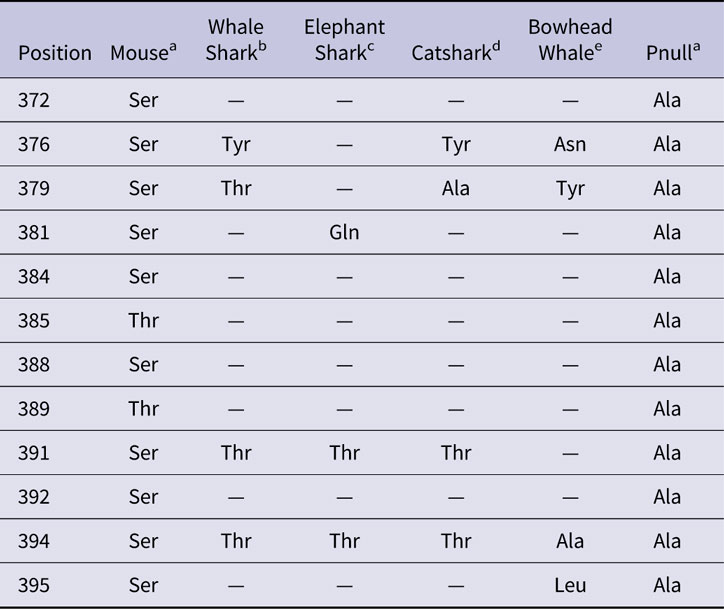
The deactivation kinetics of Opn4 have been previously described in the mouse, along with the phosphorylation sites involved, and the resulting pupillary light reflex (PLR) when these sites are mutated (Blasic et al., Reference Blasic, Brown and Robinson2012; Blasic et al., Reference Blasic, Matos-Cruz, Ujla, Cameron, Hattar, Halpern and Robinson2014; Somasundaram et al., Reference Somasundaram, Wyrick, Fernandez, Ghahari, Pinhal, Richardson, Rupp, Cui, Wu and Brown2017). To better understand the PLR of the whale shark, we examined 12 potential phosphorylation sites located within the carboxyl terminal domain (CTD; Fig. 2 and Table 3). When examining these 12 potential phosphorylation sites, whale shark Opn4 possesses a single non-serine/threonine amino acid substitution (Tyr376) compared with wild type mouse Opn4. Likewise, elephant shark Opn4m-1 possesses a single non-serine/threonine amino acid substitution (Gln381) within these 12 sites. Unlike the whale shark and elephant shark, the cloudy catshark and bowhead whale possess non-serine/threonine amino acid substitutions at more than one position with regard to these phosphorylation sites. The cloudy catshark possesses two substitutions (Tyr376 and Ala379), whereas the bowhead whale possesses four substitutions (Asn376, Tyr379, Ala 394, and Leu395).
Naturally occurring Opn4 carboxyl termini phosphorylation site substitutions
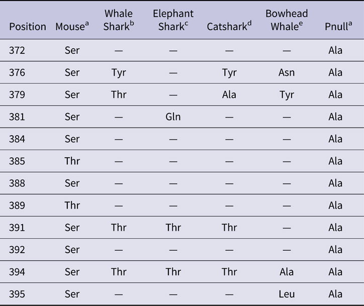
Note: Blank spaces indicate conservation of residue found in mouse Opn4.
a Sites 388–395 previously identified and described (Blasic et al., Reference Blasic, Brown and Robinson2012; Blasic et al., Reference Blasic, Matos-Cruz, Ujla, Cameron, Hattar, Halpern and Robinson2014).
b Accession number XM_020511520 and Curated gene ID Rhity2000050 from Hara et al., Reference Hara, Yamaguchi, Onimaru, Kadota, Koyanagi, Keeley, Tatsumi, Tanaka, Motone and Kageyama2018.
c Accession number NM_001292116.
d Curated gene ID Scyto2000059 from Hara et al., Reference Hara, Yamaguchi, Onimaru, Kadota, Koyanagi, Keeley, Tatsumi, Tanaka, Motone and Kageyama2018.
e Identifier number bmy_16888 from Keane et al., Reference Keane, Semeiks, Webb, Li, Quesada, Craig, Madsen, Van Dam, BraWand and Marques2015.
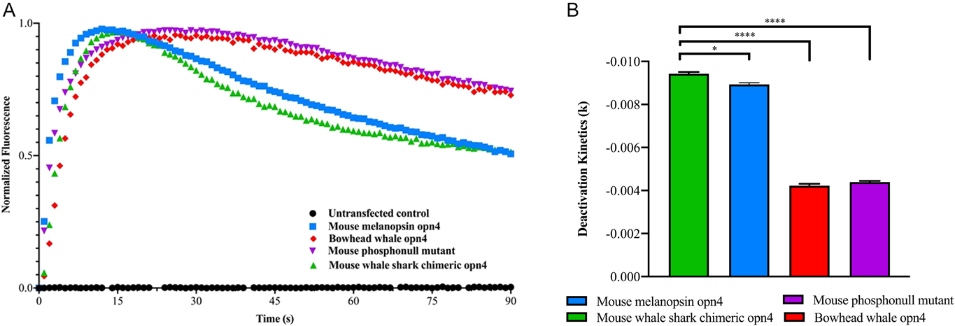
To examine the deactivation kinetics of the whale shark Opn4 pigment and the potential effect of the Tyr376 substitution compared with mouse controls, calcium imaging assays were performed that compared the deactivation rates of the mouse–whale shark Opn4 chimera with the deactivation rates from wild type mouse and bowhead whale Opn4. A mouse Opn4 mutant, where all putative serine/threonine phosphorylation sites in the CTD were replaced with alanine residues (mouse phosphonull mutant: Pnull), was also used for comparison. The deactivation kinetics of the mouse–whale shark Opn4 chimera is more similar to that of wild type mouse Opn4 than to the deactivation kinetics of either the bowhead whale Opn4 or Pnull mutant (Fig. 9A). When the rates of deactivation were examined (Fig. 9B), mouse-whale shark Opn4 chimera and mouse Opn4 deactivation rates are similar to each other and significantly faster than both bowhead whale and mouse Pnull Opn4 deactivation rates (P < 0.0001).
Deactivation kinetics of Opn4 constructs. (A) In vitro Opn4 signaling kinetics based on calcium imaging data of mouse Opn4 (blue trace), mouse–whale shark chimeric Opn4 (green trace), bowhead whale (red trace), and mouse Opn4 lacking CTD phosphorylation sites (mouse phosphonull mutant - purple trace). Negative control of untransfected HEK293 cells (black trace) is shown for comparison. (B) Deactivation rates between mouse Opn4 and mouse–whale shark chimeric Opn4 are significantly different from the deactivation rates of bowhead whale Opn4 and the mouse phopshopnull mutant. All replicates across three transfections were pooled and averaged, and the deactivation phase was fitted to the exponential function y = y(0) × e (k×x), where K is the rate constant. All rates were compared with mouse–whale shark opn4 chimera deactivation rate. Unpaired two tailed t-test was used to calculate the P value. * and **** correspond to values of <0.05 and <0.0001, respectively. Error bars denote standard error of the mean.

Discussion
This study examined three retinal opsins identified in the whale shark (Rhincodon typus) genome: the rod (Rh1), long-wavelength sensitive (LWS) cone visual pigments, and the retinal pigment melanopsin (Opn4). Whale shark Rh1 and LWS opsins were characterized with regard to spectral tuning properties to estimate absorbance maxima important for visual foraging. The spectral tuning properties of whale shark Opn4 were investigated to determine if the absorbance maximum of this pigment diverged from the Opn4 pigments of previously studied vertebrates. The deactivation kinetics of expressed whale shark Opn4 was examined, and the rates of deactivation were compared with those from vertebrates possessing either a duplex retina or a rod-only retina. The results from this study found that the whale shark Rh1 and LWS pigments are predicted to maximally absorb light around 496 nm and 500 nm, respectively. This phenotype has most likely been selected for by the dominant wavelengths of available light at or near the surface of the water used for foraging under either photopic or scotopic light conditions. The deactivation rate of the whale shark Opn4 pigment suggests rapid mydriasis, or pupillary dilation, and would be consistent with the pupil response from an animal possessing a duplex retina possessing both functional rod and cone photoreceptors. Although other opsin classes have been identified in the whale shark genome (see Hara et al., Reference Hara, Yamaguchi, Onimaru, Kadota, Koyanagi, Keeley, Tatsumi, Tanaka, Motone and Kageyama2018), we focused on Rh1, LWS, and Opn4, as these three pigments are critical to visually guided tasks, including foraging. We initially believed the whale shark to be a rod monochromat as no cone opsins were identified in the available Read et al. (Reference Read, Petit, Joseph, Alam, Weil, Ahmad, Bhimani, Vuong, Haase and Webb2017) whale shark genome. However, the short sequence reads from the genome provided by Hara et al. (Reference Hara, Yamaguchi, Onimaru, Kadota, Koyanagi, Keeley, Tatsumi, Tanaka, Motone and Kageyama2018) resulted in the presence of a whale shark LWS gene. Thus, a duplex retinal structure consisting of a single rod and single LWS cone class appears to be present in the whale shark. This duplex retinal design is not unique in vertebrates and is in fact quite common in shark species (Hart et al., Reference Hart, Theiss, Harahush and Collin2011).
The spectral tuning properties of the whale shark Rh1 and LWS cone pigments, like the visual pigments from other species, rely on specific amino acid substitutions at key positions to modulate the absorbance spectrum of the chromophore, most likely 11-cis retinal in the whale shark (Douglas & Partridge, Reference Douglas and Partridge1997; Cronin et al., Reference Cronin, Johnsen, Marshall and Warrant2014). The whale shark Rh1 opsin possesses amino acids at critical spectral tuning positions that are most similar to those found in ornate wobbegong Rh1 (λ max = 498 nm) and elephant shark (λ max = 496 nm) and is predicted to share a similar absorbance maximum around 496 nm. By contrast, a recent study investigating shark genomes has placed the λ max of whale shark Rh1 at 478 nm (Hara et al., Reference Hara, Yamaguchi, Onimaru, Kadota, Koyanagi, Keeley, Tatsumi, Tanaka, Motone and Kageyama2018), a value that is significantly blue-shifted from the predicted whale shark Rh1 λ max reported here. The pigment reported in the Hara et al. (Reference Hara, Yamaguchi, Onimaru, Kadota, Koyanagi, Keeley, Tatsumi, Tanaka, Motone and Kageyama2018) study, however, is a member of the peropsin family of retinal pigments and was described by the authors as a member of the retinal pigment epithelium-derived rhodopsins, or RRhs, which typically possess λ max values in the blue region of the spectrum between 470–485 nm (Hao & Fong, Reference Hao and Fong1996; Koyanagi et al., Reference Koyanagi, Terakita, Kubokawa and Shichida2002). The whale shark RRh sequence from Hara et al. (Reference Hara, Yamaguchi, Onimaru, Kadota, Koyanagi, Keeley, Tatsumi, Tanaka, Motone and Kageyama2018) sorts with other shark RRh opsins (shown in Fig. 3) and is most likely a member of this family of retinal opsins and not an Rh1 opsin. Rh1s with λ max values around 500 nm are found in terrestrial mammals, as well as surface foraging fish (Losey et al., Reference Losey, Mcfarland, Loew, Zamzow, Nelson and Marshall2003; Taylor et al., Reference Taylor, Loew and Grace2011; Schweikert et al., Reference Schweikert, Caves, Solie, Sutton and Johnsen2018). We believe that the amino acid substitutions associated with the spectral tuning properties of whale shark Rh1 were selected for by the broad-spectrum light available at the surface of the water, where foraging has frequently been documented, and not by the available light found at depth, which typically results in Rh1 absorbance spectra that are significantly blue-shifted relative to 500 nm. The spectral tuning of whale shark Rh1 appears to share the ancestral trait within the subgroup of galean sharks: Lamniformes, Carcharhiniformes, Orectolobiformes, and Heterodontiformes (Maisey, Reference Maisey1984; Naylor et al., Reference Naylor, Caira, Jensen, Rosana, Straube, Lakner, Carrier, Musick and Heithaus2012). Although no Rh1s have been characterized from lamniform sharks, it is apparent that the ancestral heterodontiforms, like the Port Jackson shark, and the orectolobiform species have retained the ancestral Rh1 phenotype possessing λ max values ≤ 500 nm, whereas many of the carcharhiniform species possess Rh1 pigments that have red-shifted relative to the ancestral state. Future experiments can test this hypothesis by isolating and sequencing the visual pigment opsin genes from these sharks and reconstructing the ancestral sequences to determine when the spectral tuning amino acid substitutions described earlier first appeared.
The whale shark LWS cone opsin possesses a novel combination of residues at five key spectral tuning positions described here and is most similar to the elephant shark LWS pigment (λ max = 499) and human Opn1-MW pigment (λ max = 532 nm) with regard to these positions. Unlike the elephant shark LWS opsin, the whale shark LWS opsin possesses Ala180 which is predicted to blue shift the whale shark LWS pigment 7 nm relative to the elephant shark LWS pigment, thus providing the whale shark with a LWS pigment with a λ max = 492 nm. Unlike the human Opn1-MW opsin, the whale shark LWS opsin possesses Ser308 which is predicted to blue shift the whale shark pigment 28 nm relative to the human Opn1-MW pigment, providing the whale shark with a LWS pigment with λ max = 504. Regardless of the exact placement of the λ max value of the whale shark LWS pigment, it is clear that this model predicts a blue-shifted LWS pigment in the whale shark that mostly resembles the spectral placement of the elephant shark LWS-1 cone pigment. It has previously been established that interactions of the PSB with negatively charged residues result in larger energy gaps between the ground and excited states and a blue shift in pigment absorbance (Sekharan et al., Reference Sekharan, Katayama, Kandori and Morokuma2012; Ernst et al., Reference Ernst, Lodowski, Elstner, Hegemann, Brown and Kandori2013; Collette et al., Reference Collette, Renger, Müh and am Busch2018). This is the case with the whale shark Ser308 substitution and is an example of the OH-site rule (Sekharan et al., Reference Sekharan, Katayama, Kandori and Morokuma2012). Likewise, negative charges located near the β-ionone ring of the chromophore energetically stabilize the excited state relative to the ground state, causing a red shift in pigment absorbance (Ernst et al., Reference Ernst, Lodowski, Elstner, Hegemann, Brown and Kandori2013). On this basis, the OH-site rule predicts the whale shark LWS cone pigment to be blue-shifted relative to elephant shark LWS-1 because of the Ala180 substitution found in whale shark LWS.
Taken together, we suggest that both the whale shark Rh1 and LWS pigments have adapted with regard to maximal spectral absorbance to the broadband light available at the surface of the water and not to the blue-shifted light available at depth. In the context of visual foraging ecology, the absorbance maximum of the whale shark LWS pigment is ideally placed in the blue-green region of the visible spectrum which maximizes the capture of available photons at or near the surface of the water. In this instance, the spectral placement of the whale shark LWS pigment would be more useful under photopic conditions than typical LWS pigments which maximally absorb at wavelengths between 530–570 nm. Although the whale shark LWS cone would be suitable for photopic underwater vision to depths up to 100–200 m, it is unlikely that the placement of the whale shark LWS cone λ max resulted from an adaptation selected for by foraging beyond these depths, as the resulting scotopic conditions are not suitable for cone-based vision. Likewise, the Rh1 pigment is also placed in the blue-green region of the visible spectrum where it would be most useful under scotopic conditions than deep-sea rhodopsins which maximally absorb wavelengths typically between ∼470–495 nm (Douglas et al., Reference Douglas, Partridge and Marshall1998), although a recent study has identified several deep-sea rhodopsins with absorbance maxima >500 nm (Musilova et al., Reference Musilova, Cortesi, Matschiner, Davies, Patel, Stieb, De Busserolles, Malmstrøm, Tørresen, Brown, Mountford, Hanel, Stenkamp, Jakobsen, Carleton, Jentoft, Marshall and Salzburger2019). Although it is possible that this adaptation in visual pigment spectral tuning is the result of foraging exclusively on deep-sea bioluminescent prey that transmit light ≈500 nm, this is unlikely as the dominant prey items of whale sharks are non-bioluminescent Calinus copepods, epipelagic crabs, mollusks, and spawn. More than likely, both the whale shark rod and cone visual pigments have adapted over their roughly 245 million years of evolution to the dominant wavelengths of solar spectral irradiance present in surface water environments: ≈510 nm (Shanmugam & Ahn, Reference Shanmugam and Ahn2007; Johnsen, Reference Johnsen2012).
A whale shark melanopsin (Opn4) gene was present in both the Read et al. (Reference Read, Petit, Joseph, Alam, Weil, Ahmad, Bhimani, Vuong, Haase and Webb2017) genome as well as in the genome analyzed by Hara et al. (Reference Hara, Yamaguchi, Onimaru, Kadota, Koyanagi, Keeley, Tatsumi, Tanaka, Motone and Kageyama2018). Although the two sequences are identical for the most part, they deviate significantly when examining the third intracellular loop (ICL-3) and CTD. The extra amino acids in the whale shark Opn4 ICL-3 from the Read et al. (Reference Read, Petit, Joseph, Alam, Weil, Ahmad, Bhimani, Vuong, Haase and Webb2017) genome were also present in the raw sequence reads of the Hara et al. (Reference Hara, Yamaguchi, Onimaru, Kadota, Koyanagi, Keeley, Tatsumi, Tanaka, Motone and Kageyama2018) genome, but were most likely removed during annotation as they are not found in any other reported Opn4 sequence. Likewise, the longer CTD associated with the whale shark Opn4-2 from Hara et al. (Reference Hara, Yamaguchi, Onimaru, Kadota, Koyanagi, Keeley, Tatsumi, Tanaka, Motone and Kageyama2018) most likely resulted from the reassembled short sequence reads. With regard to whale shark Opn4 spectral tuning, we predict that the absorbance spectrum of whale shark Opn4 should be similar to other vertebrate Opn4 pigments where the absorbance maxima have been experimentally determined (≈480 nm) (Koyanagi et al., Reference Koyanagi, Kubokawa, Tsukamoto, Shichida and Terakita2005; Panda et al., Reference Panda, Nayak, Campo, Walker, Hogenesch and Jegla2005; Qiu et al., Reference Qiu, Kumbalasiri, Carlson, Wong, Krishna, Provencio and Berson2005; Torii et al., Reference Torii, Ojima, Okano, Nakamura, Terakita, Shichida, Wada and Fukada2007; Bailes & Lucas, Reference Bailes and Lucas2013). This conclusion is based on conserved charged and polar residues surrounding the chromophore-binding pocket observed in whale shark Opn4 when compared with other vertebrates. Although several nonconserved residues were identified in whale shark Opn4, their additive effects are predicted to result in only a slight red shift from the conserved vertebrate Opn4 absorbance maxima values that fall around 480 nm (Koyanagi et al., Reference Koyanagi, Kubokawa, Tsukamoto, Shichida and Terakita2005; Panda et al., Reference Panda, Nayak, Campo, Walker, Hogenesch and Jegla2005; Qiu et al., Reference Qiu, Kumbalasiri, Carlson, Wong, Krishna, Provencio and Berson2005; Torii et al., Reference Torii, Ojima, Okano, Nakamura, Terakita, Shichida, Wada and Fukada2007; Bailes & Lucas, Reference Bailes and Lucas2013). The conserved nature of the Opn4 absorbance maxima across taxa is most likely the result from selection pressures on these pigments to maximally absorb the dominant short-wavelength spectral irradiance associated with dawn and dusk, illuminating both terrestrial environments and surface water in a similar fashion (Johnsen et al., Reference Johnsen, Kelber, Warrant, Sweeney, Widder, Lee and Hernandez-Andres2006; Sweeney et al., Reference Sweeney, Boch, Johnsen and Morse2011). With regard to deactivation kinetics, the deactivation rate of the expressed mouse–whale shark Opn4 chimera is similar to that of a species possessing a duplex retina (mouse) and is significantly different from that of a species lacking cone photoreceptors (bowhead whale). This provides initial evidence of the role of melanopsin with regard to the PLR in a duplex retina when compared with the PLR from a rod monochromat, most likely resulting in relatively rapid mydriasis (pupillary dilation) in the former and slower in the latter. Thus, this result supports the hypothesis that the whale shark possesses an intact and functional LWS cone photoreceptor. This is relevant in that the identification of an intact coding region does not predispose functionality. The bowhead whale, for example, has retained the LWS cone soma with continued transcription of the LWS opsin pseudogene, but in a retinal cell that is no longer functional in detecting light (Schweikert et al., Reference Schweikert, Fasick and Grace2016). In the context of visual ecology, rapid mydriasis in a duplex retina allows for rapid adaptation when transitioning from photopic to scotopic environments as occurs during the rapid descents often observed with whale sharks. In this instance, rod monochromats, such as the mysticete whales, would rely on extended pupillary constriction in response to likely photobleaching of the all-rod retina when moving back and forth between photopic and scotopic conditions. Alternatively, the relatively rapid pupillary dilation demonstrated in species with duplex retinae, such as proposed for whale shark, would be advantageous by providing vastly greater numbers of photons required for photoreceptor signaling when transitioning from photopic to scotopic conditions. The cloudy catshark offers an opportunity to test this hypothesis as this species is a bottom dweller found at depths in excess of 200 m, but has also been observed along the shoreline in less than 1 m (Compagno, Reference Compagno1984). Whereas both the whale and elephant sharks possess duplex retinae, the cloudy catshark and bowhead whale possess rod-only retinae and lack cones (Meredith et al., Reference Meredith, Gatesy, Emerling, York and Springer2013; Schweikert et al., Reference Schweikert, Fasick and Grace2016; Hara et al., Reference Hara, Yamaguchi, Onimaru, Kadota, Koyanagi, Keeley, Tatsumi, Tanaka, Motone and Kageyama2018).
In summary, we have examined three putative retinal pigments identified from previously published whale shark genomes with regard to their spectral tuning properties and deactivation kinetics. The deduced amino acid sequences from both the whale shark rod and cone opsin genes predict visual pigments with similar absorbance maxima around 500 nm. This supports the hypothesis that the visual system of this species has adapted for visually guided tasks at the surface of the water and not at depth. It should be noted that although whole genome data were critical in the analyses and predictions put forth here, neither gene expression data nor retina inspection was included in this study. Future work should include in vitro expression experiments of whale shark Rh1 and LWS pigments or microspectrophotometry of whale shark retinal photoreceptors to confirm the predicted absorbance maxima presented here. The deactivation kinetics of Opn4, which is directly tied to the rate of mydriasis, or pupillary dilation, was examined to determine if the rate of deactivation of whale shark Opn4 was more like that of an animal with a duplex retina or a rod monochromat. Our results support the presence of a duplex retina in whale shark because of the relatively fast deactivation rate of whale shark Opn4. It is our hope that this work provides new insight and understanding of the visual processes of the whale shark and how this species has adapted to its underwater photic environment, as well as to conservation efforts of this species, particularly to fisheries bycatch reduction.
Acknowledgments
We thank T. Cronin, D. Huber, and T. McMahon for input into this project and reviewing the draft manuscript. We thank J. Schmidt for whale shark genomic DNA. We also thank M. Carastro for assistance with DNA preparation and J. Valdez for assistance with LOMETS and PyMOL. We thank N. Belfiore for her assistance with phylogenetic analyses. Funding for this work was provided by a Research Innovation and Scholarly Excellence Award/Dana Foundation Grant (J.I.F.) and Biology Summer Research and Honors Undergraduate Research Fellowships (K.M.S.) from The University of Tampa. P.R.R. was supported by National Institutes of Health grant R01 EY027202-02. Special thanks to F. and A. Fasick for their encouragement with this project.