Highlights
-
The KIDScoreTM D3 scoring system is a promising predictive tool for embryo screening.
-
The KIDScoreTM D3 scoring system could be a good predictor for patients < 35 years old.
Introduction
Evaluating and selecting high-quality embryos has always been an active and difficult issue in in vitro fertilization (IVF) and embryo transplantation-related studies (Alpha Scientists in Reproductive Medicine and ESHRE Special Interest Group of Embryology, 2011; Cohen et al., Reference Cohen, Alikani and Bisignano2012). Traditional static morphological observation under an optical microscope suffers from one-sided static evaluation and the subjective influence of viewing by different embryologists (Paternot et al., Reference Paternot, Wetzels, Thonon, Vansteenbrugge, Willemen, Devroe, Debrock, D’Hooghe and Spiessens2011; Zaninovic et al., Reference Zaninovic, Irani and Meseguer2017). Since 2011, time-lapse imaging (TLI) technology has provided new methods for more accurate evaluation and selection of high-quality embryos by recording and analyzing the dynamic parameters of embryo development and the specific phenomena occurring during the development process (Wong et al., Reference Wong, Loewke, Bossert, Behr, De Jonge, Baer and Reijo Pera2010; Meseguer et al., Reference Meseguer, Herrero, Tejera, Hilligsøe, Ramsing and Remohí2011; Basile et al., Reference Basile, Caiazzo and Meseguer2015a).
Although TLI can dynamically and continuously monitor embryos without affecting the embryo culture conditions and improve the clinical outcomes compared with traditional morphological assessment, its clinical application value is still controversial (Milewski et al., Reference Milewski, Kuć, Kuczyńska, Stankiewicz, Łukaszuk and Kuczyński2015; Liu et al., Reference Liu, Chapple, Feenan, Roberts and Matson2016; Paulson et al., Reference Paulson, Reichman, Zaninovic, Goodman and Racowsky2018). In recent studies, TLI analysis has been challenged, leading to conflicting results (Armstrong et al., Reference Armstrong, Bhide, Jordan, Pacey and Farquhar2018a, Reference Armstrong, Bhide, Jordan, Pacey and Farquhar2018b). At present, there is no international consensus on the standards of high-quality embryos observed by the TLI system. Therefore, an accurate and efficient screening tool to avoid embryos with low implantation potential is desirable.
The KIDScoreTM D3 (KID3) scoring system is a decision support tool for day 3 (D3) embryo selection developed by the Vitrolife company and it is a model based on the morphodynamic characteristics of 3300 transplanted and implanted day 3 embryos. The KID3 algorithm was developed with a large multicentre dataset, including different patient groups, culture conditions and fertilization methods (Petersen et al., Reference Petersen, Boel, Montag and Gardner2016). In addition, other morphokinetic algorithms have also emerged for the prediction of embryonic development potential or clinical outcomes. However, the studies developing these algorithms often had differences in patient characteristics, culture conditions and so on, which resulted in a lack of general consensus and a failure to achieve the desired universal forecasting capabilities (Kaser and Racowsky, Reference Kaser and Racowsky2014).
It has been reported that KID3 analysis may be suitable for the prediction of embryo implantation potential and blastocyst formation (Petersen et al., Reference Petersen, Boel, Montag and Gardner2016). This model is mainly used to help avoid the transfer of embryos with low developmental potential in clinical treatment, thereby reducing the number of embryos needed for transfer and freezing per patient (Van Royen et al., Reference Van Royen, Mangelschots, De Neubourg, Valkenburg, Van De Meerssche, Ryckaert, Eestermans and Gerris1999). For the observation of embryos and the prediction of a live birth, the KID3 scoring model may surpass the predictive power of morphology (Adolfsson et al., Reference Adolfsson, Porath and Andershed2018). The Vitrolife’s TLI system has been used in many clinics, but there have been few reports on the effectiveness of the KID3 scoring system.
In this study, we found that the KID3 scoring system can better reflect the further development potential of embryos in comparison with the traditional static scoring system, and the KID3 scoring system may be an available tool for predicting embryonic development potential.
Methods
Research object and grouping
A retrospective analysis was conducted on the data of patients who had undergone TLI observation during conventional IVF/intracytoplasmic sperm injection (ICSI) in Hengyang Nanhua-Xinghui Reproductive Health Hospital from January 2019 to June 2020. Inclusion criteria for the patients: (1) fresh oocyte retrieval cycle; and (2) patients with timely remedial ICSI or ICSI were observed via short-term IVF. Exclusion criteria: none.
In this study, in total, 477 embryos were collected for blastocyst culture. These embryos assessed by the TLI system were also scored by KID3, and the blastocyst development potential of embryos with different scores was analyzed retrospectively. In addition, the differences between excellent embryos and non-excellent embryos screened by the KID3 score and the traditional static score were compared, and the accuracy of KID3 for predicting the developmental potential of embryos observed by the TLI system was investigated. Furthermore, the patients were grouped according to their ages to explore the predictive value of the KID3 scoring system on the embryonic development potential for patients of different ages.
According to the KID3 scores, the embryos were divided into five groups: score 1 (n = 81), score 2 (n = 77), score 3 (n = 52), score 4 (n = 122) and score 5 (n = 145). Then, the embryos were divided into four groups based on whether the selection of the two screening methods was consistent. Group A: the evaluation results of both methods showed that the embryos were excellent (n = 91); Group B: the results of KID3 evaluation indicated that the embryos were excellent, while the results of the traditional method showed that the embryos were not excellent (n = 54); Group C: the evaluation results were contrary to those of group C (n = 99); Group D: the results of the two methods both found that the embryos were not excellent (n = 233). On the basis of age, the embryos were divided into four groups: 20–29 years old (n = 268), 30–34 years old (n = 191), 35–39 years old (n = 95) and ≥ 40 years old (n = 40).
Time-lapse imaging system observation
Primo Vision EVO+ (Vitrolife, Sweden) was used for embryo observation combined with incubation and inverted microscopy. Embryos (one in each well) were cultured in a 16-well microporous dish (Vitrolife). Images were taken every 10 min and the dynamic parameters of embryo development were collected by analysis of the images.
Gamete collection and embryo culture
A routine ovulation induction programme was used and follicle growth was monitored by B-ultrasound. When the diameter of the dominant follicle reached 18–20 mm, the 10,000 IU hCG (Livzon, Zhuhai, China) trigger was applied. After 34–36 h, vaginal ultrasound-guided follicular puncture was used for oocyte retrieval. Short-term IVF or ICSI insemination was performed according to the patients’ condition. The duration of short-term IVF insemination was 39 h after hCG. The sperm were coincubated with the eggs at a concentration of 1.5 × 10⁶/ml and degranulation was observed after 5 h. If the second polar bodies were extruded, the eggs were transferred to the TLI system immediately. If after 6 h of observation the second polar bodies were not discharged, they underwent remedial ICSI followed by observation and culture in the TLI system. The duration of ICSI insemination was 41 h after hCG. After microinjection with a single sperm, the metaphase II (MII) oocytes were transferred to the TLI system. The embryos were cultured in cleavage embryo medium (Cook, USA) and blastocyst medium (Cook) in an incubator with 6% CO2 and 5% O2 at 37°C.
Embryo assessment
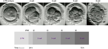
The KID3 scoring system is based on five time points of embryonic development (Figure 1): time of pronuclei fading (tPNf) and 2-, 3-, 5- and 8-cell division time (t2), (t3), (t5) and (t8), respectively. The KID3 score ranges from 1 to 5 and is based on five parameters: (1) t3 − tPNf ≥ 11.48 h; (2) t3 ≤ 42.91 h; (3) (t5 − t3)/(t5 − 52) ≥ 0.3408; (4) (t5 − t3)/(t5 − 52) < 0.5781; and (5) t8 < 66 h. The static morphology score is based on the standard for high-quality embryos in the Istanbul Consensus (on the third day after fertilization, the number of cells is 7–9, the cell size is in line with the development stage, the fragmentation degree is less than 10% and the embryo is without multinucleation). To predict the formation of blastocysts, that is to predict the embryonic development potential through embryo score, blastocysts were observed on days 5 or 6. The blastocysts were graded according to Gardner’s criteria: the size of the blastocyst cavity, the inner cell mass and the ectotrophoblast. The blastocysts can be divided into six levels by the size of the blastocyst cavity: Grade 1 (the blastocyst cavity is less than half of the embryo volume); Grade 2 (the blastocyst cavity is more than half of the embryo volume); Grade 3 (the blastocyst cavity almost fills the entire embryo); Grade 4 (the blastocyst volume and cavity is expanded, the zona pellucida is thinned); Grade 5 (some ectotrophoblast cells have begun to hatch from the zona pellucida); and Grade 6 (the blastocyst has completely hatched from the zona pellucida and separated from it). For the cell mass score: Grade A (the number of cells is large, there are compact clusters and obvious protuberances); Grade B (few cells, scattered or clustered); Grade C (the number of cells is very small, difficult to identify the obvious structure of the inner cell clusters). For the trophoblast score: Grade A (Grade 3 blastocyst trophoblast cells ≥ 10, Grades 4–6 blastocyst trophoblast cells ≥ 15, the cells are continuous, plump and well demarcated); Grade B (10 > Grade 3 blastocyst trophoblast cell number ≥ 5, 15 > Grades 4–6 blastocyst trophoblast cells ≥ 10); Grade C (Grade 3 blastocyst trophoblast cell number < 5, Grades 4–6 blastocyst trophoblast cells < 10). Blastocysts with a score of 3 BB or more were of high quality. Abnormal development is defined as follows: nondiploid cleavage (one blastomere cleaves directly into two or more cells), multinucleation (at least one blastomere with two or more nuclei during the period from 2-cells to 4-cells), asymmetric division (the blastomeres divide into two cells of unequal size with a one-fifth or greater diameter difference), fragmentation (the fragmentation rate is more than 30% during cleavage), fusion (two cleaved blastomeres fuse into one blastomere) and vacuoles (necrotic vacuoles appear in the cytoplasm).
Embryo development was observed using the EmbryoScope™ with the corresponding time points used for the KID3 score.

Transplantation and pregnancy outcome determination
In the transfer cycle, embryos with higher KID3 scores were preferentially transferred. Four weeks after the embryo transfer, the presence of embryo sacs in the uterine cavity observed using B-ultrasound was regarded as a clinical pregnancy.
Statistical analysis
Statistical analysis was performed with GraphPad Prism 5.0 and Fisher’s exact test was used for comparisons between groups. A P-value < 0.05 was considered to be statistically significant.
Results
Correlation between embryonic KID3 score and blastocyst formation
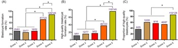
First, we investigated the relationship of the KID3 score with blastocyst formation. As shown in Figure 2(A), there was no significant difference in the blastocyst formation rates among the score 1, score 2 and score 3 groups. However, the formation rates of blastocysts in the score 4 and 5 groups were increased in comparison with the score 1–3 groups. Moreover, the blastocyst formation rate in the score 5 group was significantly higher than that in the score 4 group. Similarly, the formation rate of high-quality blastocysts also showed marked upregulation in the score 4 and 5 groups but no obvious change in the score 1–3 groups (Figure 2B). Compared with the score 1–4 groups (no obvious change), the ratio of high-quality blastocysts to total formed blastocysts in the score 5 group exhibited an obvious increase (Figure 2C). Taken together, these results supported the hypothesis that embryos with high KID3 scores are more likely to form high-quality blastocysts.
Correlation between embryonic KID3 score and blastocyst formation. (A) Blastocyst formation rate of embryos with scores of 1–5. (B) High-quality blastocyst formation rate of embryos with scores of 1–5. (C) Proportion of high-quality blastocysts was assessed. *P < 0.05.

Comparison of the KID3 score and traditional morphological score for screening high-quality embryos
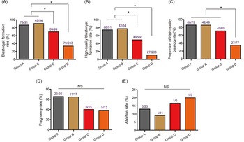
Next, we compared the difference between the KID3 score and the traditional morphological score for embryo screening. We found that the blastocyst formation rates in the A and B groups were distinctly higher than that in the C and D groups (Figure 3A). In addition, the formation rates of high-quality blastocysts in the A and B groups were also enhanced compared with those in the C and D groups (Figure 3B). Furthermore, the proportion of high-quality blastocysts in group D was decreased and there was no visible change in the A, B and C groups (Figure 3C). However, there was no significant difference between the KID3 score and traditional morphological score screened embryos for pregnancy and abortion rates (Figure 3D,E). In summary, the excellent embryos screened by the KID3 score had higher blastocyst and high-quality blastocyst formation rates compared with the excellent embryos screened by the traditional morphological score.
Comparison of the KID3 score and the traditional morphological score for screening high-quality embryos. (A) Blastocyst formation rate of embryos in groups A–D. (B) High-quality blastocyst formation rate of embryos in groups A–D. (C) Proportion of high-quality blastocysts in groups A–D. (D, E) Rates of pregnancy and abortion of embryos from groups A–D. *P < 0.05; NS: No significance.

KID3 scoring system used for the comparison of blastocyst formation for patients of different ages
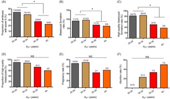
Then, we explored the effect of age on the selection of high-quality embryos with the KID3 score. As shown in Figure 4(A), the proportion of embryos with a score of 5 in patients over 35 years old was markedly lower than that in patients under 35 years old. Consistently, the blastocyst formation rate (Figure 4B) and high-quality blastocyst formation rate (Figure 4C) showed the same trend. Additionally, the proportion of high-quality blastocysts (Figure 4D), pregnancy rate (Figure 4E) and abortion rate (Figure 4F) had no significant correlation with age. While the pregnancy rate of patients over 35 years old showed a downward trend, the abortion rate increased with age. These results suggested that the ability to form score 5 high-quality embryos was decreased for patients over 35 years old and the ability of blastocyst formation and high-quality blastocyst formation in their score 5 embryos was also significantly decreased. Collectively, the embryos of young patients under 35 years old may have good developmental potential.
The KID3 scoring system was used for the comparison of blastocyst formation in patients of different ages. (A) Proportion of embryos from patients of different ages with a score of 5. (B–D) Blastocyst formation rate, high-quality blastocyst formation rate and proportion of high-quality blastocysts in embryos from patients of different ages. (E, F) Rates of pregnancy and abortion in embryos from patients of different ages. *P < 0.05; NS: No significance.

Comparison of abnormal embryogenesis for patients of different ages
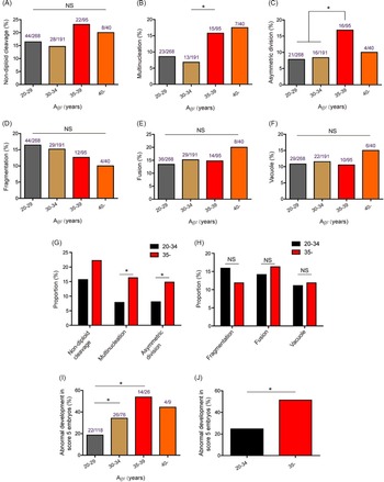
Finally, the relationship between age and abnormal embryo development was investigated. The incidences of multinucleation (Figure 5B) and asymmetric division (Figure 5C) in embryos from patients 35–39 years old were significantly higher than those in embryos from patients 30–34 years old. Nevertheless, the incidence of abnormal phenomena, such as nondiploid cleavage (Figure 5A), fragmentation, fusion and vacuoles, was not apparently correlated with the age of the patients (Figure 5D–F). Consistently, the incidences of multinucleation and asymmetric division in embryos from patients 20–34 years old were markedly decreased compared with ≥ 35-year-old patient embryos (Figure 5G,H). In addition, the incidence of score 5 embryo abnormal development in patients ≥ 35 years old was markedly increased compared with that in patients 20–34 years old. Taken together, patients over 35 years old may be more prone to abnormal embryonic development.
Comparison of abnormal embryogenesis in patients of different ages. (A–C) Incidences of nondiploid cleavage, multinucleation and asymmetric division in embryos from patients of different ages (20–29, 30–34, 35–39, ≥ 40 years old). (D–F) Incidence of fragmentation, fusion and vacuoles in embryos from patients of different ages. (G, H) Incidences of nondiploid cleavage, multinucleation and asymmetric division in embryos from patients 20–34 years old and ≥ 35 years old. (I, J) Incidences of abnormal development of score 5 embryos in patients of different ages. *P < 0.05; NS: No significance.

Discussion
The D3 high-quality embryo rate is a widely used reference index for internal quality control in embryo laboratories; however, the morphological definition of D3 high-quality embryos has always been controversial (Zhu et al., Reference Zhu, Zhang, Fadlalla, Wang, Geng and Liu2014; Tong et al., Reference Tong, Sheng, Sun, Li, Li, Zhang and Chen2017). At present, the definition of the D3 high-quality embryo standard in the Istanbul Consensus is the most widely used. This standard is based on one or several limited time points for observation, which has great limitations. It is easy to miss some important developmental phenomena during the process of embryo development, such as the appearance and disappearance of abnormal pronuclei, abnormal cleavage mode, multinucleation phenomenon, etc. In addition, this assessment is also easily affected by the evaluators’ subjectivity and is difficult to check and confirm (Alpha Scientists in Reproductive Medicine and ESHRE Special Interest Group of Embryology, 2011).
TLI observation makes up for the limitations of traditional morphological evaluation by collecting a large amount of dynamic development information during the early development of embryos and provides more abundant and objective data for embryo evaluation (Adamson et al., Reference Adamson, Abusief, Palao, Witmer, Palao and Gvakharia2016; Barrie et al., Reference Barrie, Homburg, McDowell, Brown, Kingsland and Troup2017; Zaninovic et al., Reference Zaninovic, Irani and Meseguer2017). To predict the embryonic development potential, researchers retrospectively analyzed blastocyst formation, blastocyst quality and the characteristic parameters of the implanted embryos and established a variety of different algorithm models (Meseguer et al., Reference Meseguer, Herrero, Tejera, Hilligsøe, Ramsing and Remohí2011; Conaghan et al., Reference Conaghan, Chen, Willman, Ivani, Chenette, Boostanfar, Baker, Adamson, Abusief, Gvakharia, Loewke and Shen2013; VerMilyea et al., Reference Vermilyea, Tan, Anthony, Conaghan, Ivani, Gvakharia, Boostanfar, Baker, Suraj, Chen, Mainigi, Coutifaris and Shen2014; Basile et al., Reference Basile, Vime, Florensa, Aparicio Ruiz, García Velasco, Remohí and Meseguer2015b; Milewski et al., Reference Milewski, Kuć, Kuczyńska, Stankiewicz, Łukaszuk and Kuczyński2015; Liu et al., Reference Liu, Chapple, Feenan, Roberts and Matson2016). Petersen et al. (Reference Petersen, Boel, Montag and Gardner2016) compared seven algorithms based on the characteristic parameters of blastocyst formation or implantation (Eeva I, Eeva II, Milewski, Meseguer, Basile, Liu and KID3) and found that the prediction of blastocyst formation and blastocyst quality by the KID3 score and Liu algorithm were significantly superior to other algorithms, suggesting that the KID3 score could be used for the prediction of embryo development potential.
In this study, we found that the KID3 score was correlated with the development potential of embryos, which is consistent with the results of Petersen et al. (Reference Petersen, Boel, Montag and Gardner2016). In addition, 85% of the blastocysts formed by embryos with a score of 5 were high-quality blastocysts, which was significantly higher than that of embryos with scores of 1–4, suggesting that the KID scoring system could be a predictive tool for screening high-quality D3 embryos.
In the clinical evaluation and screening of TLI-cultured embryos, screening methods based on cleavage mode and embryo dynamics parameters often produce evaluation results inconsistent with those of traditional morphological evaluation methods. These inconsistencies will interfere with embryologists’ selection of superior embryos and introduce challenges in doctor–patient communication. In the present study, we conducted a retrospective analysis of embryos with inconsistent selection between the two evaluation methods. From the comparison between group A and group B, it can be seen that, when the results of the KID3 assessment are excellent, whether the embryos are rated as excellent by traditional morphology or not, it does not affect the probability of high-quality blastocyst formation and a successful pregnancy. The embryos in group B were assessed by KID3 as superior embryos but not by traditional methods. The cleavage mode in most of these embryos was normal, but the number of cells exceeded nine during D3 observation, or more fragments (≥10%) appeared in the development process due to the fast development rate, which led to the non-excellent rating in the traditional evaluation. This result suggested that embryos with a normal cleavage pattern and a faster development rate still had good potential for further development. The developmental potential of group C (KID3 assessment: non-excellent; conventional morphological evaluation: excellent) was significantly lower than that of the former two groups. There may be an abnormal cleavage pattern in the development of these embryos. For example, nondiploid cleavage will produce more cells in a short period of time and this is accompanied by stagnation of the development of some blastomeres. Alternatively, the larger cell fragments produced in the early irregular division were mistaken for blastomeres and included in the count by the observers, resulting in the number of cells and the fragmentation rate reaching the standard of traditional excellent embryos during D3 observation. Previous studies have shown that nondiploid cleavage may be related to chromosomal abnormalities. Athayde Wirka et al. (Reference Athayde Wirka, Chen, Conaghan, Ivani, Gvakharia, Behr, Suraj, Tan and Shen2014) found that the blastocyst formation rate (11.7%) and implantation rate (3.7%) of this kind of embryo were lower.
TLI can identify embryos with normal morphology and abnormal cleavage patterns; for example, one blastomere directly splits into three blastomeres (Omidi et al., Reference Omidi, Khalili, Halvaei, Montazeri and Kalantar2020). Aneuploidy or gene abnormality are the main causes of abnormal cleavage (Athayde Wirka et al., Reference Athayde Wirka, Chen, Conaghan, Ivani, Gvakharia, Behr, Suraj, Tan and Shen2014). If such embryos are transferred, the abortion rate will be increased, which indicates that the cleavage pattern has an important effect on embryo development outcomes (Werner et al., Reference Werner, Reh, Grifo and Perle2012). Embryos with a low blastomere number, abnormal cleavage pattern and high fragmentation rate were found in group D, which had the lowest developmental potential among the four groups. Previous studies have found that the incidence of chromosomal abnormalities in embryos with developmental delay is higher than that in embryos with normal development and the rate of embryo fragmentation is significantly associated with chimerism and other postfertilization abnormalities (Alikani et al., Reference Alikani, Calderon, Tomkin, Garrisi, Kokot and Cohen2000; Munné et al., Reference Munné, Colls, Garrisi, Zheng, Cekleniak, Lenzi, Hughes, Fischer, Garrisi, Tomkin and Cohen2007). The probability of normal blastocyst formation in embryos with a fragmentation rate greater than 15% is significantly lower than that in embryos with a fragmentation rate between 0 and 15% (Alikani et al., Reference Alikani, Calderon, Tomkin, Garrisi, Kokot and Cohen2000). The chromosome abnormality rate of embryos with a fragmentation rate > 35% increased to 70–90%, which may be the main factor leading to the decline of embryonic development potential (Munné et al., Reference Munné, Colls, Garrisi, Zheng, Cekleniak, Lenzi, Hughes, Fischer, Garrisi, Tomkin and Cohen2007).
The KID3 score can accurately screen out high-quality embryos with development potential, but the predictive value of the KID3 score for patients of different ages has not been reported. In this study, we found that more than 80% of high-quality embryos screened by the KID3 score for young patients (under 35 years old) could form high-quality blastocysts, while the blastocyst development potential and blastocyst quality of score 5 high-quality embryos for older patients (over 35 years old) were significantly reduced. This may be related to the KID3 scoring algorithm, which is based on five time parameters and does not include abnormal cleavage mode or multinucleation phenomenon in the scoring system. The incidence of nondiploid cleavage, multinucleation and asymmetric division in embryos from patients > 35 years old is significantly higher than that in younger patients, suggesting that the higher frequency of abnormal development may be the reason for the reduced KID3 score 5 embryonic development potential. Therefore, the inclusion of these abnormal embryonic development phenomena into the evaluation system is one of the aspects that can be improved in the future. Additionally, a deficiency of this study lies in the small sample size of embryo transfer, which leads to an obvious trend but no significant difference. We will continue to expand the sample size in future research. Also, the present study is an academically interesting assessment of the ability of KID3 to predict blastulation, not an implantation study.
In this study, the KID3 scoring system may be a better predictive tool for embryo screening than the traditional morphological score. It could be a good predictor for patients < 35 years old. For patients > 35 years old, we should consider abnormal embryo development to conduct a more comprehensive evaluation of the embryos.
Author contributions
Jing Zhou: guarantor of integrity of the entire study; study design; experimental studies. Jun Zhou: literature research; data analysis. Mei-Qing Li: clinical studies. Rou Li: experimental studies. Yu-Lin Nie: statistical analysis. Hong-Qing Liao: manuscript editing; manuscript review.
Financial support
None.
Conflicts of interest
The authors declare no competing financial interests.
Ethical standards
The procedure and protocol were reviewed and approved by the ethics committee of our hospital. All patients have consented to the study.