Introduction
Zebrafish as a reproductive model
Zebrafish are excellent models for studying vertebrate reproduction, development and toxicology (Segner, Reference Segner2009; Lessman and Carver, Reference Lessman and Carver2014). A considerable amount of genomic and physiological information exists regarding zebrafish, especially for embryonic development. However relatively less information is known about zebrafish reproduction, this has received more attention recently, for example puberty in zebrafish has been reported in two studies (Chen and Ge, Reference Chen and Ge2013; Jones and Lessman, Reference Jones, Lessman, Lessman and Carver2014). Further study of this fundamental aspect of reproductive biology will help to provide the basis for understanding the processes leading to the next generation in fish and other vertebrates. Zebrafish are practical test animals because they are cost effective to raise and maintain compared with some other vertebrates. Also, they produce large quantities of transparent embryos useful for toxicological and other development assays (Hill et al., Reference Hill, Teraoka, Heideman and Peterson2005; Duan et al., Reference Duan, Zhu, Zhu, Kun and Zhu2008). A growing number of endocrine-disrupting chemicals (EDCs) has been tested in assays of reproductive output in zebrafish, including the oestrogenic mimic diethylstilbestrol (DES), which generally decreases reproduction (Hill and Janz, Reference Hill and Janz2003; Hill et al., Reference Hill, Teraoka, Heideman and Peterson2005; Segner, Reference Segner2009). DES is used in this present study as a ‘proof of principle’ to test whether endocrine disruption can be detected in vivo as evidenced by gonad changes related to puberty.
Why Casper?
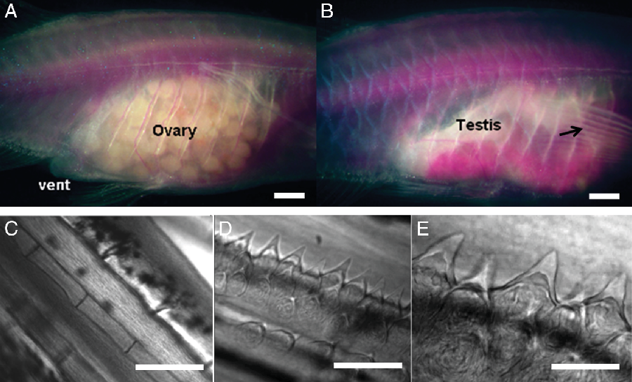
Various mutations in genes of pigment cells have yielded strains of transparent or ‘see-through’ medaka (Wakamatsu et al., Reference Wakamatsu, Pristyazhnyuk, Kinoshita, Tanaka and Ozato2001) and zebrafish (White et al., Reference White, Sessa, Burke, Bowman, LeBlanc, Ceol, Bourque, Dovey, Goessling and Burns2008; Takatsu et al., Reference Takatsu, Miyaoku, Roy, Murono, Sago, Itagaki, Nakamura and Tokumoto2013). These fish strains allow the study of internal organs in vivo that normally are obscured by an opaque pigment. The transparent Casper zebrafish has great potential for studying reproduction, gonad development and sexual maturation, as the reproductive organs as well as many other internal organs are visible. Casper is a double mutant and is transparent because it lacks functional iridophores and melanocytes (Lister et al., Reference Lister, Robertson, Lepage, Johnson and Raible1999; White et al., Reference White, Sessa, Burke, Bowman, LeBlanc, Ceol, Bourque, Dovey, Goessling and Burns2008; D’Agati et al., Reference D’Agati, Beltre, Sessa, Burger, Zhou, Mosimann and White2017). Therefore, the gonad can be studied in the live animal over extended periods of time (i.e. longitudinally) and without specialized equipment. A standard dissecting microscope fitted with a digital camera is all that is required to document changes in the gonads (Fig. 1). Casper is ideal for observing the formation of gonads, onset of puberty and gonad dynamics in vivo. Prior to the generation of the Casper zebrafish, sex determination and puberty studies were conducted using wild-type zebrafish that become pigmented and opaque well before gonad maturation (Maack and Segner, Reference Maack and Segner2003; Wang et al., Reference Wang, Bartfai, Sleptsova-Freidrich and Orban2007; Chen and Ge, Reference Chen and Ge2013). These former studies used histological data to determine at which point primordial germ cells became male or female gametes. As the animals in these previous studies had to be sacrificed to obtain the data, their subsequent reproductive events, such as spermatogenesis, oocyte growth, ovulation, oviposition and atresia, could not be determined. Our study will compliment previous work by providing data on weeks post fertilization (WPF) when the Casper zebrafish become sexually mature and develop secondary sex characteristics such as breeding tubercles in males and vent enlargement (genitalia differentiation) in females (Fig. 1).
Adult female (A, left) and male (B, right) imaged in vivo, note the gravid ovary and milky-white testes, respectively. The larger genital vent is seen in the female while the breeding tubercles appear as bright thickenings of pectoral fin rays in the male (arrow); ×8 total magnification; bars (A, B) represents 1 mm. Representative breeding tubercle montage (C–E). From left to right: adult female fin at ×100 magnification without tubercles (C), bar represents 200 μm; adult male fin at ×100 magnification showing pointed tubercles (D), bar represents 200 μm; ×400 magnification of the same male fin from middle frame (E), bar represents 50 μm.

Zebrafish sexual dimorphism
While male and female zebrafish exhibit minimal obvious sexual dimorphism, male zebrafish have testosterone-dependent epidermal specializations called breeding tubercles (Fig. 1) that are found especially on pectoral fins of sexually mature males (McMillan et al., Reference McMillan, Xu, Zhang, Teh, Korzh, Trudeau and Akimenko2013, Reference McMillan, Geraudie and Akimenko2015). Also the enlarged genital vent of sexually mature females is another secondary sex characteristic useful for sexing zebrafish (Yossa et al., Reference Yossa, Sarker, Proulx, Saxena, Ekker and Vandenberg2013). In some fish species the vent (urogenital pore or ovipositor) is under hormonal control, for example the extensive ovipositor of the Japanese Rose Bitterling, used to deposit eggs into bivalve mantles, has been shown to be oestrogen dependent and capable of changing length in sync with the ovarian cycle (Asahina et al., Reference Asahina, Hanyu, Aida, Nishina, Kobayashi, Loft and Holmes1985; Shirai, Reference Shirai1962). This evidence, along with our preliminary data, suggests that the female Casper zebrafish vent could be under hormonal control as well. With DES potentially causing endocrine disruption or changes in the developing gonad, it is also useful to simultaneously observe the secondary sex characteristics between males and females for possible effects of DES.
The transparent Casper zebrafish will allow in vivo visualization of the reproductive organs and therefore, the effects of DES on gonad maturation. This will allow visualization of reproductive changes over time in a living vertebrate animal, in vivo, including puberty onset and secondary sex characteristics associated with puberty in Casper. The rapidly growing field of zebrafish research indicates that this is a potent vertebrate model for elucidation of answers to a variety of biological problems. Casper extends this usefulness to in vivo studies of reproduction.
Materials and Methods
Zebrafish
A juvenile transparent zebrafish line, the Casper zebrafish (roy –/–;nacre –/–), was generously donated by Ryan Gebert at St. Jude Children’s Research Hospital, Memphis, TN, USA. The Casper zebrafish is named for its ghost-like appearance because it lacks melanocytes and iridophores and, therefore, is transparent (White et al., Reference White, Sessa, Burke, Bowman, LeBlanc, Ceol, Bourque, Dovey, Goessling and Burns2008). The Casper zebrafish line is derived from two mutated zebrafish lines. The first, nacre –/– zebrafish, lacks melanocytes due to a mitfa gene mutation (Lister et al., Reference Lister, Robertson, Lepage, Johnson and Raible1999). The second, roy orbison (roy –/–) zebrafish, lacks iridophores associated with an Mpv17 mutation (D’Agati et al., Reference D’Agati, Beltre, Sessa, Burger, Zhou, Mosimann and White2017). Crosses of these two mutants yield a transparent animal (White et al., Reference White, Sessa, Burke, Bowman, LeBlanc, Ceol, Bourque, Dovey, Goessling and Burns2008) that permits clear visualization of the gonad in vivo. The study was conducted under an IACUC protocol (number 0714) for physiology in zebrafish.
A second Casper line, Tg(Glut1b:mCherry; Fli1:GFP), was reared in the Lessman laboratory from two lines kindly provided by Bensheng Ju and Michael Taylor at St. Jude Children’s Research Hospital. The Tg(Glut1b:mCherry) line on the AB background shows expression in the blood–brain barrier vessels (Umans et al., Reference Umans, Henson, Mu, Parupalli, Ju, Peters, Lanham, Plavicki and Taylor2017) and in adult females the ovary shows expression (pers. commun.: Bensheng Ju and Michael Taylor). This line was crossed with Tg(Fli-:GFP) Casper (Lawson and Weinstein. Reference Lawson and Weinstein2002) expressing GFP in vasculature to give the double mutant, double transgenic. This strain was used to allow confocal microscopic optical sectioning in vivo of the mCherry-expressing ovary with the potential of observing GFP-expressing follicle wall blood vessels. This latter objective was obscured somewhat by the dense body wall vasculature. However, in favourable views, association of follicle vasculature could be seen. Therefore this strain may be a useful tool in studying puberty and developing ovarian follicle vasculature in future work.
The Casper zebrafish were reared at 28°C on a 14-h photoperiod cycle, and fed a combination mixture of tropical fish flake, artaemia (freeze-dried brine shrimp), and earthworm flakes daily (Aquaticeco.com). Tank water was changed daily. For spawning studies, spawning tanks were checked daily and embryos were collected.
Bright field microscopy
Juveniles were imaged in clear plastic bags (7.5 × 14 cm, Amazon.com) containing sufficient 0.04% tricaine methanesulfonate (TMS), (0.1 mg in 250 ml) in egg water (reverse osmosis purified water; dechlorinated tap water (1:1) containing three drops 1% methylene blue per 4 litres) to allow ventilation. Routinely, fish were in the bags less than 3 min while five images were taken: (1) a full body image taken against a ruler with a Canon digital camera; and (2) right and left trunk images in both transmitted and reflected illumination using a Zeiss Stemi SR microscope with an Amscope 9MP eyepiece camera. None of the animals imaged this way, once per week, showed any adverse effects. To image pectoral fins of juveniles, animals were anaesthetised in 0.04% TMS. The fish were placed in an agarose-lined Petri dish containing a pre-formed trough to position fish and filled with TMS and egg water to allow for respiration. The animals were imaged individually on each side using a Zeiss Stemi SR microscope with an Amscope 9MP eyepiece camera focused on the mid-trunk containing the gonad and vent. Images were taken using both reflected and transmitted lighting. For some experiments, animals were propped in the agarose trough with pectoral fins splayed for imaging. After images of right and left sides and fins were captured, the animals were placed in continuously aerated fresh water for recovery from the anaesthesia.
Confocal microscopy
Confocal images were captured using a Nikon A1r confocal with inverted Ti Nikon microscope and Nikon XYZ motorized stage with standard detector filters cubes: 450/50 nm, 485/40 nm, 525/50 nm, 545/40 nm and 600/50 nm equipped with lasers: 405 nm, 457 nm, 477 nm, 488 nm, 514 nm, 561 nm, 640 nm with four standard photomultipliers (PMTs) detectors and Nikon A1 spectral detector (32 PMT array; wavelength range: 400–770 nm). Standard image acquisition speed was 1 frame/s (512 × 512) and z axis stacks of 5 μm optical sections were made of anaesthetized Casper zebrafish with added 3% methyl cellulose to minimize movement during image capture. Image stacks were analyzed using Nikon NIS Elements Acquisition Software and ImageJ software.
Analysis methods
Bright field digital images were downloaded from the Amscope camera as.jpg files. These files were analyzed using ImageJ analysis software. ImageJ was downloaded from http://rsb.info.nih.gov/ij/. For quantification of ovarian follicle diameters, a stage micrometer image was captured and used for calibration in ImageJ software
Ovarian follicle diameter measurement: female puberty
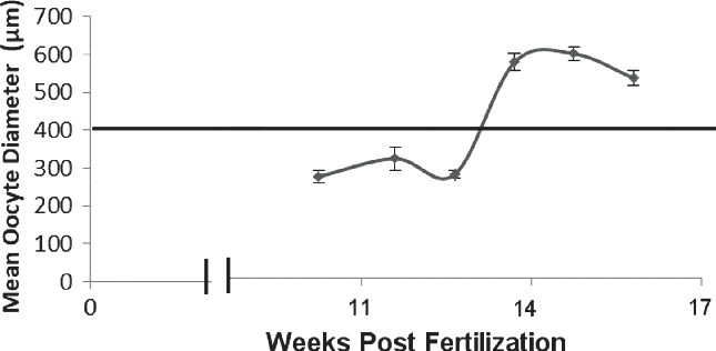
All visible ovarian follicles with distinct outlines were measured for the left and right images of the animal. Generally, the reflected mode image was found most suitable for these measurements and follicles less than 200 μm were usually not easily visualized as they are previtellogenic and transparent. Previous reports suggested stage II follicles indicate puberty in female zebrafish (Chen and Ge, Reference Chen and Ge2013) and farmed species (Taranger et al., Reference Taranger, Carrillo, Schulz, Fontaine, Zanuy, Felip, Weltzien, Dufour, Karlsen and Norberg2010) these follicles in zebrafish range from 0.14–0.34 mm in diameter and are transparent (Selman et al., Reference Selman, Wallace, Sarka and Qi1993). However, these small, clear follicles are difficult to image in vivo due to the lack of contrast. Instead we opted to use the stage III follicles that are opaque due to vitellogenin uptake, therefore readily imaged, and larger ranging from 0.34–0.69 mm (Selman et al., Reference Selman, Wallace, Sarka and Qi1993). In ImageJ software, a calibrated line tool was used to calculate the height and width of the individual follicles. These line lengths were recorded in Microsoft® Excel and averaged as ovarian follicle diameters. Puberty onset in females was defined here as the larger size classes of ovarian follicles attaining an average diameter of 400 μm, which is at lower end of stage III size range (Fig. 2). Follicles are vitellogenic at this size and indicate significant oestrogen presence (Selman et al., Reference Selman, Wallace, Sarka and Qi1993). As images and therefore measurements were made weekly, graphs of the follicle diameters were used to estimate puberty onset by determining the intercept between the graphed data and the 400 μm y-axis scale of oocyte diameter (Fig. 2).
Graph of mean oocyte diameter of juvenile Casper female over time. All visible oocytes were measured using ImageJ and plotted. Puberty is defined as occurring when the ovary contains oocytes that are >400 µm. The intersection of the 400 μm line and graph line indicate puberty (13.6 WPF). Data presented as x ± standard error of the mean (SEM) of oocytes from both left and right ovaries. WPF, weeks post fertilization.

Diethylstilbestrol treatment
In this study, the effects of exogenous diethylstilbestrol (DES) on puberty, gonad morphology and spawning were determined. DES was purchased from Sigma Aldrich. DES was added directly to tank water at 0.01, 0.1, 1.0, or 10.0 µg/l (3.73E–5 µM, 3.73E–4 µM, 3.73E–3 µM, or 0.037 µM, respectively) from concentrated stocks dissolved in vehicle (EtOH:propylene glycol, 1:1). DES treatment lasted for specific times that the protocols describe above. Tank water was changed daily and fresh DES added. Animals were imaged before, during and after DES treatment to assess the effects in vivo.
Statistical analysis
In this study, statistical analysis was performed in MS Excel; Student’s t-test was used to determine the P-value of DES-treated and non-DES-treated animals. Two arrays were selected to compare two groups of data selected (i.e. DES females verses non-DES females).
Results
Puberty in control and diethylstilbestrol (DES)-treated Casper juveniles
Juvenile sibling Casper zebrafish (n = 63; 43 control and 20 DES treated) were imaged weekly beginning at 6 WPF until 14 WPF. The control group of 43 juveniles was randomly chosen from the larger sibling pool and divided into groups of 10–15 animals. An additional 20 animals were divided into two groups of 10, each receiving 1.0 or 10.0 µg/l of DES with staggered treatment times to allow imaging. All animals were housed individually in uniquely numbered 500 ml tanks. DES treatment occurred during the 8–9 WPF after sex determination, but prior to sexual maturation as reported in wild-type zebrafish (Chen and Ge, Reference Chen and Ge2013). DES was applied directly to the tank water of each group and fresh DES was added every other day when the tank water was changed for a total incubation of six days.
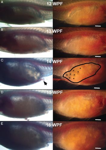
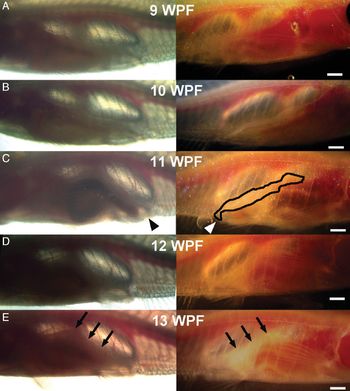
As imaging was done weekly, the data are reported as weeks of development. Groups of 10–15 animals were imaged each weekday and the groups staggered to spread imaging over the 5-day week. Full body images were taken for all animals each week to determine standard length (i.e. snout to caudal peduncle). In addition the mid-trunk of both sides of each animal was imaged by dissecting microscope-mounted camera. For males, testes and vent (i.e. genitalia) were imaged; for females, ovary and vent were imaged. The images were analyzed using ImageJ software to determine the qualitative opacity of testes in males and ovarian follicle diameters were measured in females using the line tool. In females, puberty was defined as the WPF when the average ovarian follicle size of the largest clutches reached 400 µm and the follicles became increasingly opaque due to vitellogenesis (Fig. 3). As follicles rapidly grow and undergo meiotic maturation, they become translucent and the vent enlarges (Fig. 3C). Males were defined as having achieved puberty when the testes became milky opaque and the tubular structure of the opaque testis extended to the vent (Fig. 4) while the vent size remains unchanged (Fig. 4E).
Representative puberty montage of a serially imaged female Casper. (A) 12 WPF. Puberty occurred at 13.6 WPF, i.e. between the panels (B) and (C), 13 WPF and 14 WPF, respectively. Left panels show transmitted light microscopy mode and right panels show reflected light microscopy mode of respective ovary. Ovary indicated by encircled area, asterisks indicate mature eggs and arrow indicates enlarged genital vent. This fish spawned for the first time at 14 DPF (16.3 WPF) which provides confirmation of the sexually mature ovary. (D) 15 WPF. (E) 16 WPF. All images were taken at a magnification of ×12. Bars represent 1 mm. WPF, weeks post fertilization.

Representative puberty montage of a serially imaged male Casper. (A) 9 WPF. (B) 10 WPF. Puberty occurred at 11 WPF panel (C). Left side shows transmitted light microscopy mode and right side shows reflected light microscopy mode. (D) 12 WPF. (E) 13 WPF. Puberty was defined here as the first time at which tubular structure of the testis was clearly formed and when the testis is an opaque, milky-white extending (area encircled in (C) and arrows in (E)) to the vent (arrowheads). All images were taken at a magnification of ×12. Bars represent 1 mm. WPF, weeks post fertilization.

Vehicle control group
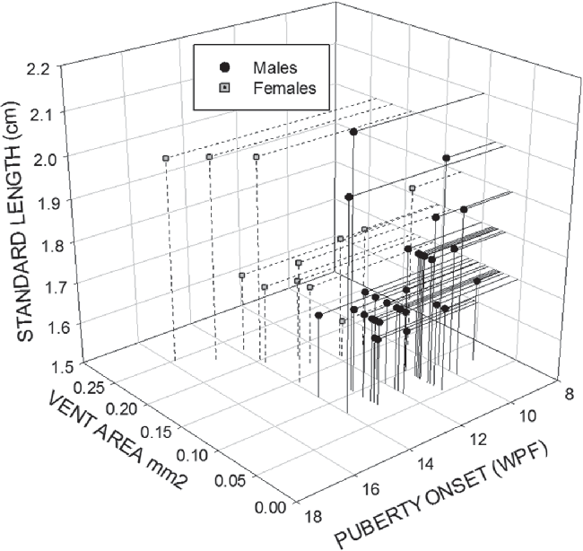
Of the 43 control animals, 12 developed ovaries and 31 developed testes (Fig. 5). Female Casper zebrafish developed mature ovaries at 13.2 ± 1.7 WPF (mean ± SD) with an average body length of 1.8 ± 0.1 cm and an average vent area of 0.18 ± 0.04 mm2 (Figs 5 and 6). Male Casper zebrafish developed mature testes at 11.7 ± 1.2 WPF with an average body length of 1.8 ± 0.1 cm and vent area of 0.09 ± 0.01 mm2 (Figs 5 and 6). Increased vent area occurs in female puberty onset, but not in males (Fig. 6).
A 3-D plot relating length, vent area and puberty onset in 31 male (solid circles) and 12 female (open circles) Casper zebrafish. Males clustered at lower end of vent area axis compared with females with larger vents at puberty. Males attained puberty about 2 weeks sooner and at a shorter standard length than females on average. WPF, weeks post fertilization.

Vent area as a function of age in juvenile Casper zebrafish. Data presented as mean ± standard error of the mean (SEM) of vent area mm2; N = 31 males and 12 females. Arrows indicate average attainment of puberty, asterisks denote significant difference P < 0.05 between males and females using Student’s t-test.

Effect of diethylstilbestrol (DES) on puberty
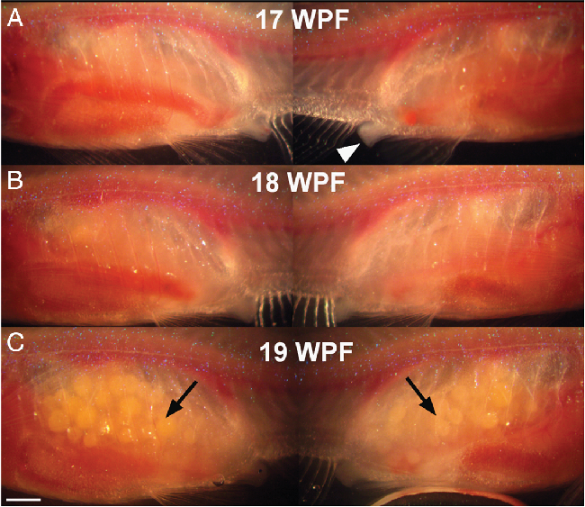
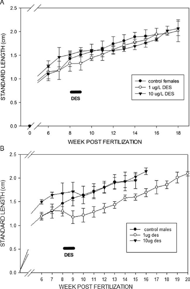
In the 10.0 µg/l group, three of 10 animals developed female gonads and reached puberty at 18.0 ± 0.0 WPF with an average body length of 2.0 ± 0.0 cm (Fig. 7A). Three of the animals developed male gonads and attained puberty at 22.3 ± 0.6 WPF with an average body length of 2.1 ± 0.1 cm (Fig. 7B). The remaining four animals died during treatment, prior to attaining puberty. Three of the animals treated with 1 μg/l DES developed enlarged, opaque follicles in their ovaries at 18.3 ± 2.1 WPF with a body length of 1.9 ± 0.2 cm (Figs 7A and 8). Five out of 10 animals developed opaque, milky-white tubular testes at 21.4 ± 0.9 WPF with an average body length of 2.1 ± 0.1 cm (Figs 7B and 9). One animal died from apparent non-DES-related injuries at 16.3 WPF and another was severely growth retarded and died at 28.9 WPF; both failed to attain puberty. DES treatment delayed puberty in females by an average of 5 weeks compared with controls (Fig. 7A) and in males by 10 weeks on average (Fig. 7B) when compared with non-DES-treated sibling groups. Surprisingly, a 1 μg/l DES-treated female achieved puberty 4 weeks ahead of the rest of the treatment group; the outlier was 1.7 cm in length at the time of puberty (Fig. 7A). These data suggest that animals must reach a critical body length before puberty onset and that the age of the animal is less critical for puberty. These results are in general agreement with those for wild-type zebrafish (Chen and Ge, Reference Chen and Ge2013). At the time of puberty, control males, had a body length of 1.8 ± 0.1 cm, while DES males had a body length of 2.1 ± 0.2 cm clustering well away from the control group (Fig. 7B). The 6-day DES treatments (8 WPF) resulted in slowed or arrested growth (Fig. 10) especially in males (Fig. 10B), then after drug withdrawal a return to growth rates similar to controls in females (Fig. 10A) and males (Fig. 10B).
Effects of diethylstilbestrol (DES) on juvenile Casper (A) female and (B) male body length compared with controls at puberty. X-axis shows weeks post fertilization (WPF) and Y-axis shows standard body length (snout to caudal peduncle) in centimetres. Control males N = 31, control females N = 12, surviving DES males 1 μg/l N = 5 and 10 μg/l N = 3, females 1 μg/l N = 3 and 10 μg/l N = 3. DES males show clear segregation from their controls, while DES females are less well segregated from control females.

Effects of 1 μg/l diethylstilbestrol (DES) treatment for 6 days at 8 WPF on puberty of female Casper juvenile. (A) 17 WPF. (B) 18 WPF. (C) 19 WPF. Each panel (A–C) shows left and right sides at ×12 total magnification imaged with reflected light microscopy. Montage shows the delayed progression of puberty due to DES treatment for 6 days 10 weeks earlier. This representative female went through puberty 45 days later than control females. Arrows indicate examples of vitellogenic follicles (>400 µm) indicative of puberty (C). Arrowhead indicates enlarging vent (A). Bar represents 1 mm. WPF, weeks post fertilization.

Effects of 1 µg/l diethylstilbestrol (DES) treatment for 6 days at 8 WPF on juvenile Casper male puberty. (A) 19 WPF. (B) 20 WPF. (C) 21 WPF. Each panel (A–C) shows left and right sides captured at ×12 magnification with reflected light microscopy. Montage shows the delayed progression of puberty due to DES treatment for 6 days 12 weeks earlier. This representative male attained puberty at 149 DPF, 11 weeks later than control males. Puberty is indicated in panel (C) by a white opaque tubular testis structure (arrows). Vent (arrowhead) (A) is reduced at puberty. Bar represents 1 mm. WPF, weeks post fertilization.

Effects of diethylstilbestrol (DES) 6 day treatment at 8 WPF (indicated by bars) on juvenile Casper female (A) and male (B) standard body length growth compared with controls at puberty. X-axis shows weeks post fertilization (WPF) and Y-axis shows standard body length (snout to caudal peduncle) in centimetres. Graphs show steady growth with time in control males and females. DES-treated males (B) showed a decrease in growth rate during treatment and a subsequent return to control growth rates after DES withdrawal while DES-treated females (A) were affected less. Control males N = 31, control females N = 12, surviving DES males 1 μg/l N = 5 and 10 μg/l N = 3, females 1 μg/l N = 3 and 10 μg/l N = 3.

Emergence of breeding tubercles at puberty
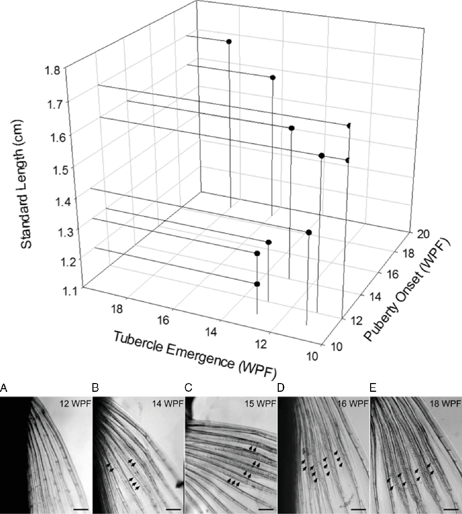
Imaging of a group of 15 juvenile Casper zebrafish Tg(Glut1b:mCherry,Fli1:GFP) siblings began at 10 WPF and was focused on the relationship between puberty and breeding tubercle emergence. The animals were imaged weekly until 2 weeks post puberty or 26 WPF. For males, testes, pectoral fins, body length and vent were imaged; for females, ovary, vent, body length and fins were imaged. Pectoral fins were included to confirm the presence or absence of breeding tubercles at puberty in both sexes. Pectoral fins of females were examined but none exhibited breeding tubercles in this study. ImageJ software was used to estimate milky whiteness (8-bit grayscale) of testes in males and ovarian follicle diameters were measured in females. In females, puberty occurred when the average ovarian follicle size of the largest clutch reached 400 µm and above. Males achieved puberty when the testes became milky opaque and the tubular structure of the opaque testis was completely patent with the vent. Of the 15 animals, three developed ovaries and 10 developed testes. Two of the animals died before puberty onset. The three female Casper zebrafish developed mature ovaries at 12.7 ± 1.2 WPF with a standard length of 2.1 ± 0.1 cm and were used for confocal analysis (see next section). Male Casper zebrafish had mature testes at 12.7 ± 3.0 WPF with standard length of 1.5 ± 0.2 cm (Fig. 11A). Breeding tubercle emergence on the pectoral fins of males occurred at 12.3 ± 2.4 WPF (Fig. 11A, B) and therefore was concurrent with puberty (Student’s paired t-test P = 0.38). No breeding tubercle emergence was seen in female pectoral fins during the study.
Upper panel: 3-D plot relating male (N = 10) Casper standard length (cm), puberty onset (WPF) and breeding tubercle emergence (WPF). Lower panel: representative control Casper male’s pectoral fin imaged at ×32 magnification from 12 WPF (A), 14 WPF (B), 15 WPF (C), 16 WPF (D) to 18 WPF (E). Bars represent 250 μm. This male attained puberty at 13.4 WPF and exhibited breeding tubercles at 14 WPF. Arrows indicated rows of tubercles. WPF, weeks post fertilization.

Confocal microscopy
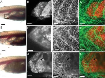
Puberty onset, in two TG (GLUT1–mCherry: Fli-1–GFP) Casper juvenile females and one TG(GLUT1–mCherry) Casper juvenile female, was determined by weekly imaging via confocal microscopy for 6 weeks. Imaging began 3 weeks before puberty and continued until all three females were confirmed to have undergone puberty as evidenced by the emergence of large, increasingly opaque follicles. Females were imaged on a stereoscope before confocal microscopy to determine ovarian status. Females were then imaged by confocal microscopy to determine the presence of small oocytes and fluorescent blood vessels in follicles surrounding oocytes (Fig. 12). Abundant, small oocytes/oogonia expressed the mCherry reporter, whereas larger oocytes (>100 µm) had reduced expression. Association of blood vessels surrounding oocytes was qualitatively assessed because the body wall blood vessels partly obscured visualization of the ovarian blood vessels. Figure 12(L) shows a large oocyte (>100 µm) associated with blood vessels expressing GFP mostly in cross-section at the follicle periphery.
Puberty onset in a TG(GLUT1-mCherry: Fli-1-GFP) Casper juvenile female. (A–D) 14 WPF, 11 days prior to puberty. (E–H) 15 WPF, 4 days prior to puberty. (I) 17 WPF, 11 days post puberty and (J–L) 16 WPF, 6 days post puberty. (A, E, I) show transmission bright field at ×12 magnification; bars represent 1 mm; black square shows region imaged by confocal. (B, F, J) show z stack (70 images; standard deviation) of GLUT1–mCherry expression in small oocytes/oogonia. Arrowheads (J–L) point to larger follicles no longer expressing reporter. Bars (B–D, F–H, J–L) represent 100 µm, taken at ×100 magnification. (C, G, K) show z stack (70 images; standard deviation) of Fli-1–GFP expression in blood vessels, some extend to larger follicles (arrowhead). (D, H, L) show merged z stacks. WPF, weeks post fertilization.

Effect of DES on post-pubescent males
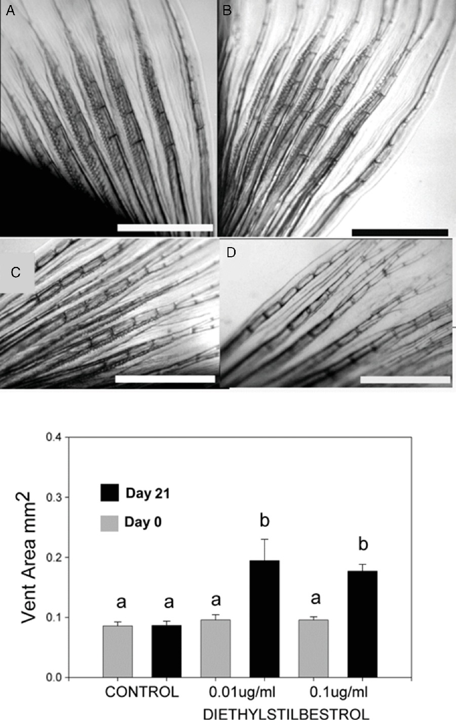
A group of 15 sexually mature Casper zebrafish sibling males was randomly selected and the males divided into three groups for a 4-week DES treatment. Males were imaged at 17 WPF (117 DPF), 1 day prior to treatment to establish testes, vent and breeding tubercle status. The next day, two treatment groups received either 0.1 or 0.01 µg/l DES and the other group received vehicle without DES to function as the control group. DES was applied directly to tank water and fresh DES was added every other day with tank water change. Treatment ended at 20 WPF (139 DPF). The effect of DES was documented by imaging the testes, vent, and breeding tubercles each week. The 0.1 µg/l dose of DES induced complete loss of breeding tubercles (Fig. 13D, upper panel) and an increase in the vent size by 20 WPF (139 DPF) (Fig. 13, lower panel). This dose did not induce complete loss of the testis. The 0.01 µg/l dose of DES induced breeding tubercle size reduction, but not total loss (Fig. 13C, upper panel). There was no change in breeding tubercles of the control males (Fig. 13A, B, upper panel). DES treatment also caused an increase in the size of the vent in both treatment groups (Fig. 13, lower panel). Statistical analysis revealed a significant (P < 0.05) increase in the male vent sizes in the treatment groups when compared with the control group. Imaging of fins at 21 WPF (146 DPF) revealed re-emergence of breeding tubercles in the 0.1 µg/l DES group (data not shown).
Effects of diethylstilbestrol (DES) on secondary sex characteristics of post-pubescent male Casper zebrafish. Upper panel: Breeding tubercle montage ×32 magnification. (A) Control male imaged at 17 WPF. (B) Control male imaged 3 weeks later (20 WPF). (C) 0.01 μg/l DES-treated male after 3 weeks. (D) 0.1 μg/l DES-treated male after 3 weeks; the latter two treatments caused reduced or complete loss of tubercles, respectively. Bars represent 0.5 mm. Lower panel: Bar graph of vent area changes with DES versus control treatment. Letter superscripts that differ indicate significant changes among treatments at P < 0.05, data presented as mean ± standard error of the mean (SEM), N = 5. WPF, weeks post fertilization.

Discussion
Puberty in transparent Casper zebrafish
A distinction should be made between sex determination and sexual maturation or puberty. The former is defined by the type of primary germ cell present in the juvenile gonad either primary oocytes or primary spermatocytes. The latter, puberty, is defined by the production of mature gametes. In the current set of experiments, transparent Casper zebrafish were utilized to determine sexual maturation or puberty in vivo. Currently, there is little information known about sexual maturation in zebrafish and some conflicting reports on the timing of sex determination. A 2003 study reported that sex determination in wild-type zebrafish occurs at 35 DPF (Maack and Segner, Reference Maack and Segner2003), while a 2007 study reported that sex determination in wild-type zebrafish occurs at 21 DPF (Wang et al., Reference Wang, Bartfai, Sleptsova-Freidrich and Orban2007). Most recently, it was reported that gonad differentiation or sex determination in females occurs at 35 DPF and at 45 DPF in males (Chen and Ge, Reference Chen and Ge2013). This latter study also reported that females begin sexual maturation or puberty at a critical body length of 1.8 cm (Chen and Ge, Reference Chen and Ge2013). Chen and Ge also indicated that differential rearing conditions could affect the growth rate of the animals (i.e. water conditions or feeding frequency). Our results have shown that the Casper zebrafish females must reach a critical length of 1.8 cm to achieve puberty and the Casper males must reach a critical length of 1.7 cm. There was less dependence on age of the animal and puberty onset. For example, one female went through puberty at 67 DPF whereas another female attained puberty at 105 DPF. Therefore, our data suggest that puberty is not strictly dependent on age and agree with a previous report (Chen and Ge, Reference Chen and Ge2013). While several studies have reported changes in gonad dynamics in other transparent lines of fish (Takatsu et al., Reference Takatsu, Miyaoku, Roy, Murono, Sago, Itagaki, Nakamura and Tokumoto2013; Wakamatsu et al., Reference Wakamatsu, Pristyazhnyuk, Kinoshita, Tanaka and Ozato2001), this paper’s central focus is on puberty or sexual maturation in Casper zebrafish and related secondary sex changes.
The ovary is composed of many developing oocytes (immature eggs) that can be in any one of five stages of development (Selman et al., Reference Selman, Wallace, Sarka and Qi1993). Previous reports used stage II oocytes assessed in histological section as a marker of puberty or sexual maturation (Taranger et al., Reference Taranger, Carrillo, Schulz, Fontaine, Zanuy, Felip, Weltzien, Dufour, Karlsen and Norberg2010; Chen and Ge, Reference Chen and Ge2013), however these small (0.14–0.34 mm), transparent cells are not reliably visualized in vivo and therefore not suitable as an in vivo marker. For puberty determination, we focused on the third stage of oocyte development encompassing oocytes 0.34–0.69 mm in diameter, the oestrogen-dependent vitellogenic stage. In this stage, oocytes are characterized by the recruitment of vitellogenin, the yolk precursor protein (Selman et al., Reference Selman, Wallace, Sarka and Qi1993). The endocytosis of this protein causes oocytes to increase in size from 340 µm to 700 µm (Selman et al., Reference Selman, Wallace, Sarka and Qi1993) and become opaque from yolk accumulation, making them readily visible and therefore measureable in live Casper. We selected oocytes at 0.4 mm in diameter as a readily measurable size but at the lower end of the stage III size range as a criterion for puberty. Oocytes at 0.4 mm, unlike the 0.34 mm diameter oocytes at the junction of the stage II and III ranges, have amassed sufficient yolk to become opaque and have high contrast for in vivo imaging. Vitellogenin is produced via the upregulation of the vitellogenin gene in the liver by oestrogen (Sumpter and Jobling, Reference Sumpter and Jobling1995; Tyler et al., Reference Tyler, Van der Eerden, Jobling, Panter and Sumpter1996). Juvenile zebrafish are too small to allow blood levels of oestrogen or other hormones to be measured without sacrificing and exsanguination of the animal. However, the appearance of vitellogenic oocytes is indicative of increased estrogen levels, and therefore is an excellent marker of puberty onset in female zebrafish (Sumpter and Jobling, Reference Sumpter and Jobling1995; Tyler et al., Reference Tyler, Van der Eerden, Jobling, Panter and Sumpter1996). In males, androgen has been implicated in the appearance of breeding tubercles and their presence may be considered a marker for male puberty (McMillan et al., Reference McMillan, Xu, Zhang, Teh, Korzh, Trudeau and Akimenko2013, Reference McMillan, Geraudie and Akimenko2015).
Effects of diethylstilbestrol (DES) on puberty in juvenile Casper zebrafish
EDCs can disturb endocrine events especially when exposed at a critical period of development (Brion et al., Reference Brion, Tyler, Palazzi, Laillet, Porcher, Garric and Flammarion2004; van der Ven et al., Reference van der Ven, van den Brandhof, Vos and Wester2007). Exposure of EDCs at the critical period causes permanent changes in the offspring’s reproductive capacity such as infertility, masculinization, feminization, or intersexed with an ovotestis (Brion et al., Reference Brion, Tyler, Palazzi, Laillet, Porcher, Garric and Flammarion2004). Critical periods include both sex determination and sexual maturation. These EDC effects are disturbing, as they could lead to a decline in the population of affected groups.
For this study, the 6-day DES treatment began at 8 WPF. Gonad images and body lengths were obtained 2 weeks before treatment and were subsequently imaged weekly until 2 weeks post puberty. In the 1 µg/l DES treatment group, puberty was delayed by 5 weeks in females and 9 weeks in males. In the 10 µg/l DES treatment group, puberty was delayed by 5 weeks in females and 11 weeks in males.
It should also be noted that several animals in both treatment groups exhibited dose-dependent signs of compromised health. These included haemorrhage, anaemia, oedema, liver enlargement, heart enlargement and death. Nevertheless, after DES treatment ended, puberty was attained, and the surviving animals were placed with non-DES-treated animals in spawning boxes and were found capable of spawning.
In zebrafish and goldfish oocytes, DES has been shown to induce oocyte maturation (Tokumoto et al., Reference Tokumoto, Tokumoto, Horiguchi, Ishikawa and Nagahama2004, Reference Tokumoto, Tokumoto and Nagahama2005). It has been reported that the mechanisms of DES maturation induction works via mimicking a naturally occurring maturation inducing hormone, 17α,20β-dihydroxy-4-pregnone-3-one (DHP) (Tokumoto et al., Reference Tokumoto, Tokumoto, Horiguchi, Ishikawa and Nagahama2004). In goldfish oocytes, oocyte maturation is induced via the novel membrane-bound progestin receptor alpha (mPRα) and DES binds to mPRα (Tokumoto et al., Reference Tokumoto, Tokumoto and Thomas2007). By binding to mPRα, DES mimics the non-genomic actions of progestins to induce oocyte maturation (Tokumoto et al., Reference Tokumoto, Yamaguchi, Ii and Tokumoto2011).
Endocrine-disrupting chemicals
It has been determined that production of functional oocytes is controlled by hormone regulation and is subject to disruption (Sumpter and Jobling, Reference Sumpter and Jobling1995; Tyler et al., Reference Tyler, Van der Eerden, Jobling, Panter and Sumpter1996). EDCs are substances capable of disturbing endocrine-regulated events in organisms, such as oogenesis and spermatogenesis. EDCs are found ubiquitously in the environment and have been shown to be deleterious to aquatic species and non-aquatic mammals (Iguchi et al., Reference Iguchi, Watanabe and Katsu2001; Kim et al., Reference Kim, Cho, Kim, Vanderford and Snyder2007). There are a variety of EDCs that can act as oestrogen agonists by eliciting a response that mimics the naturally occurring oestrogens (Sumpter and Jobling, Reference Sumpter and Jobling1995; Tyler et al., Reference Tyler, Van der Eerden, Jobling, Panter and Sumpter1996; Brion et al., Reference Brion, Tyler, Palazzi, Laillet, Porcher, Garric and Flammarion2004). EDCs can hinder hormone synthesis and steady-state levels (Brion et al., Reference Brion, Tyler, Palazzi, Laillet, Porcher, Garric and Flammarion2004) and can have the greatest effect when exposure occurs at a critical period in sexual or steroid-dependent development (Weber et al., Reference Weber, Hill and Janz2003; Brion et al., Reference Brion, Tyler, Palazzi, Laillet, Porcher, Garric and Flammarion2004). Exposure at this critical period may cause permanent changes in the reproductive capacity of the offspring, such as infertility, masculinization, feminization, or intersex state with an ovotestis (Hill and Janz, Reference Hill and Janz2003; Brion et al., Reference Brion, Tyler, Palazzi, Laillet, Porcher, Garric and Flammarion2004). During these critical periods, hormones are capable of permanently turning on or off genes for hormone receptors in certain cells and could potentially lead to a change in sex and fertility (vom Saal et al., Reference vom Saal, Timms, Montano, Palanza, Thayer, Nagel, Dhar, Ganjam, Parmigiani and Welshons1997).
DES is a potent synthetic oestrogen that binds to oestrogen receptor alpha (ERα) in humans and shares structural similarity to many known xenoestrogens (Reed and Fenton. Reference Reed and Fenton2013). DES can be found in aquatic environments with concentrations in the µg/l range (Yang et al., Reference Yang, Lin, Weng, Feng and Luan2008; Campinho and Power, Reference Campinho and Power2013). DES concentrations of 10.0 µg/l have been shown to modify the testis of male carp (Yang et al., Reference Yang, Lin, Weng, Feng and Luan2008). DES is lipid soluble and is readily absorbed into the body (Reed and Fenton, Reference Reed and Fenton2013). As DES is relatively water insoluble, it tends to accumulate at high concentrations in non-polar areas of the environment (Yang et al., Reference Yang, Lin, Weng, Feng and Luan2008). DES regularly enters aquatic environments via run-off from the chemical and medical industry (Yang et al., Reference Yang, Lin, Weng, Feng and Luan2008).
DES non-specific effects
In humans, it has been shown that DES compromises the ability of blood platelets to clot properly (Dobrydneva et al., Reference Dobrydneva, Williams, Katzenellenbogen, Ratz and Blackmore2003). DES is a calcium channel blocker in blood platelets and acts by blocking calcium channels and leads to an inability for platelet activation and blood clotting (Dobrydneva et al., Reference Dobrydneva, Williams and Blackmore2010). These reports may explain the haemorrhage, oedema and anaemia observed in some DES-treated Casper. Exposure of DES did not induce irreversible morphological effects on the gonads when applied for 6 days during late pre-pubescence. Also, DES exposure did not inhibit eventual spawning capability or appear to effect sex determination. As DES application began at 8 WPF, when sex determination has already occurred, it did not appear to be disturbed (Chen and Ge, Reference Chen and Ge2013). Puberty however, was obviously delayed. As mentioned earlier, this could be attributed, at least in part, to the toxicological effects of DES on growth and health.
Acknowledgements
We are grateful to Michael Taylor and Wilson Clements as well as Ryan Gebert for providing Casper zebrafish for this study. We also thank Michael Taylor and Bensheng Ju for their gift of transgenic zebrafish lines. NK was funded by a Graduate Assistantship awarded by the University of Memphis Graduate School. General supplies and fish husbandry items were procured by a grant-in-aid to CAL by the Department of Biological Sciences at the University of Memphis.
Financial Support
This research received no specific grant from any external funding agency, commercial or not-for-profit sectors.
Statement of Interest
The authors have no conflicts of interest regarding this work.
Ethical Standards
The authors assert that all procedures contributing to this work complied with the ethical standards of the relevant national and institutional guidelines on the care and use of laboratory animals.