INTRODUCTION
It is a key characteristic of excitable cells (neurons but not astrocytes) that potassium ions (K+) flow out of the cell during the depolarizing phase of the action potential, and an increase in extracellular K+ ([K+]o) during brain activity has been known for almost 40 years (Krnjevic and Morris, Reference Krnjevic and Morris1972; Vyklický et al., Reference Vyklický, Syková, Kriz and Ujec1972). Since neuronal function is influenced by [K+]o (reviewed by Walz and Hertz, Reference Walz and Hertz1982), efficient clearance of K+ from the extracellular space has high priority and is a major energy-requiring process in the brain (Hertz et al., Reference Hertz, Peng and Dienel2007). An important role for the astrocytic Na+,K+-ATPase in this process is well accepted (Somjen et al., Reference Somjen, Kager and Wadman2008), but information is lacking about density in each cell type of different Na+,K+-ATPase subtypes, and their turnover rates, which determine not only initial K+ uptake but also its indispensable return to neurons. These key parameters are investigated in the present study.
The catalytic units of the Na+,K+-ATPase are the α isoforms, of which the α2 and α3 are potently inhibited by ouabain, whereas especially in mice and rats the affinity of the α1 isoform for ouabain is much lower (Marks and Seeds, Reference Marks and Seeds1978; Urayama and Nakao, Reference Urayama and Nakao1979; Blanco and Mercer, Reference Blanco and Mercer1998). Immunochemical studies by McGrail et al. (Reference McGrail, Phillips and Sweadner1991) found that most α1 subunits of the Na+,K+-ATPase in the ventral horn of the rat spinal cord were expressed in glial processes that co-labeled for GFAP, i.e. were astrocytic, and that astrocytes also expressed the α2 subunit. Later work by the same group indicated that the α3 isoform is strictly neuronal both in the brain in vivo and in cultured cells (Peng et al., Reference Peng, Martin-Vasallo and Sweadner1997); moreover, α3 mRNA is restricted to a neuronal cell fraction obtained by fluorescence-activated cell sorting of cell acutely isolated from the mouse brain (Cahoy et al., Reference Cahoy, Emery, Kaushal, Foo, Zamanian and Christopherson2008). Mouse astrocyte cultures express both α2 and α1 subtypes, and among the β subunits β1 and β2, but virtually no β3 (Peng et al., Reference Peng, Arystarkhova and Sweadner1998). The glutamatergic cerebellar granule neuron cultures express α1 and α3 subunits (Peng et al., Reference Peng, Martin-Vasallo and Sweadner1997).
Even though both α1 and α2 subunits are strongly expressed in cultured astrocytes and α1 and α3 subunits in cultured cerebellar granule neurons, neither the relative nor the absolute densities of their binding sites can be determined directly from immunochemical data. However, the density of both sites can be determined by measuring B max for high- and low-affinity ouabain binding, because there is stoichiometry between Na+,K+-ATPase and ouabain binding sites (Clausen, Reference Clausen2003), and the Na+,K+-ATPase in rodent brain has two separate affinities for ouabain, one with a Ki of ~1 × 10⁻⁷ M and the other with a Ki of ~1 × 10⁻⁴ M (Marks and Seeds, Reference Marks and Seeds1978; Urayama and Nakao, Reference Urayama and Nakao1979; Blanco and Mercer, Reference Blanco and Mercer1998), corresponding to the combined α2 and α3 subunits and the α1 subunit, respectively (Sweadner, Reference Sweadner1979; Juhaszova and Blaustein, Reference Juhaszova and Blaustein1997). That α2 is the isoform, which is inhibited by low-concentration ouabain in cultured mouse astrocytes, has been confirmed by Golovina et al. (Reference Golovina, Song, James, Lingrel and Blaustein2003). The different sensitivity to ouabain is reflected in the concentration/response curve for inhibition of K+ uptake into dibutyryl cyclic AMP (dBcAMP)-treated astrocytes (Walz and Hertz, Reference Walz and Hertz1982) and cerebellar granule neurons (Atterwill et al., Reference Atterwill, Cunningham and Balázs1984). In both cell types, it is extremely broad with some inhibition occurring at 100–300 nM and full inhibition requiring a concentration of 0.3 to >1 mM. It is also distinctly biphasic, with similar inhibition at 1 and 10 µM ouabain (astrocytes) or 100 nM and 10 µM (neurons), allowing determination of K+ uptake catalyzed by each subtype. Comparison of uptake rates and density of ouabain binding sites makes estimates of the turnover rate possible for each subtype.
The affinity of the Na+,K+-ATPase for ouabain is also determined by the ionic composition of the incubation medium. Thus, the presence of K+ interferes with ouabain binding, generally by potently reducing it (Landowne and Ritchie, Reference Landowne and Ritchie1970; Noël et al., Reference Noël, Fagoo and Godfraind1990; Crambert et al., Reference Crambert, Hasler, Beggah, Yu, Modyanov and Horisberger2000), although Maki et al. (Reference Maki, Baskin and Stahl1992) found a large fraction of ouabain binding sites in brain which was not inhibited by elevated [K+]o, and Crambert et al. (Reference Crambert, Hasler, Beggah, Yu, Modyanov and Horisberger2000) described one isozyme combination that was relatively insensitive to low millimolar concentrations of [K+]o. There are also tissue and species differences. One reason for this is a regulation by FXYD domain-containing ion transport regulator family (Sweadner and Rael, Reference Sweadner and Rael2000), among which FXYD7, is selectively expressed in brain, where its presence has been reported in both neurons and astrocytes (Béguin et al., Reference Béguin, Crambert, Monnet-Tschudi, Uldry, Horisberger and Garty2002), apparently with considerable astrocytic contribution. FXYD7 decreases the apparent affinity of the rat α1 isozyme for external K+ ~two-fold and reduces pump activity by more than one-third (Béguin et al., Reference Béguin, Crambert, Monnet-Tschudi, Uldry, Horisberger and Garty2002). Moreover, endogenous steroids with ouabain-like activity are present in brain (Fedorova et al., Reference Fedorova, Zhuravin, Agalakova, Yamova, Talan and Lakatta2007) and in cultured astrocytes (Kala et al., Reference Kala, Kumarathasan, Peng, Leenen and Hertz2000).
OBJECTIVE
The objective of the present study is to determine ouabain binding in primary cultures of mouse astrocytes and cerebellar granule neurons over a wide concentration range by non-linear regression analysis, assuming two binding sites. This analysis will indicate kinetics for high-affinity binding, i.e. density of the α2 (astrocytic) and α3 (neuronal) subtypes of the Na+,K+-ATPase, and for low-affinity binding, i.e. density of its α1 subtype (both astrocytes and neurons). Comparison with known rates of K+ uptake by each individual site will allow estimates of turnover rates of subunits in either cell type. In addition, the effect of K+ on ouabain binding and FXYD gene expression will be determined in either cell type and previous data on ‘endogenous ouabain’ discussed in order to better comprehend the calculated turnover rates. Knowledge of these parameters will facilitate understanding of the relative importance of neuronal and astrocytic Na+,K+-ATPase activity in the regulation of [K+]o in cerebral cortex of the brain in vivo.
MATERIALS AND METHODS
Materials
Most chemicals, including those used for preparation of cell culture medium were from Sigma Chemical Company, St. Louis, MO. [3H(G)]ouabain (~30 Ci/mmol and 99% radiochemical purity) was the PerkinElmer product NET-211.
Cell cultures
Cultures of astrocytes, without any neuronal contamination, were prepared in Falcon Primaria culture dishes as described by Hertz et al. (Reference Hertz, Juurlink, Fosmark, Schousboe and Pfeiffer1982, Reference Hertz, Peng and Lai1998): the parts of the cerebral hemispheres above the lateral ventricles (i.e. including all cerebral cortical areas plus the subventricular zone) were dissected out of the brains from newborn Swiss mice, and mechanically dissociated after removal of meninges, olfactory bulb and hippocampus. The cultures were grown for 2 weeks under a CO2/air (5%/95%) atmosphere in a tissue culture medium with 7.5 mM glucose and 5.4 mM K+, 26 mM bicarbonate, no pyruvate, glutamate or antibiotics, which is virtually identical to Dulbecco's low glucose medium. It was fortified with horse serum (initially 20% and later declining to 10%). After 2 weeks the culturing was continued for another 2–3 weeks in a similar medium, which in addition contained 0.25mM dBcAMP, a procedure known to evoke a pronounced morphological (including process extension) and functional differentiation of the cells (Meier et al., Reference Meier, Hertz and Schousboe1991; Schubert et al., Reference Schubert, Morino, Miyazaki, Ogata, Nakamura and Marchini2000) without leading to the formation of reactive astrocytes (Wandosell et al., Reference Wandosell, Bovolenta and Nieto-Sampedro1993).
Cerebellar granule neurons with slight astrocytic contamination were cultured as described by Peng et al. (Reference Peng, Juurlink and Hertz1991). Briefly, 7-day-old mouse pups were rapidly decapitated and the brains taken out. The cerebella were aseptically separated from the remainder of the brain, and after removal of the meninges, the tissue was cut into cubes of ~0.4 mm side dimensions, exposed to trypsin in a calcium-magnesium-free salt solution, reintroduced into tissue culture medium, passed through nylon sieves and seeded into polylysine-coated standard Falcon 35-mm tissue culture dishes, using one cerebellum per culture dish. The cultures were grown in the modified Dulbecco's medium in which the glucose concentration was increased to 30 mM and the K+ concentration to 24.5 mM, the glutamine concentration was decreased to 0.8 mM and 7% horse serum was added. The elevation of the K+ concentration is necessary for normal development of the cells (Gallo et al., Reference Gallo, Kingsbury, Balázs and Jørgensen1987; Peng et al., 1991), they survive better with 0.8 mM glutamine than with the standard concentration of 2 mM, and the increase of the glucose concentration enables culturing without medium change, which is not well tolerated. After 2 days, cytosine arabinoside was added to the medium to a final concentration of 40 µM to curtail the number of astrocytes that develop in the cultures. The cells were used at the age of at least 8 days, at which time they have reached maturity (Peng et al., Reference Peng, Juurlink and Hertz1991).
Ouabain binding
For determination of total binding of [3H]ouabain to intact cultures each 60-mm culture dish was washed three times with phosphate-buffered saline (PBS) (pH 7.3) at 37°C and incubated in a atmosphere of 5% CO2 in air for 2 h (which in preliminary experiments using 1 µM [3H]ouabain had been found suitable for steady-state labeling of the cells) in 1 ml of tissue culture medium containing different concentrations of [3H]ouabain (≥0.5 µCi/ml) and, unless otherwise specified, no K+. Following the incubation the cultures were quickly rinsed five times with ice-cold PBS, which was found sufficient for release of radioactivity not bound to the Na+,K+-ATPase. The cells were scraped off the culture dishes in 1.0 M NaOH solution and left at room temperature for 1 day. Part of the solution was then used for determination of radioactivity by a RackBeta liquid scintillation counter (LKB Wallac 1217–001), whereas another part was used for protein determination by the conventional Lowry technique (Lowry et al., Reference Lowry, Rosebrough, Farr and Randall1951). From the DPM per culture, the specific activity of the medium and the protein content of the culture total binding per mg protein was determined. Non-specific binding, measured with 10 mM of non-labeled ouabain, was over a wide concentration range of [3H]ouabain determined to account for 5% of total binding in astrocytes and 20% in neurons, which were subtracted from the total counts. Subtraction of a fixed fraction introduces a small error at the lowest concentrations, but the effect of this on the computed constants is negligible.
Expression of mRNA for FXYD determined by reverse transcription-polymerase chain reaction (RT-PCR)
For determination of mRNA expression of FXYD7 by RT-PCR, a cell suspension was prepared from cultured astrocytes or cerebellar granule neurons by discarding the culture medium, adding Trizol to cultures on ice and scraping the cells off the culture dish. A similar suspension was made by homogenization of cerebral or cerebellar tissue from adult mice, similar to that used for preparation of the cultures. An RNA pellet was precipitated with isopropanol, washed with 70% ethanol and dissolved in 10 µl sterile, distilled water, and an aliquot was used for determination of the amount of RNA (Kong et al., Reference Kong, Peng, Chen, Yu and Hertz2002).
RT was initiated by a 5 min incubation at 65°C of 1 µg RNA extract with Random Hexamer at a final concentration of 12.5 ng/l and deoxy-ribonucleoside triphosphates (TaKaRa Biotechnology Co., Ltd, Dalian, China) at a final concentration of 0.5 mM. The mixture was rapidly chilled on ice and briefly spun, and 4 µl 5X First-Strand Buffer, 2 µl 0.1 M dithiothreitol and 1 µl RNaseOUT Recombinant RNase Inhibitor (40 U/μl) were added. After the mixture had been incubated at 42°C for 2 min, 1 µl (200 U) of Superscript II (Gibco Life Technology Invitrogen, Grand Island, NY, USA) was added, and the incubation at 42°C continued for another 50 min. Subsequently, the reaction was inactivated by heating to 70°C for 15 min, and the mixture was chilled and briefly centrifuged. PCR amplification was performed in a Robocycler thermocycler with sense (5′-GCCTCAGCATTCAGCAACC-3′) and antisense (5′-CAGGGACAGCAGTGGAAATCT-3′) for FXYD7 (Garcia-Rudaz et al., Reference Garcia-Rudaz, Deng, Matagne, Ronnekleiv, Bosch and Han2009) and sense (5′-AAAAGGCAGAGCTGGAGTCGG-3′) and antisense (5′-TGTACGAAGGGCTAACAACGG-3′) for TATA-binding protein (TBP) (el-Marjou et al., Reference el-Marjou, Delouvée, Thiery and Radvanyi2000), used for house keeping. Initially, the template was denatured by heating to 94°C for 2 min, followed by 40 amplification cycles for FXYD7 and 30 for TBP, each consisting of three periods, the first at 94°C, the second at 60°C for FXYD7 or at 55°C for TBP and the third at 72°C. The final step was extension at 72°C for 10 min. The PCR products were separated by 1.5% agarose gel electrophoresis, and captured by Fluorchem 5500 (Alpha Innotech Corporation, San Leandro, CA, USA).
Statistics
GraphPad Prism non-linear regression analysis of concentration dependence of [3H]ouabain binding, assuming the presence of two binding sites were used to analyze the results, construct concentration-dependent binding graphs based on least square statistics and provide K D and B max values with their standard errors and 95% confidence limits for each of two binding sites. Standard errors were often of considerable magnitude, reflecting overlap between high-affinity and low-affinity sites, and between the latter and an additional very low-affinity component, specifically in astrocytes, which may represent binding to cells damaged by ouabain. Effects of different K+ concentrations on [3H]ouabain binding were analyzed by GraphPad non-linear regression analysis, assuming one binding site.
RESULTS
Concentration dependence
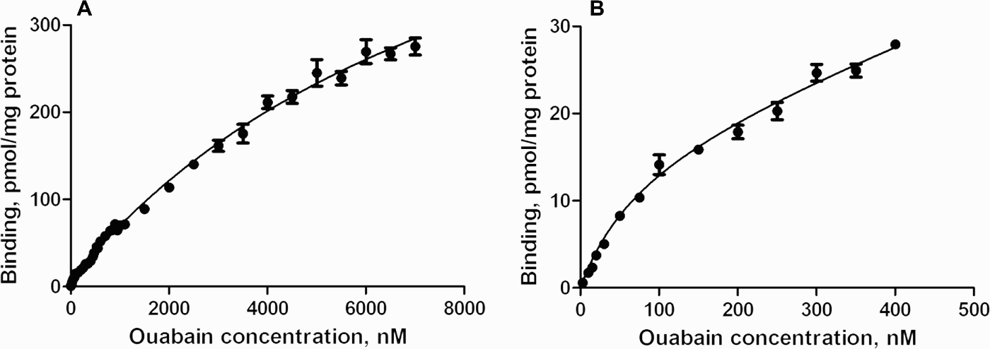
Specific binding to astrocytes was determined during incubation in tissue culture medium at 32 different concentrations of ouabain between 3 nM and 7 µM after subtraction of non-specific binding, measured with 10 mM non-labeled ouabain. Number of individual experiments at different concentrations varied between 3 and 26. In each individual experiment, only a minor part of the entire concentration range was determined in triplicates of cultures from the same batch. Although the general courses of these graphs appeared to show a similar pattern, some batch-to-batch variation was observed. The binding at 1 µM [3H]ouabain was determined to be 70.0 ± 5.4 pmol/mg protein, and binding at 1 µM in each individual experiment was normalized to this value in the non-linear regression analysis. Therefore, the standard errors in Figs 1 and 2 only apply to the kinetics, whereas each individual value has an additional uncertainty of 5.4/70 or 7.7%. At most concentrations the SEM values shown in the graphs are much smaller than this value, indicating that the actual values have an uncertainty below ~10%. The added uncertainty of the absolute binding at 1 µM [3H]ouabain does not affect the K D values obtained from the Graph-Pad analysis, but should be added to the calculated B max values. This has been carried out in Table 1 and below.
(A) Ouabain binding to mouse astrocytes in primary cultures determined within the total concentration range 3 nM–7 µM. The curve is determined by least squares non-linear regression analysis for two binding sites, and after subtraction of a non-specific binding of 5%. The figure shows means ± SEM for the kinetic analysis of individual results based on normalization of all results to a preset value for binding at 1 µM. However, for the absolute amount of binding at each concentration there is the additional uncertainty that the absolute value at 1 µM ouabain was 70 ± 5.4% (SEM) nmol/mg protein. This uncertainty is not included in the Figure. n = 3–26. (B) Ouabain binding determined as in A, and showing the same data, but exclusively for the concentration range 3–400 nM. n = 3–15.

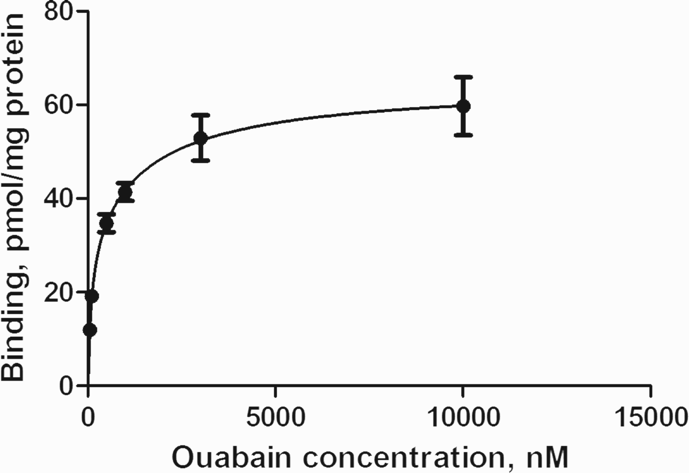
Ouabain binding to mouse cerebellar granule neurons in primary cultures determined within the concentration range 50 nM–10 µM as described above after subtraction of a non-specific binding of 20%. The data are absolute values without any additional source of uncertainty. The figure shows means ± SEM values. n = 4 for the 3 µM value and otherwise 6–10.

K D and B max values in mouse astrocytes and cerebellar granule neurons in primary cultures for the high-affinity ouabain binding site (to the α2 and the α3 isozyme, respectively) and for the low-affinity ouabain binding site (to the α1 isozyme).

K D and B max values (means ± SEM) were determined for binding of [3H]ouabain to primary cultures of mouse astrocytes and of cerebellar granule neurons by non-linear regression analysis on the assumption of two binding sites and based on least square statistics. Results for the low-affinity sites were obtained from the whole concentration ranges shown in Figs 1A and 2, but for the high-affinity sites they were obtained by analysis selectively in the range 3–400 nM (astrocytes – Fig. 1B) or 50 nM–1 µM (neurons – Fig. 2).
Figure 1A shows the binding of [3H]ouabain in the entire concentration range (3 nM–7 µM). Non-linear regression analysis assuming two binding sites indicated the presence of a large low-affinity site with a B max of 645 ± 73 pmol/mg protein and K D of 9.3 ± 1.4 µM (Table 1). To ascertain that this was not an artifact introduced by a wrong estimate of non-specific binding, a separate non-linear regression analysis was made covering only the range from 3 to 1100 nM, a concentration chosen because of a slight tendency towards saturation at this concentration (Fig. 1). This analysis provided B max and K D values that were not statistically significantly different from those obtained from the total range, but had larger uncertainties; an attempt to identify a binding site with intermediate affinity by selectively analyzing binding between 450 nM and 7 µM and assuming two binding sites did also not reveal any such site (results not presented). Above 7 µM ouabain an apparent disproportionately large increase in binding occurred, perhaps due to astrocyte-specific cell damage by ouabain (Bignami and Palladini, Reference Bignami and Palladini1966; Cornog et al., Reference Cornog, Gonatas and Feierman1967), which prevented the use of ouabain concentrations above 7 µM.
In addition to the low-affinity site, Fig. 1 shows a much less dense high-affinity site, approaching saturation at a binding of approximately 20 pmol/mg protein. The kinetics of this site were more exactly determined within the range 3–400 nM (Fig. 1B), where interference from the low-affinity site was less. This analysis yielded a B max value of 17.6 ± 5.3 pmol/mg protein and a K D value of 77 ± 35.7 nM (Table 1), values that were not significantly different from those for the high-affinity binding site that could be obtained by analysis of binding in the entire Fig. 1A (results not presented).
Corresponding binding to cerebellar granule neurons in the concentration range 50 nM to 10 µM is shown in Fig. 2. With the exception of the 3 µM value, which is the average of four individual experiments, all values are the averages of six to ten individual experiments. Analyzing only results within the range 50 nM–1 µM for determination of the kinetics of the high-affinity site, its B max was computed to be 38 ± 8.4 pmol/mg protein and its K D value to be 111 ± 76 nM. The means were not significantly changed if the entire concentration range was analyzed, but the uncertainties increased. For the low-affinity site the B max value, determined from the entire concentration range, was 33.5 ± 30.5 pmol/mg protein and the K D value 1.5 ± 3.7 µM. The SEM values of some of these values is rather large, but it should be emphasized that the upper 95% confidence limit for the low-affinity binding site is just above 100 pmol/mg protein, whereas the corresponding binding site in the astrocytic cultures has a lower 95% confidence limit for the same site of ~500 pmol/mg protein.
Effects of K+
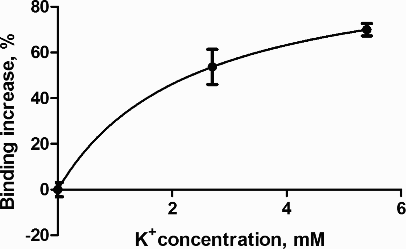
In contrast to the usually observed inhibition of ouabain binding when K+ is present in the medium, the binding at 1.0 µM ouabain to astrocytes was increased by ~50% in the presence of 2.7 mM K+ and, if anything, even more enhanced when the [K+]o was further raised to 5.4 mM (Fig. 3). A one-site non-linear regression analysis suggested a peak increase of 101 ± 26.4% and a half-maximum effect at 2.4 ± 1.7% mM. However, at still higher concentrations the magnitude of the stimulation decreased, and at 20 mM it amounted to 54 ± 8.9%. A stimulation of ouabain binding by 5.4 mM K+ was also seen at 10 µM ouabain (results not presented). Binding in the neuronal cultures showed a similar tendency as in the astrocytes, although the increase for 0–5.4 mM [K+]o was only from 50.7 to 59.9 nmol/mg protein, and the binding at 20 mM was similar to that at in the absence of extracellular K+. The slight stimulation might reflect a small astrocytic contamination. If this is the case a difference between a stimulation of ~70% in astrocytes and an apparent stimulation of ~18% in the neuronal cultures would suggest that one-fifth of the low-affinity binding in the cerebellar granule neurons might occur to the astrocytic population in these cultures. The astrocytic contamination is much less, but the high density of the astrocytic low-affinity binding site can explain the relatively large astrocytic contribution to ouabain binding in the neuronal cultures.
K+-mediated percentage increase in ouabain binding to primary cultures of astrocytes at 1.0 µM [3H]ouabain at 2.7 and 5.4 mM K+ as percent of control value in K+-free medium (0%) and analyzed by least squares non-linear regression analysis for one binding sites. Values are means ± SEM with n = 6.

Expression of FXYD7 mRNA
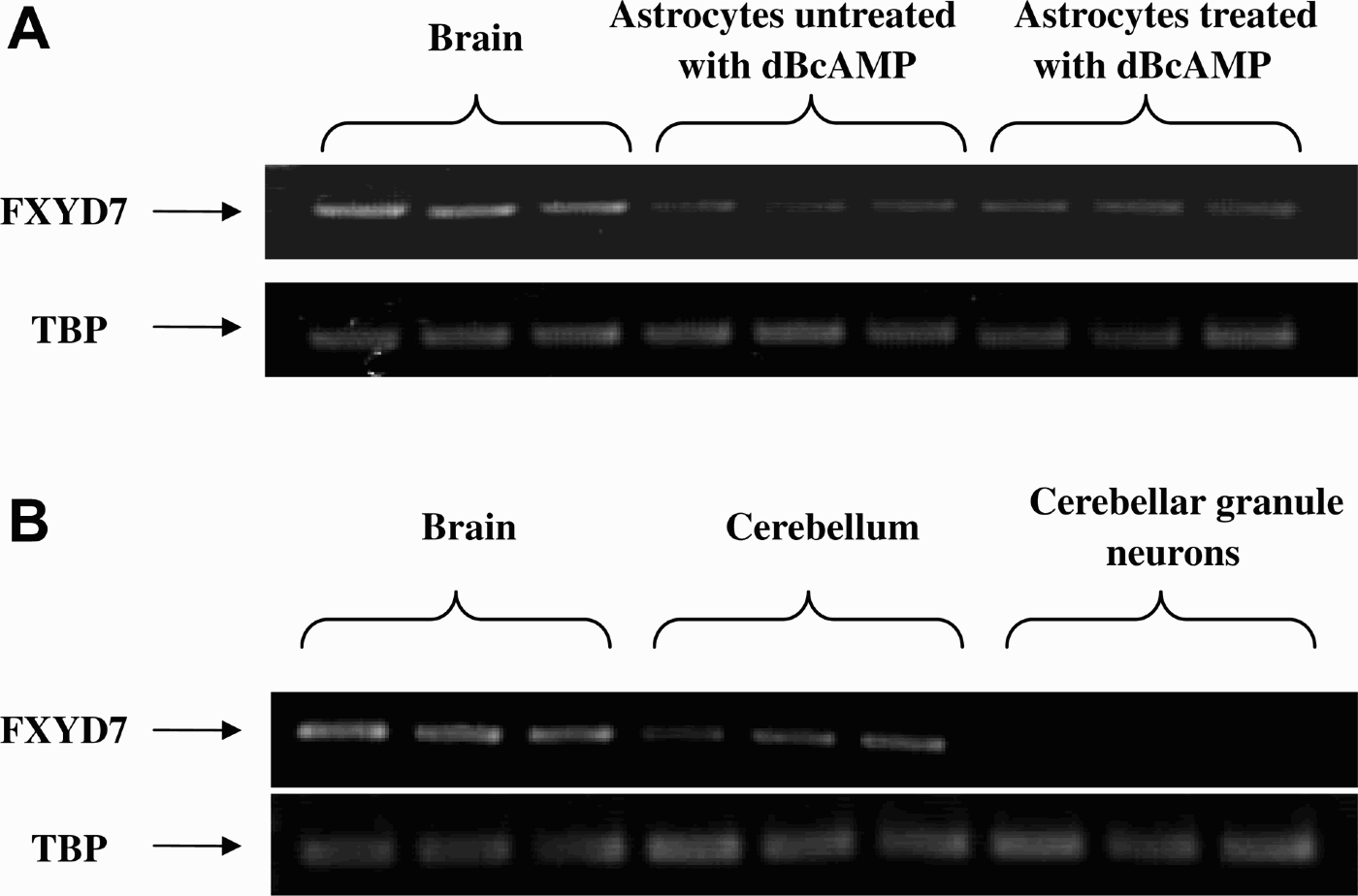
As shown in Fig. 4, mRNA of FXYD7 was consistently expressed both in the brain in vivo and in cultured astrocytes but not at all in cerebellar granule neurons. However, the level of expression was higher in brain samples than in astrocytes, and in astrocytes treated with dBcAMP the expression of FXYD7 was slightly higher than in untreated cells.
mRNA expression of FXYD7 in cerebral or cerebellar tissue from adult mice, similar to that used for preparation of the cultures, in primary cultures of untreated astrocytes or astrocytes treated with dBcAMP and in primary cultures of cerebellar granule neurons. (A) Astrocytes compared to brain (cerebral cortex); (B) cerebellar granule neurons compared to cerebral cortex and cerebellum. TBP: TATA box protein, used as housekeeping gene. The sizes of the gene products are 309 bp for FXYD7 and 236 bp for TBP. n = 3.

CONCLUSIONS
Astrocytes in primary cultures express a high-affinity binding site for ouabain with a relatively low B max, representing the α2-binding site of the Na+,K+-ATPase. They express a low-affinity binding site for ouabain with a very high B max, indicating the α1-binding site of the Na+,K+-ATPase, which shows a low affinity for K+, probably at least partly due to the presently demonstrated expression of FXYD7. The high B max and a low K+ affinity of the Na+,K+-ATPase endows astrocytes with a high capacity for K+ uptake, which is stimulated at above-normal [K+]o. In contrast to most observations in the literature, ouabain binding to the cultured astrocytes was not reduced by increased [K+]o, but showed a stimulation.
Cerebellar granule neurons in primary cultures express a high-affinity binding site for ouabain with a B max that may be slightly higher than in astrocytes, and indicates the α3-binding site of the Na+,K+-ATPase, as well as low-affinity binding site for ouabain, representing the α1-subunit, which is of much lower density (at the very least five times and probably more) than in astrocytes. A tendency towards a small stimulation of ouabain binding at 5.4 mM [K+]o may represent contamination of the neuronal cultures with astrocytes.
Binding affinities in the cultured cells are consistent with data for ouabain binding and ouabain effects in brain, but the very high density of the astrocytic α1 site and the increase in ouabain binding in the presence of K+ are novel observations.
DISCUSSION
High-affinity sites
The present study determined kinetics for both high-affinity and low-affinity sites for ouabain binding in astrocytes, and information is available about ouabain effects on K+ uptake in similar cells (Walz and Hertz, Reference Walz and Hertz1982). Ouabain binding to the high-affinity site corresponding to the α2 binding site (Sweadner, Reference Sweadner1979; Matsuda et al., Reference Matsuda, Murata, Kawamura, Hayashi, Tamada and Takuma1993; Golovina et al., Reference Golovina, Song, James, Lingrel and Blaustein2003) accounts for only ~2.6% of its total specific binding. Yet, Walz and Hertz (Reference Walz and Hertz1982) found ~30% of the uptake to be inhibited within the lower range of ouabain concentrations, and this component was virtually completely inhibited of at a concentration ten times the presently determined K D value for the binding site; nevertheless, a concentration above the K D value for the binding was required to exert a measurable inhibition. A similar discrepancy has previously been seen by Lobaugh and Lieberman (Reference Lobaugh and Lieberman1987) in cultured cardiac myocytes, where the apparent lack of effect of ouabain could be explained by an ouabain-induced increase in intracellular Na+, which enhanced the transport activity of remaining uninhibited transporter sites (Stimers et al., Reference Stimers, Lobaugh, Liu, Shigeto and Lieberman1990).
The total ouabain-sensitive K+ uptake was previously measured to be ~250 nmol/mg protein per min (~4 nmol/mg protein per sec), peaking at slightly elevated K+ concentrations. However, this represented an underestimate by a factor of up to 2 (Walz and Hertz, Reference Walz and Hertz1982), partly because the uptake of K+ does not remain strictly linear during the 2 min incubation time used for the uptake determination (Hertz, Reference Hertz1979). An uptake rate corresponding to the potently inhibited 30% of the total uptake may therefore equal ~2.0 nmol/mg protein per sec. Since there is a stoichiometric relationship between ouabain binding sites and Na+,K+-ATPase sites, and two molecules of K+ are taken up for each molecule of ATP converted to ADP (Blanco and Mercer, Reference Blanco and Mercer1998), this uptake rate combined with the presently observed B max of 17.6 picomol/mg protein, allows the calculation of a turnover rate of the Na+,K+-ATP-ase of almost 60/s (2000/[2 × 17.6]). This rate was measured under conditions which may not have provided maximum stimulation of the Na+,K+-ATPase, as suggested by an increase in metabolic activity, when similar cells are exposed to both an elevated K+ concentration and treated with the Na+ ionophore monensin, increasing intracellular Na+ (Peng et al., Reference Peng, Zhang and Hertz1994). The turnover rate therefore compares well to a maximum turnover rate for the Na+,K+-ATPase of 8000/min or 133/s (Plesner and Plesner, Reference Plesner and Plesner1981) and to that of ~75/s (at 30°C) in Na+-loaded muscle (Clausen et al., Reference Clausen, Everts and Kjeldsen1987).
Two kinetically different ouabain binding sites were also found in primary cultures of the glutamatergic cerebellar granule cell neurons, cells in which we have not determined K+ uptake rates. In similar cultures Atterwill et al. (Reference Atterwill, Atkinson, Bermudez and Balázs1985) concluded that only one binding site existed, with a K D of ~400 nM. We might have reached a similar conclusion, had we not performed an analysis based on the presence of two sites, and the data of Table 1 suggest that the high- and low-affinity sites may each account for about one-half of total ouabain binding to cerebellar granule neurons. In the cultures used by Atterwill et al. (Reference Atterwill, Atkinson, Bermudez and Balázs1985) the total binding amounted to somewhat less than in our cultures, i.e. 17.3 nmol/mg protein, possibly reflecting that their cells had been grown in serum-free medium. From this value, together with an ouabain-resistant uptake of K+ (measured by the aid of 86Rb) of 32 nmol/min (533 pmol/s) per mg protein in their cells a turnover rate for the Na+,K+-ATPase of 15.4/s (533/[17.3 × 2]) can be calculated. This is a not a high turnover rate, but a monensin-induced increase in intracellular Na+, could cause a five-fold increase in K+ uptake (Atterwill et al., Reference Atterwill, Atkinson, Bermudez and Balázs1985), which may raise the turnover rate to above 75/s. A large stimulus-induced increase in turnover rate is analogous to observations in muscle (Clausen, Reference Clausen2003). Thus in both neurons and astrocytes the B max values for high-affinity binding sites seem to provide meaningful estimates of the density of Na+,K+-ATPase sites.
Low-affinity sites
The low-affinity site, which corresponds to the α1 binding site (Matsuda et al., Reference Matsuda, Murata, Kawamura, Hayashi, Tamada and Takuma1993), was expressed to a much higher degree in astrocytes than in neurons. The high density of this site has not been acknowledged earlier, although Golovina et al. (Reference Golovina, Song, James, Lingrel and Blaustein2003) concluded that the α2-isoform can account for no more than ~20% of the total subunits in mouse astrocytes, based on 20% decline in the expression of an α isoform-non-selective antibody in astrocytes in which the α2 subunit was knocked out without upregulation of the α1 subunit. In spite of the fact that the highest ouabain concentrations, which could be used, corresponded to only about 75% of the K D value, the non-linear regression analysis was able to determine the density of the astrocytic binding site (B max) with a relatively small SEM value. The affinity (a K D of in the micromolar range) is consistent with ouabain effects on K+ uptake in rodent brain tissue in vitro (Walz and Hertz, Reference Walz and Hertz1982) and in vivo (Marks and Seeds, Reference Marks and Seeds1978; Bianco and Mercer, 1998), and a large and increasing ouabain binding in the low micromolar range is consistent with ouabain binding in samples of rat (Schmidt et al., Reference Schmidt, Larsen and Kjeldsen1992) and human brain cortex (Schmidt et al., Reference Schmidt, Hasselbalch, Larsen, Bundgaard, Juhler and Kjeldsen1996), but not with the findings in cerebellar granule cells. Thus, our observations suggest that the large ouabain binding occurring in the brain in vivo mainly occurs to astrocytes. This is consistent with the immunohistochemical data for the α1 subunit by McGrail et al. (Reference McGrail, Phillips and Sweadner1991).
On the assumption that twice the estimated uptake rate of K+ at the site with high affinity, i.e. ~4 nmol/mg protein per second occurred at the site with low affinity for ouabain (Walz and Hertz, Reference Walz and Hertz1982), the determination of a B max of 645 pmol/mg protein suggests a turnover rate of the Na+,K+-ATPase of only 3.1/s. This is lower than reported values for any naturally occurring combinations of α and β subunits of the Na+,K+-ATP-ase and only 2–3% of that of 6700/min or 133/s known for human α1–β1 expressed in yeast cells (Müller-Ehmsen et al., Reference Müller-Ehmsen, Juvvadi, Thompson, Tumyan, Croyle and Lingrel2001). The expression of FXYD7, which associates specifically with Na,K-ATPase α1-β1 isoforms after co-expression in Xenopus oocytes and also with α1-β1 isoforms in native brain tissue, can explain a lowering of the activity, but only by ~30% (Béguin et al., Reference Béguin, Crambert, Monnet-Tschudi, Uldry, Horisberger and Garty2002). Thus, the Na+,K+-ATPase must in the experiments by Walz and Hertz (Reference Walz and Hertz1982) have worked much below its capacity. Part of the explanation for this is that the astrocytes were at rest, and energy metabolism in cultured astrocytes, which to a large degree is driven by ion transport, is doubled when [K+]o is increased to 12 mM and intracellular Na+ at the same time is elevated by the addition of monensin (Peng et al., Reference Peng, Zhang and Hertz1994). However, the main reason for the low uptake rate might be that cultured astrocytes similar to those used in the present study produce and release large amounts of endogenous steroids with ouabain-like activity, which might affect K+ uptake. The production rate is high enough that the concentration of these steroids in the entire incubation medium reaches >1 nmol/mg cell protein after 4 days (Kala et al., Reference Kala, Kumarathasan, Peng, Leenen and Hertz2000). With a water space of 4 µl/mg protein in these cells (Chen et al., Reference Chen, McNeill, Hajek and Hertz1992), this amounts to >200 nmol/g wet wt., i.e. at least 2 nmol/g wet wt./hr. Endogenous steroids with ouabain-like activity include the cardenolide, endogenous ouabain and the bufadienolide, marinobufagenin (Bagrov and Fedorova, 2009), which are found in equal amounts (~40 nmol/g wet wt.) in brain cortex (Fedorova et al., Reference Fedorova, Zhuravin, Agalakova, Yamova, Talan and Lakatta2007). The rat (and thus probably also the mouse) α1 subunit is orders of magnitude more sensitive to marinobufagenin (IC50 for purified marinobufagenin of 70 nM for inhibition of rat kidney, i.e. α1-β1, Na+,K+-ATPase), than to either endogenous or plant-derived ouabain (Fedorova et al., Reference Fedorova, Kolodkin, Agalakova, Lakatta and Bagrov2001; Bagrov et al., Reference Bagrov, Shapiro and Fedorova2009). Assuming that one-half of the steroids with ouabain-like effect released from cultured astrocytes (Kala et al., Reference Kala, Kumarathasan, Peng, Leenen and Hertz2000) is marinobufagenin, i.e. a similar fraction as in brain (Fedorova et al., Reference Fedorova, Zhuravin, Agalakova, Yamova, Talan and Lakatta2007), Na+,K+-ATPase activity might be inhibitable within minutes and possibly within seconds of its release. Competitive inhibition of ouabain binding by endogenous steroids will also tend to displace the ouabain binding curve to the right, with a resulting increase in apparent K D, but B max will not be affected. It would be of great importance to establish whether any specific conditions (e.g. previous exposure to K+-free medium, as used by Walz and Hertz (Reference Walz and Hertz1982) to prevent channel-mediated K+ homoexchange) lead to release of steroids that inhibit Na+,K+-ATPase activity, since such a release might favor neuronal reuptake, i.e. return of astrocytically accumulated K+ to neurons. Dis-inhibition of the huge amount of Na+,K+-ATPase sites represented by low-affinity ouabain binding would, on the other hand, endow astrocytes with an enormous capacity for rapid K+ accumulation.
K+ effects
An apparent anomaly of the low-affinity binding site in astrocytes was that the generally observed K+-induced inhibition of ouabain binding (Landowne and Ritchie, Reference Landowne and Ritchie1970; Gardner and Conlon, Reference Gardner and Conlon1972; Noël et al., Reference Noël, Fagoo and Godfraind1990; Crambert et al., Reference Crambert, Hasler, Beggah, Yu, Modyanov and Horisberger2000) was replaced by K+ stimulation. At 1 µM ouabain, where most of the binding must have occurred to the low-affinity binding site, 5.4 mM K+ stimulated, rather than inhibited ouabain binding, and there was no inhibition, but a stimulation, even at 20 mM K+. Lack of inhibition by low millimolar concentrations of K+ has previously been observed, because Crambert et al. (Reference Crambert, Hasler, Beggah, Yu, Modyanov and Horisberger2000) showed that out of nine cloned combinations of α and β subunits of the human Na+,K+-ATPase, one showed no inhibition in this range of K+ concentrations. Moreover, the lack of inhibition of ouabain binding by 20 mM [K+]o is consistent with the observation by Maki et al. (Reference Maki, Baskin and Stahl1992) that at 1.5 µM ouabain 40% of the [3H]ouabain binding sites were insensitive to 15–90 mM K+. Since B max for the low-affinity ouabain binding site in astrocytes was at the very least five times larger than in neurons most of this binding is likely to have occurred to astrocytes, although they only account for 20% of the volume of brain cortex (reviewed by Hertz, Reference Hertz2008). This conclusion is consistent with higher Na+,K+-ATPase activity in cultured or bulk-separated astrocytes than in neurons (Henn et al., Reference Henn, Haljamäe and Hamberger1972; Grisar et al., Reference Grisar, Frère and Franck1979; Hajek et al., Reference Hajek, Subbarao and Hertz1996).
FXYD7 expression
The present study showed that gene expression of FXYD7 was present in astrocytes but absent in cultured cerebellar granule neurons. Astrocytic expression is in agreement with in vivo observations by Béguin et al. (Reference Béguin, Crambert, Monnet-Tschudi, Uldry, Horisberger and Garty2002), but they found both astrocytes and neurons to express FXYD7. Only one type of neurons was used in the present study, and additional experiments are required for determination of cellular expression of this important regulator of Na+,K+-ATPase activity. It has already been mentioned that FXYD7 decreases the turnover rate of the α1–β1 isoform. In addition, it decreases the apparent affinity of the rat α1 isozyme for external K+ ~2-fold from ~0.7 to 1.35 mM at −90 mV (Béguin et al., Reference Béguin, Crambert, Monnet-Tschudi, Uldry, Horisberger and Garty2002). This K D value is not significantly different from the K m value of 1.9 mM for K+-induced stimulation of the astrocytic Na+,K+-ATPase, determined in homogenates of mouse astrocytes (Hajek et al., Reference Hajek, Subbarao and Hertz1996). Nor is the apparent K D value of 2.4 mM for stimulation of ouabain binding to cultured astrocytes found in the present study significantly different from 1.9 mM, perhaps suggesting a possible, but as yet unknown, correlation between low K+ affinity of the Na+,K+-ATPase and maintained ouabain binding in the presence of K+. That such an association may exist is also suggested by the observation that the combination of subunits of human Na+,K+-ATPase that is relatively resistant to inhibition of ouabain binding by K+ also shows a remarkably low affinity for K+ (Crambert et al., Reference Crambert, Hasler, Beggah, Yu, Modyanov and Horisberger2000).
Low K+ affinity for Na+,K+-ATPase activation has also been found in cultured rat astrocytes by Moonen and Franck (Reference Moonen and Franck1977) and Mercado and Hernandez (Reference Mercado and Hernández1992) and in rabbit astrocytes obtained by gradient centrifugation by Henn et al. (Reference Henn, Haljamäe and Hamberger1972) and Grisar et al. (Reference Grisar, Frère and Franck1979). This low affinity allows stimulation of the enzyme by above-normal extracellular K+ concentrations, decreasing intracellular Na+ (Rose and Ransom, Reference Rose and Ransom1996), and facilitating accumulation of excess extracellular K+. Thus, astrocytes are especially well suited for clearance of an elevated [K+]o, whereas neuronal uptake may be facilitated by the higher K+ affinity of the neuronal Na+,K+-ATPase (Hajek et al., Reference Hajek, Subbarao and Hertz1996), once a resting [K+]o level has been re-established or [K+]o has been reduced below its normal level during the undershoot. Thus, FXYD7 expression in neurons might, if anything, hamper K+ re-accumulation in neurons following its initial uptake into astrocytes.
Concluding remarks
The results for both ouabain kinetics and FXYD7 expression in cultured astrocytes and neurons are consistent with intense K+ uptake capacity in astrocytes, especially when its extracellular concentration is increased. The expression of FXYD7 in astrocytes modifies Na+,K+-ATPase activity to enhance this capacity, whereas it would hinder K+ re-accumulation in neurons, once elevated K+ has been brought back to (or below) normal. An important question is whether an endogenous Na+,K+-ATPase inhibitor might normally inhibit the high-density α1 subunit in astrocytes.
ACKNOWLEDGEMENTS
This study was supported by the Saskatchewan Heart and Stroke Foundation, Saskatoon, Canada and by Grants Nos. 30711120572 and 30770667 from the National Natural Science Foundation of China. Ting Du, MD, Department of Clinical Pharmacology, China Medical University, Shenyang, P.R. China is cordially thanked for help with the graphics.
Statement of interest
None