INTRODUCTION
Neospora caninum is a heteroxenous, cyst-forming coccidian closely related to Toxoplasma gondii that has been recognized as a major cause of abortion and reproductive failure in cattle (reviewed by Dubey et al. Reference Dubey, Schares and Ortega-Mora2007). At present, bovine neosporosis is an important worldwide concern because of its wide geographic distribution (reviewed by Dubey et al. Reference Dubey, Schares and Ortega-Mora2007 and Dubey and Schares, Reference Dubey and Schares2011) and economic impact (Trees et al. Reference Trees, Davison, Innes and Wastling1999; Reichel et al. Reference Reichel, Alejandra Ayanegui-Alcerreca, Gondim and Ellis2013). Serologic prevalence of N. caninum antibodies in cattle indicates that there are considerable differences in prevalence of infection among countries, within countries, between regions and between beef and dairy cattle (reviewed by Dubey and Schares, Reference Dubey and Schares2011). Major economic losses in livestock operations are associated with lengthened calving interval, reduced stock value and increased culling rate (Trees et al. Reference Trees, Davison, Innes and Wastling1999). In a recent review and economic analysis by Reichel et al. (Reference Reichel, Alejandra Ayanegui-Alcerreca, Gondim and Ellis2013), the impact of N. caninum abortions in just 10 countries was estimated to be, on average, US$ 1 billion.
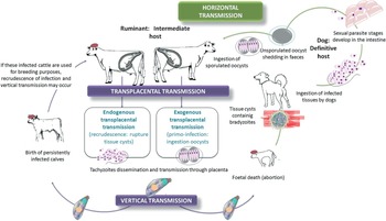
The life cycle of N. caninum has three infectious stages: tachyzoites, tissue cysts bearing bradyzoites, and oocysts (Fig. 1). Intermediate hosts harbour intracellular tachyzoites and bradyzoites. Tissue cysts are primarily found in the central nervous system of intermediate hosts, but this stage has also been detected in cattle muscle (Peters et al. Reference Peters, Lutkefels, Heckeroth and Schares2001). Definitive hosts excrete environmentally resistant oocysts in an unsporulated stage. The finding of N. caninum oocysts in the faeces of naturally infected dogs and grey wolves has allowed identifying them as natural definitive hosts for the parasite (Basso et al. Reference Basso, Venturini, Venturini, Hill, Kwok, Shen and Dubey2001; Dubey et al. Reference Dubey, Jenkins, Rajendran, Miska, Ferreira, Martins, Kwok and Choudhary2011). The shedding of oocysts in faeces has also been proven in experimentally infected dogs, coyotes and dingoes (reviewed by Dubey et al. Reference Dubey, Chapman, Rosenthal, Mense and Schueler2006 and Dubey and Schares, Reference Dubey and Schares2011). Serological evidence in domestic, wild and zoo animals indicates that many species have been exposed to this parasite, but viable N. caninum has only been isolated from a few host species such as cattle, sheep, water buffalo, dog, horse, bison and white-tailed deer (reviewed by Dubey and Schares, Reference Dubey and Schares2011).
Transmission routes of Neospora caninum infection in cattle.

The routes of N. caninum transmission in cattle include transplacental infection from the dam to the foetus during gestation through tachyzoites (vertical transmission) and ingestion of sporozoite-containing oocysts shed by a definitive host (horizontal transmission). Exogenous transplacental transmission occurs following primary oocyst-derived infection of pregnant dams, while endogenous transplacental transmission occurs following recrudescence of infection in persistently infected cows during pregnancy (Trees and Williams, Reference Trees and Williams2005). Transmission of the parasite from dam to offspring seems to be highly efficient on the basis of precolostral serological data, ranging from 48–95% (Schares et al. Reference Schares, Peters, Wurm, Barwald and Conraths1998; reviewed by Dubey et al. Reference Dubey, Schares and Ortega-Mora2007). To date, cow to cow horizontal transmission of N. caninum has not been observed and the ingestion of sporulated N. caninum oocysts from the environment is the only demonstrated natural mode of infection of cattle after birth (McCann et al. Reference McCann, McAllister, Gondim, Smith, Cripps, Kipar, Williams and Trees2007). Post-natal transmission is responsible for less than 15% of infections in the herd (Hall et al. Reference Hall, Reichel and Ellis2005), although horizontal transmission could be important in some herds in high prevalence areas (Eiras et al. Reference Eiras, Arnaiz, Alvarez-Garcia, Ortega-Mora, Sanjuanl, Yus and Dieguez2011). In this study, a large sample size was investigated showing high herd (80·6%) and within herd (25·1%) seroprevalence and a significant increase of seroprevalence with age (11, 14·4 and 27·5 in animals younger than 24 months, 25–36 months and higher than 36 months, respectively). Seropositivity is assumed to persist for life (reviewed by Dubey and Schares, Reference Dubey and Schares2006), although fluctuation in antibody responses may result in some animals becoming seronegative again (Pereira-Bueno et al. Reference Pereira-Bueno, Quintanilla-Gozalo, Perez-Perez, Espi-Felgueroso, Alvarez-Garcia, Collantes-Fernandez and Ortega-Mora2003). Seropositive animals have a higher risk of abortion, ranging between 1·7- and 7·4-fold but the risk decreases over time with increasing parity (Pare et al. Reference Pare, Thurmond and Hietala1997; Thurmond and Hietala, Reference Thurmond and Hietala1997; Wouda et al. Reference Wouda, Moen and Schukken1998). Lactogenic and venereal routes are other suggested modes of transmission. Neospora caninum DNA has been detected in milk and colostrum (Moskwa et al. Reference Moskwa, Pastusiak, Bien and Cabaj2007) and neonatal calves may become infected after ingestion of milk experimentally spiked with tachyzoites (Davison et al. Reference Davison, Guy, McGarry, Guy, Williams, Kelly and Trees2001), but the lactogenic transmission of N. caninum was not demonstrated in natural conditions (Dijkstra et al. Reference Dijkstra, Barkema, Eysker and Wouda2001a , Reference Dijkstra, Eysker, Schares, Conraths, Wouda and Barkema b ). Although the presence of N. caninum DNA has been detected in semen (Ortega-Mora et al. Reference Ortega-Mora, Ferre, del-Pozo, Caetano-da-Silva, Collantes-Fernandez, Regidor-Cerrillo, Ugarte-Garagalza and Aduriz2003), dams naturally bred with experimentally infected bulls failed to seroconvert (Osoro et al. Reference Osoro, Ortega-Mora, Martinez, Serrano-Martinez and Ferre2009).
Control measures for bovine neosporosis such as management strategies, chemotherapy and vaccination have been reviewed in Dubey et al. (Reference Dubey, Schares and Ortega-Mora2007) and Reichel and Ellis (2002). However, a general strategy to control neosporosis worldwide is not applicable because of regional differences in the epidemiology of bovine neosporosis. Standard biosecurity measures are paramount to avoid the introduction of infection in N. caninum-free herds. In N. caninum-infected herds, control programmes are based on decreasing the vertical transmission in herd by reduction of the number of seropositive cattle and/or decreasing the risk of horizontal transmission of N. caninum mainly by controlling the definitive host population as a source of oocyst contamination (reviewed by Dubey et al. Reference Dubey, Schares and Ortega-Mora2007). Test and cull of N. caninum-infected cattle seems to be an effective but costly option. Other practical alternatives for the control of neosporosis would be the selective breeding of only seronegative cows, breeding of seropositive cows only to beef, and the culling of those cows that actually aborted. In high prevalence areas, the difficulties that need to be overcome in establishing a control programme for neosporosis include the limited number of seronegative animals available for breeding replacement animals, reducing selection by genetic merit and an increase in replacement costs (Eiras et al. Reference Eiras, Arnaiz, Alvarez-Garcia, Ortega-Mora, Sanjuanl, Yus and Dieguez2011).
Initiatives for model harmonization in N. caninum infection
During the 2011 meeting of the World Association for the Advancement of Veterinary Parasitology held in Buenos Aires, a workshop dealing with ‘Perspectives for control for cattle reproductive diseases caused by protozoans’ stressed the urgency to standardize pre-clinical and clinical models for N. caninum. These models are essential to study host-pathogen interactions, host immunity at the local and systemic level, and for evaluating vaccine candidates and therapeutics (Campero and Ortega-Mora, personal communication). The following year during the first ‘Apicowplexa’ meeting in Lisbon (Apicomplexans in farm animals, http://www.apicowplexa.net/), a presentation including ‘pros’ and ‘cons’ of in vitro and in vivo models used for N. caninum vaccine testing was made. The necessity of consensus guidelines including isolates/strains of N. caninum used, challenge dose, time and route of challenge, preparation of inoculum, animal model (mice vs cattle; sheep vs cattle) and other parameters were discussed and a proposal to develop international standard guidelines that would be an aid to researchers in this field and gain acceptance worldwide was put forward (reviewed by Ortega-Mora et al. Reference Ortega-Mora, Regidor-Cerrillo, Rojo-Montejo, Collantes-Fernandez and Alvarez-Garcia2012).
Several recent reviews have discussed in vitro and in vivo laboratory animal (mainly mice) models of N. caninum infection. In vitro culture systems represent powerful tools for the study of apicomplexan parasites like Neospora and have enabled studies on host-parasite interactions including pathogen virulence, host factors involved in innate resistance, parasite stage conversion and differentiation, genetics and transfection technology and vaccine candidates and drug effectiveness (reviewed by Muller and Hemphill, Reference Muller and Hemphill2013). Notably, a recent comparative analysis of the invasive and proliferative capacities of bovine N. caninum isolates obtained from asymptomatic calves revealed intra-species variability, which was associated with disease severity in a pregnant mice model (Regidor-Cerrillo et al. Reference Regidor-Cerrillo, Gomez-Bautista, Sodupe, Aduriz, Alvarez-Garcia, Del Pozo and Ortega-Mora2011). In vitro invasion rate and tachyzoite yield were confirmed as traits associated with the virulence phenotype for these N. caninum isolates (Regidor-Cerrillo et al. Reference Konrad, Moore, Crudeli, Caspe, Cano, Leunda, Lischinsky, Regidor-Cerrillo, Odeon, Ortega-Mora, Echaide and Campero2012).
Mouse models are a suitable tool for an initial screening of vaccine candidates to assess their in vivo efficacy (reviewed by Innes and Vermeulen, Reference Innes and Vermeulen2006). Indeed, cerebral and congenital mouse models of N. caninum infections associated with exogenous transplacental transmission have been successfully developed (Collantes-Fernandez et al. Reference Collantes-Fernandez, Lopez-Perez, Alvarez-Garcia and Ortega-Mora2006; Lopez-Perez et al. Reference Lopez-Perez, Risco-Castillo, Collantes-Fernandez and Ortega-Mora2006, Reference Lopez-Perez, Collantes-Fernandez, Aguado-Martinez, Rodriguez-Bertos and Ortega-Mora2008) and employed in vaccine efficacy assays (Aguado-Martinez et al. Reference Aguado-Martinez, Alvarez-Garcia, Fernandez-Garcia, Risco-Castillo, Marugan-Hernandez and Ortega-Mora2009; Rojo-Montejo et al. Reference Rojo-Montejo, Collantes-Fernandez, Regidor-Cerrillo, Rodriguez-Bertos, Prenafeta, Gomez-Bautista and Ortega-Mora2011). However, low rates of reactivation of N. caninum infection in both persistently and congenitally infected mice confirm that the mouse is not an appropriate or suitable species for inducing the endogenous transplacental transmission that frequently occurs in naturally infected cattle (Jimenez-Ruiz et al. Reference Jimenez-Ruiz, Alvarez-Garcia, Aguado-Martinez and Ortega-Mora2013a , Reference Jimenez-Ruiz, Alvarez-Garcia, Aguado-Martinez and Ortega-Mora b ).
Ruminant challenge models are critical to evaluate potential vaccine candidates to help confront bovine neosporosis and to study pathogenesis and host responses to infection. Several research groups have developed ruminant models of N. caninum infection independently of others, resulting in a high degree of variability due to the use of different species of animals, breeds, strain/isolate of N. caninum, doses, route and time of inoculation. Standardization is needed to advance research in a more collaborative, timely and efficient manner. These models will be useful to study host-pathogen interactions, host immunity at the local and systemic level, and for evaluating diagnostics, vaccines and therapeutics. In the absence of widely accepted international guidelines, this article serves to summarize and discuss the different models and parameters currently in use. Methodologies essential for the development of non-pregnant and pregnant ruminant models are outlined and the main knowledge gaps are identified. This information could act as the basis to develop a consensus for internationally accepted standard guidelines for ruminant models of neosporosis that would be helpful for researchers in this field worldwide.
NEOSPORA CANINUM INFECTION DYNAMICS IN CATTLE
Abortion is the main clinical sign of bovine neosporosis in both dairy and beef cattle. Despite many studies in experimentally infected pregnant cows, only limited information is available in naturally occurring infection. It is unclear which factors play a major role in determining the outcome of the infection; these include parasite virulence, routes of Neospora transmission (vertical or horizontal), type of infection (primary infection, recrudescence and re-infection), the efficacy of maternal and foetal immune response and the stage of gestation at which the dam is infected.
Cows of any age may abort from 3 months of gestation to term, with most abortions observed at 5–7 months of gestation (reviewed by Dubey et al. Reference Dubey, Schares and Ortega-Mora2007 and Almeria and Lopez-Gatius, Reference Almeria and Lopez-Gatius2013). Persistent N. caninum infection prior to pregnancy is not responsible for abortion during the early foetal period, but does exert a significant abortifacient effect after 90 days of pregnancy (Lopez-Gatius et al. Reference Lopez-Gatius, Pabon and Almeria2004). Neospora-induced abortions may occur year-round. Both dairy and beef cattle with antibodies to N. caninum can have a 7·4-fold increased risk of abortion compared with seronegative cows (Pare et al. Reference Pare, Thurmond and Hietala1997; Thurmond and Hietala, Reference Thurmond and Hietala1997; Wouda et al. Reference Wouda, Moen and Schukken1998). Several studies showed that N. caninum antibody titres were significantly correlated with abortion in parous cows (Thurmond and Hietala, Reference Thurmond and Hietala1997; Lopez-Gatius et al. Reference Lopez-Gatius, Garcia-Ispierto, Santolaria, Yaniz, Lopez-Bejar, Nogareda and Almeria2005a , Reference Lopez-Gatius, Santolaria, Yaniz, Garbayo and Almeria b ). Irrespective of the herd level of Neospora-seroprevalence, the individual serum antibody titre against N. caninum is a good indicator of the risk of abortion (Quintanilla-Gozalo et al. Reference Quintanilla-Gozalo, Pereira-Bueno, Seijas-Carballedo, Costas and Ortega-Mora2000). Increasing N. caninum antibody titres in the second and third trimester of gestation have been associated with abortion and birth of congenitally infected calves, respectively (Quintanilla-Gozalo et al. Reference Quintanilla-Gozalo, Pereira-Bueno, Seijas-Carballedo, Costas and Ortega-Mora2000; Stenlund et al. Reference Stenlund, Kindahl, Magnusson, Uggla and Bjorkman1999; Williams et al. Reference Williams, Guy, McGarry, Guy, Tasker, Smith, MacEachern, Cripps, Kelly and Trees2000).
Foetuses in Neospora-infected dams may die in utero and be reabsorbed, mummified, autolysed or stillborn. Infections after mid-pregnancy may produce calves born alive but with clinical signs or apparently clinically normal but persistently infected with the parasite. In aborted foetuses, the most common lesions are multifocal non-suppurative necrotizing encephalitis and myocarditis (Wouda et al. Reference Wouda, Moen, Visser and van Knapen1997). In addition, multifocal areas of necrosis surrounded by inflammatory cells may be observed in the spinal cord, liver, lung and placenta (Barr et al. Reference Barr, Conrad, Dubey and Anderson1991). It has been indicated that the age of foetus is an important factor which appears to influence the histological features and distribution of lesions. Frequency and severity of lesions appear to be higher in foetal tissues during the first and second trimesters of gestation rather than the last trimester, and this is likely to be a consequence of the ontogeny of the foetal immune response which progressively matures throughout gestation (Maley et al. Reference Maley, Buxton, Rae, Wright, Schock, Bartley, Esteban-Redondo, Swales, Hamilton, Sales and Innes2003; Macaldowie et al. Reference Macaldowie, Maley, Wright, Bartley, Esteban-Redondo, Buxton and Innes2004; Gibney et al. Reference Gibney, Kipar, Rosbottom, Guy, Smith, Hetzel, Trees and Williams2008; Benavides et al. Reference Benavides, Katzer, Maley, Bartley, Canton, Palarea-Albaladejo, Purslow, Pang, Rocchi, Chianini, Buxton and Innes2012).
In cattle, N. caninum is an abortifacient and abortions follow two main patterns: endemic and epidemic abortions. The epidemic abortions show a storm-like pattern with a large proportion (>10%) of at-risk cows (in-calf) aborting over a short period of time (reviewed by Dubey et al. Reference Dubey, Schares and Ortega-Mora2007). In contrast, endemic abortion persists in the herd for several months or years. Epidemic and endemic abortion patterns are related to horizontal and vertical routes of transmission of N. caninum infections, respectively. The immunological context of the initial priming of the immune system by the parasites may have profound consequences for the risk of disease and transmission of N. caninum. It appears that infection in cattle originally infected in utero when their immune system is still developing behave differently from infection in post-natally infected cattle (reviewed by Innes and Vermeulen, Reference Innes and Vermeulen2006 and Williams et al. Reference Williams, Hartley, Bjorkman and Trees2009). Cattle infected in utero (transplacental transmission) remain persistently infected and can experience recrudescence of infection (Williams et al. Reference Williams, Guy, Smith, Guy, McGarry, McKay and Trees2003). Heifers can abort or transmit the infection in successive gestations and immunity after in utero infection protects against an exogenous challenge (concomitant immunity) (McAllister et al. Reference McAllister, Bjorkman, Anderson-Sprecher and Rogers2000; Innes et al. Reference Innes, Wright, Maley, Rae, Schock, Kirvar, Bartley, Hamilton, Carey and Buxton2001; Williams and Trees, Reference Williams and Trees2006; Williams et al. Reference Williams, Guy, Smith, Ellis, Bjorkman, Reichel and Trees2007). With post-natal transmission, some animals seem to clear the infection and they do not present abortion or transmission in successive gestations (or rarely), developing a good protective immunity against an exogenous challenge (McCann et al. Reference McCann, McAllister, Gondim, Smith, Cripps, Kipar, Williams and Trees2007; Dijkstra et al. Reference Dijkstra, Lam, Bartels, Eysker and Wouda2008). These facts are important for vaccine development.
RUMINANT MODELS FOR BOVINE NEOSPOROSIS
Purposes and endpoints of the models
Ruminant experimental models have been developed to study the main consequences of N. caninum infection: foetal death or parasite transmission to the offspring. These models have been used to understand the host-pathogen interactions, enabling the study of immune responses and disease pathogenesis during gestation. In addition, in vivo models enable studies to compare virulence between isolates, therapeutics and to test the ability of vaccine formulations to prevent abortion or transmission by immunization trials and challenge.
Different endpoints have been evaluated to provide a reliable way to monitor foetopathy or parasite dissemination and transmission, which include clinical observations, detection of N. caninum presence, lesions and immune responses. With regard to clinical observations, foetal viability is the most significant parameter to be evaluated in the abortion model and this can be monitored by trans-rectal ultrasonography during gestation. If foetal death occurs, this is normally observed between 26 and 56 days after intravenous (iv) inoculation of live tachyzoites at 70 days of gestation infection (Williams et al. Reference Williams, Guy, McGarry, Guy, Tasker, Smith, MacEachern, Cripps, Kelly and Trees2000, Reference Williams, Guy, Smith, Guy, McGarry, McKay and Trees2003, Reference Williams, Guy, Smith, Ellis, Bjorkman, Reichel and Trees2007; Macaldowie et al. Reference Macaldowie, Maley, Wright, Bartley, Esteban-Redondo, Buxton and Innes2004; Gibney et al. Reference Gibney, Kipar, Rosbottom, Guy, Smith, Hetzel, Trees and Williams2008; Rojo-Montejo et al. Reference Rojo-Montejo, Collantes-Fernandez, Blanco-Murcia, Rodriguez-Bertos, Risco-Castillo and Ortega-Mora2009, Reference Rojo-Montejo, Collantes-Fernandez, Perez-Zaballos, Rodriguez-Marcos, Blanco-Murcia, Rodriguez-Bertos, Prenafeta and Ortega-Mora2013; Caspe et al. Reference Caspe, Moore, Leunda, Cano, Lischinsky, Regidor-Cerrillo, Alvarez-Garcia, Echaide, Bacigalupe, Ortega Mora, Odeon and Campero2012; Regidor-Cerrillo et al. Reference Regidor-Cerrillo, Arranz-Solis, Benavides, Gomez-Bautista, Castro-Hermida, Mezo, Pére, Ortega-Mora and González-Warleta2014). However, abortion can also be observed later (about 80 days pi) (Rojo-Montejo et al. Reference Rojo-Montejo, Collantes-Fernandez, Perez-Zaballos, Rodriguez-Marcos, Blanco-Murcia, Rodriguez-Bertos, Prenafeta and Ortega-Mora2013). These differences could be due to the inoculum size, inoculation route or differences in virulence between parasite isolates. Additionally, another clinical parameter associated with the infection is rectal temperature. In accordance with previous reports, a transient rise in the body temperature was recorded after infection during the first week pi, which is likely to be due to tachyzoite inoculation and the first cycles of replication of the parasite in host tissues (Williams et al. Reference Williams, Guy, McGarry, Guy, Tasker, Smith, MacEachern, Cripps, Kelly and Trees2000; Innes et al. Reference Innes, Wright, Maley, Rae, Schock, Kirvar, Bartley, Hamilton, Carey and Buxton2001; Maley et al. Reference Maley, Buxton, Rae, Wright, Schock, Bartley, Esteban-Redondo, Swales, Hamilton, Sales and Innes2003; Macaldowie et al. Reference Macaldowie, Maley, Wright, Bartley, Esteban-Redondo, Buxton and Innes2004; Caspe et al. Reference Caspe, Moore, Leunda, Cano, Lischinsky, Regidor-Cerrillo, Alvarez-Garcia, Echaide, Bacigalupe, Ortega Mora, Odeon and Campero2012; Regidor-Cerrillo et al. Reference Regidor-Cerrillo, Arranz-Solis, Benavides, Gomez-Bautista, Castro-Hermida, Mezo, Pére, Ortega-Mora and González-Warleta2014). A biphasic temperature response, with a second lower temperature peak at day 7 pi, was also recorded in some studies (Maley et al. Reference Maley, Buxton, Rae, Wright, Schock, Bartley, Esteban-Redondo, Swales, Hamilton, Sales and Innes2003; Macaldowie et al. Reference Macaldowie, Maley, Wright, Bartley, Esteban-Redondo, Buxton and Innes2004; Benavides et al. Reference Benavides, Katzer, Maley, Bartley, Canton, Palarea-Albaladejo, Purslow, Pang, Rocchi, Chianini, Buxton and Innes2012; Regidor-Cerrillo et al. Reference Regidor-Cerrillo, Arranz-Solis, Benavides, Gomez-Bautista, Castro-Hermida, Mezo, Pére, Ortega-Mora and González-Warleta2014).
Other key indicators are the dissemination of the parasite to different locations, measured by PCR or immunohistochemistry; parasite loads, assessed by quantitative real-time PCR; or presence of lesions, by histopathological evaluation, which can be categorized using a severity scoring system. In models where abortion is studied, the occurrence of severe pathological changes with simultaneous parasite detection and high parasite DNA levels in target foetal tissues (brain, heart and liver) and placenta are thought to play a role in foetal death (Barr et al. Reference Barr, Rowe, Sverlow, BonDurant, Ardans, Oliver and Conrad1994; Macaldowie et al. Reference Macaldowie, Maley, Wright, Bartley, Esteban-Redondo, Buxton and Innes2004; Maley et al. Reference Maley, Buxton, Macaldowie, Anderson, Wright, Bartley, Esteban-Redondo, Hamilton, Storset and Innes2006; Gibney et al. Reference Gibney, Kipar, Rosbottom, Guy, Smith, Hetzel, Trees and Williams2008; Rojo-Montejo et al. Reference Rojo-Montejo, Collantes-Fernandez, Blanco-Murcia, Rodriguez-Bertos, Risco-Castillo and Ortega-Mora2009, Reference Rojo-Montejo, Collantes-Fernandez, Perez-Zaballos, Rodriguez-Marcos, Blanco-Murcia, Rodriguez-Bertos, Prenafeta and Ortega-Mora2013; Regidor-Cerrillo et al. Reference Regidor-Cerrillo, Arranz-Solis, Benavides, Gomez-Bautista, Castro-Hermida, Mezo, Pére, Ortega-Mora and González-Warleta2014). When the goal is to study vertical transmission, this is normally identified in calves after birth by the presence of Neospora-specific antibodies in pre-colostral blood and detection of parasite DNA or lesions in tissue samples from calves. However, in newborn calves, parasite DNA is sometimes difficult to detect and histological lesions are rare, mild and limited to brain tissue from these calves, which is probably due to the control of parasite multiplication by a competent foetal immune system which develops between mid to late gestation (Innes et al. Reference Innes, Wright, Maley, Rae, Schock, Kirvar, Bartley, Hamilton, Carey and Buxton2001; Gibney et al. Reference Gibney, Kipar, Rosbottom, Guy, Smith, Hetzel, Trees and Williams2008; Almeria et al. Reference Almeria, Araujo, Tuo, Lopez-Gatius, Dubey and Gasbarre2010; Rosbottom et al. Reference Rosbottom, Gibney, Kaiser, Hartley, Smith, Robinson, Kipar and Williams2011; Benavides et al. Reference Benavides, Katzer, Maley, Bartley, Canton, Palarea-Albaladejo, Purslow, Pang, Rocchi, Chianini, Buxton and Innes2012; Bartley et al. Reference Bartley, Katzer, Rocchi, Maley, Benavides, Nath, Pang, Canton, Thomson, Chianini and Innes2013; Rojo-Montejo et al. Reference Rojo-Montejo, Collantes-Fernandez, Perez-Zaballos, Rodriguez-Marcos, Blanco-Murcia, Rodriguez-Bertos, Prenafeta and Ortega-Mora2013). Additional measurements which could provide complementary data are detection of N. caninum DNA in dam blood and microsatellite analysis in foetal tissues. The detection of circulating parasite in the maternal blood is difficult as can be the interpretation of parasitaemia results, mainly because of low parasite numbers or short periods of detectable parasitaemia (Maley et al. Reference Maley, Buxton, Rae, Wright, Schock, Bartley, Esteban-Redondo, Swales, Hamilton, Sales and Innes2003; Macaldowie et al. Reference Macaldowie, Maley, Wright, Bartley, Esteban-Redondo, Buxton and Innes2004; Serrano et al. Reference Serrano, Ferre, Osoro, Aduriz, Mateos-Sanz, Martinez, Atxaerandio, Hidalgo and Ortega-Mora2006; Rojo-Montejo et al. Reference Rojo-Montejo, Collantes-Fernandez, Blanco-Murcia, Rodriguez-Bertos, Risco-Castillo and Ortega-Mora2009; Benavides et al. Reference Benavides, Katzer, Maley, Bartley, Canton, Palarea-Albaladejo, Purslow, Pang, Rocchi, Chianini, Buxton and Innes2012). Microsatellite analysis was very useful to distinguish the endogenous infection from any experimentally administered challenge (Benavides et al. Reference Benavides, Katzer, Maley, Bartley, Canton, Palarea-Albaladejo, Purslow, Pang, Rocchi, Chianini, Buxton and Innes2012) and to rule out the involvement of the vaccine isolate in foetopathy during vaccine assays (Rojo-Montejo et al. Reference Rojo-Montejo, Collantes-Fernandez, Perez-Zaballos, Rodriguez-Marcos, Blanco-Murcia, Rodriguez-Bertos, Prenafeta and Ortega-Mora2013).
Immune responses have also been evaluated in bovine models but currently are not precise enough to be used as definitive indicators for disease or protection. Bovine experimental models have been used to study early innate and adaptive immune responses and those involved in immunopathology, which are critical to understand foetal losses, parasite transmission or protective immunity. Standard tools (such as commercially available ELISA or in-house ELISA tests) have been used to evaluate humoral immune responses (specific IgG or IgG1, IgG2 serotypes) (Williams et al. Reference Williams, Guy, McGarry, Guy, Tasker, Smith, MacEachern, Cripps, Kelly and Trees2000; Bartley et al. Reference Bartley, Kirvar, Wright, Swales, Esteban-Redondo, Buxton, Maley, Schock, Rae, Hamilton and Innes2004; Macaldowie et al. Reference Macaldowie, Maley, Wright, Bartley, Esteban-Redondo, Buxton and Innes2004; Serrano et al. Reference Serrano, Ferre, Osoro, Aduriz, Mateos-Sanz, Martinez, Atxaerandio, Hidalgo and Ortega-Mora2006; Rojo-Montejo et al. Reference Rojo-Montejo, Collantes-Fernandez, Blanco-Murcia, Rodriguez-Bertos, Risco-Castillo and Ortega-Mora2009; Regidor-Cerrillo et al. Reference Regidor-Cerrillo, Arranz-Solis, Benavides, Gomez-Bautista, Castro-Hermida, Mezo, Pére, Ortega-Mora and González-Warleta2014). In addition, avidity N. caninum ELISA tests (Bjorkman et al. Reference Bjorkman, Alvarez-Garcia, Conraths, Mattsson, Ortega-Mora, Sager and Schares2006; Williams et al. Reference Williams, Guy, Smith, Ellis, Bjorkman, Reichel and Trees2007) and stage-specific antigen-based ELISAs employing recombinant NcGRA7 and NcSAG4 proteins, can also be used for discrimination between primoinfection and persistent infection, and also detection of recrudescence, even at the individual level (Aguado-Martinez et al. Reference Aguado-Martinez, Alvarez-Garcia, Fernandez-Garcia, Risco-Castillo, Arnaiz-Seco, Rebordosa-Trigueros, Navarro-Lozano and Ortega-Mora2008). Several techniques have also been employed to analyse N. caninum-associated cellular immune responses in cattle, such as phenotypic analysis of lymphocyte populations by flow cytometry, lymphoproliferative responses following antigen-specific stimulation, as well as the measurement of bovine cytokines in cell free supernatants by ELISA tests or cytokine gene expression by real-time RT-PCR in both peripheral blood mononuclear cells, dam and foetal tissues (spleen and lymph nodes) or at the foetal-maternal interface (Almeria et al. Reference Almeria, De Marez, Dawson, Araujo, Dubey and Gasbarre2003, Reference Almeria, Araujo, Darwich, Dubey and Gasbarre2011; Bartley et al. Reference Bartley, Kirvar, Wright, Swales, Esteban-Redondo, Buxton, Maley, Schock, Rae, Hamilton and Innes2004, Reference Bartley, Wright, Maley, Macaldowie, Nath, Hamilton, Katzer, Buxton and Innes2012, Reference Almeria and Lopez-Gatius2013; Rosbottom et al. Reference Rosbottom, Guy, Gibney, Smith, Valarcher, Taylor and Williams2007, Reference Rosbottom, Gibney, Guy, Kipar, Smith, Kaiser, Trees and Williams2008; Gibney et al. Reference Gibney, Kipar, Rosbottom, Guy, Smith, Hetzel, Trees and Williams2008; Regidor-Cerrillo et al. Reference Regidor-Cerrillo, Arranz-Solis, Benavides, Gomez-Bautista, Castro-Hermida, Mezo, Pére, Ortega-Mora and González-Warleta2014). Little is known regarding the role of innate immune responses in bovine neosporosis. Cytotoxic NK cells seem to play a role in the control of the disease through both cytotoxic and interferon gamma (IFNγ)-mediated mechanisms (Boysen et al. Reference Boysen, Klevar, Olsen and Storset2006; Klevar et al. Reference Klevar, Kulberg, Boysen, Storset, Moldal, Bjorkman and Olsen2007). Recently, it has been shown that the immune response in inoculated dams at early gestation appeared to have led to superior priming of a cell-mediated immune response in dams carrying live foetuses vs dams carrying dead foetuses (Bartley et al. Reference Bartley, Wright, Maley, Macaldowie, Nath, Hamilton, Katzer, Buxton and Innes2012) and there was also evidence of innate immunity, with significant increases in expression of Toll-like receptors 2 and 9 in spleen and lymph nodes in cattle challenged at day 210 of gestation (Bartley et al. Reference Bartley, Katzer, Rocchi, Maley, Benavides, Nath, Pang, Canton, Thomson, Chianini and Innes2013). Intracellular protozoan parasites normally induce and are controlled by cellular immune responses. Data from several studies have demonstrated that experimental infection in cattle is accompanied by lymphocyte proliferation and IFNγ responses (Lunden et al. Reference Lunden, Marks, Maley and Innes1998; Marks et al. Reference Marks, Lunden, Harkins and Innes1998; Andrianarivo et al. Reference Andrianarivo, Barr, Anderson, Rowe, Packham, Sverlow and Conrad2001; Williams et al. Reference Williams, Guy, Smith, Guy, McGarry, McKay and Trees2003), and that these responding immune cells tend to be CD4+ T lymphocytes (Marks et al. Reference Marks, Lunden, Harkins and Innes1998; Staska et al. Reference Staska, McGuire, Davies, Lewin and Baszler2003; Tuo et al. Reference Tuo, Fetterer, Jenkins and Dubey2005; Rosbottom et al. Reference Rosbottom, Guy, Gibney, Smith, Valarcher, Taylor and Williams2007; Rocchi et al. Reference Rocchi, Bartley, Inglis, Collantes-Fernandez, Entrican, Katzer and Innes2011), simultaneously with a strong anti-N. caninum humoral response (Williams et al. Reference Williams, Guy, McGarry, Guy, Tasker, Smith, MacEachern, Cripps, Kelly and Trees2000; Bartley et al. Reference Bartley, Kirvar, Wright, Swales, Esteban-Redondo, Buxton, Maley, Schock, Rae, Hamilton and Innes2004; Macaldowie et al. Reference Macaldowie, Maley, Wright, Bartley, Esteban-Redondo, Buxton and Innes2004; Serrano et al. Reference Serrano, Ferre, Osoro, Aduriz, Mateos-Sanz, Martinez, Atxaerandio, Hidalgo and Ortega-Mora2006; Rojo-Montejo et al. Reference Rojo-Montejo, Collantes-Fernandez, Blanco-Murcia, Rodriguez-Bertos, Risco-Castillo and Ortega-Mora2009, Reference Rojo-Montejo, Collantes-Fernandez, Perez-Zaballos, Rodriguez-Marcos, Blanco-Murcia, Rodriguez-Bertos, Prenafeta and Ortega-Mora2013; Regidor-Cerrillo et al. Reference Regidor-Cerrillo, Arranz-Solis, Benavides, Gomez-Bautista, Castro-Hermida, Mezo, Pére, Ortega-Mora and González-Warleta2014). Additionally, an increase of both Th1 and Th2 cytokine expression in placenta has been associated with foetal death after infection in the first trimester of gestation (Maley et al. Reference Maley, Buxton, Macaldowie, Anderson, Wright, Bartley, Esteban-Redondo, Hamilton, Storset and Innes2006; Rosbottom et al. Reference Rosbottom, Gibney, Guy, Kipar, Smith, Kaiser, Trees and Williams2008; Regidor-Cerrillo et al. Reference Regidor-Cerrillo, Arranz-Solis, Benavides, Gomez-Bautista, Castro-Hermida, Mezo, Pére, Ortega-Mora and González-Warleta2014). On the other hand, minor differences in expression of these cytokines were detected after experimental infection during the second and third trimesters of gestation, when parasite transmission to foetus occurs but the calf is born apparently healthy (Rosbottom et al. Reference Rosbottom, Gibney, Guy, Kipar, Smith, Kaiser, Trees and Williams2008; Almeria et al. Reference Almeria, Araujo, Darwich, Dubey and Gasbarre2011). Therefore, bovine immunological studies have shown that immune cytokines may be either beneficial or detrimental to the host depending on their concentration, pregnancy period or tissue location and could have a role in the transplacental transmission of the parasite and/or mediate tissue damage, specifically at the dam–foetal interface.
Pregnant ruminant models of N. caninum infection
Ruminant species
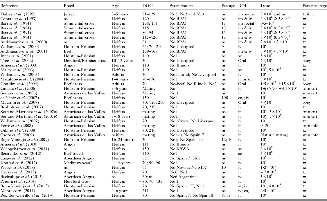
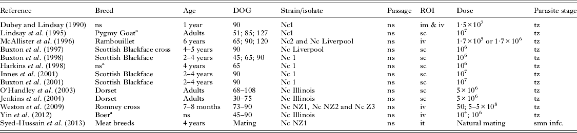
Although there is serological evidence of natural contact with N. caninum in a wide range of species, the isolation of the viable agent and experimental reproduction of the disease have been made mostly in ruminants (reviewed by Dubey et al. Reference Dubey, Schares and Ortega-Mora2007 and Dubey and Schares, Reference Dubey and Schares2011). The establishment of exogenous N. caninum infection and the occurrence of vertical transmission to the foetus or abortion have been mainly reproduced in cattle but also, to a lesser extent, in sheep, goat and water buffalo (Tables 1 and 2). The descriptions of the clinical presentation and consequences of experimental exogenous neosporosis are similar in all these species, mainly consisting of abortions or the in utero vertical transmission of the infection to the foetus. Congenitally infected animals may be born clinically normal or show weakness and clinical signs involving the CNS. The target organs in the foetus (placenta and foetal brain, heart, lung, liver or muscle) and histological lesions, characterized by multifocal necrosis and non-suppurative inflammation, are also similar in all ruminant species, although it has been suggested that focal leukomalacia would be more frequent in the ovine foetuses than in the other species. However, it is not known whether this reflects different pathogenesis of the lesions depending on the species (Buxton et al. Reference Buxton, Maley, Wright, Thomson, Rae and Innes1998).
Published studies based on Neospora caninum infections in pregnant cattle and buffalo

a Study carried out in water buffalo.
DOG, day of gestation when infected; ROI, route of inoculation; ns, not stated; iv, intravenous; im, intramuscular; sc, subcutaneous; it, intrauterine; cnj, conjuntival; tz, tachyzoites; tc, tissue cysts; oocy, sporulated oocysts; smn cnt, semen spiked with tachyzoites; smn infc, semen from infected bulls.
Published studies based on Neospora caninum infections in pregnant goats and sheep

a Study carried out in goats.
DOG, day of gestation when infected; ROI, route of inoculation; ns, not stated; iv, intravenous; im, intramuscular; sc, subcutaneous; it, intrauterine; tz, tachyzoites; smn infc, semen from infected rams.
Breed
Epidemiological studies, based on serological evaluation of a high number of naturally infected cows, have suggested that certain breeds, mainly dairy (e.g. Holstein-Friesian), are more susceptible to infection than others, e.g. Limousin (Lopez-Gatius et al. Reference Lopez-Gatius, Garcia-Ispierto, Santolaria, Yaniz, Lopez-Bejar, Nogareda and Almeria2005a , Reference Lopez-Gatius, Santolaria, Yaniz, Garbayo and Almeria b ; Armengol et al. Reference Armengol, Pabon, Santolaria, Cabezon, Adelantado, Yaniz, Lopez-Gatius and Almeria2007). However, there is no experimental evidence for a different susceptibility to infection or disease depending on the breed of the host, and the differences observed may be related to the different production systems used for a variety of breeds (Bartels et al. Reference Bartels, Arnaiz-Seco, Ruiz-Santa-Quitera, Bjorkman, Frossling, von Blumroder, Conraths, Schares, van Maanen, Wouda and Ortega-Mora2006). Similarly, other epidemiological studies have found that management practices have more influence in N. caninum infection than genetic background (Pan et al. 2004). On the other hand, using beef bull semen has been epidemiologically proven to reduce the risk of suffering abortion by dairy dams when compared with dairy bull semen (Lopez-Gatius et al. Reference Lopez-Gatius, Garcia-Ispierto, Santolaria, Yaniz, Lopez-Bejar, Nogareda and Almeria2005a , Reference Lopez-Gatius, Santolaria, Yaniz, Garbayo and Almeria b ). Bovine neosporosis has been reproduced in both beef and dairy breeds, although never in parallel in a comparative study. Ovine breeds used experimentally have been mainly those for meat or mixed production, while only Pygmy or Boer goats have been used for reproducing caprine neosporosis (Table 2).
Stage of gestation
There is a significant relationship between the stage of gestation when infection occurs and the clinical consequences of infection. This has been experimentally proved, with similar outcomes in the four ruminant species where exogenous neosporosis has been reproduced (Lindsay et al. Reference Lindsay, Rippey, Powe, Sartin, Dubey and Blagburn1995; Buxton et al. Reference Buxton, Maley, Wright, Thomson, Rae and Innes1998; Maley et al. Reference Maley, Buxton, Rae, Wright, Schock, Bartley, Esteban-Redondo, Swales, Hamilton, Sales and Innes2003; Gondim et al. Reference Gondim, McAllister, Anderson-Sprecher, Bjorkman, Lock, Firkins, Gao and Fischer2004; Macaldowie et al. Reference Macaldowie, Maley, Wright, Bartley, Esteban-Redondo, Buxton and Innes2004; McCann et al. Reference McCann, McAllister, Gondim, Smith, Cripps, Kipar, Williams and Trees2007; Rosbottom et al. Reference Rosbottom, Guy, Gibney, Smith, Valarcher, Taylor and Williams2007; Benavides et al. Reference Benavides, Katzer, Maley, Bartley, Canton, Palarea-Albaladejo, Purslow, Pang, Rocchi, Chianini, Buxton and Innes2012; Konrad et al. Reference Konrad, Moore, Crudeli, Caspe, Cano, Leunda, Lischinsky, Regidor-Cerrillo, Odeon, Ortega-Mora, Echaide and Campero2012). Reactivation of infection and abortion after exogenous, experimental infection before pregnancy has been attempted in cattle and goats but this has not yet been achieved (Lindsay et al. Reference Lindsay, Rippey, Powe, Sartin, Dubey and Blagburn1995; McCann et al. Reference McCann, McAllister, Gondim, Smith, Cripps, Kipar, Williams and Trees2007).
Infection during early gestation (first trimester) is generally associated with parasite colonization of the placenta and the development of severe necrotic and inflammatory lesions, with subsequent death and reabsorption, autolysis or mummification of the foetus (Dubey et al. Reference Dubey, Lindsay, Anderson, Davis and Shen1992; Barr et al. Reference Barr, Rowe, Sverlow, BonDurant, Ardans, Oliver and Conrad1994; Williams et al. Reference Williams, Guy, McGarry, Guy, Tasker, Smith, MacEachern, Cripps, Kelly and Trees2000; Macaldowie et al. Reference Macaldowie, Maley, Wright, Bartley, Esteban-Redondo, Buxton and Innes2004; Rojo-Montejo et al. Reference Rojo-Montejo, Collantes-Fernandez, Blanco-Murcia, Rodriguez-Bertos, Risco-Castillo and Ortega-Mora2009). However, infection of cattle at this stage of pregnancy does not always result in foetal death and dams infected during the first trimester may carry live foetuses, with no lesions, detectable parasite or serological evidence of infection in the foetus at the time of calving, while other animals, infected with the same dose and strain of N. caninum, abort (Barr et al. Reference Barr, Rowe, Sverlow, BonDurant, Ardans, Oliver and Conrad1994; Williams et al. Reference Williams, Guy, McGarry, Guy, Tasker, Smith, MacEachern, Cripps, Kelly and Trees2000; Gondim et al. Reference Gondim, McAllister, Anderson-Sprecher, Bjorkman, Lock, Firkins, Gao and Fischer2004; Macaldowie et al. Reference Macaldowie, Maley, Wright, Bartley, Esteban-Redondo, Buxton and Innes2004; McCann et al. Reference McCann, McAllister, Gondim, Smith, Cripps, Kipar, Williams and Trees2007; Caspe et al. Reference Caspe, Moore, Leunda, Cano, Lischinsky, Regidor-Cerrillo, Alvarez-Garcia, Echaide, Bacigalupe, Ortega Mora, Odeon and Campero2012). The fact that infection during the first trimester resulted in either foetal death or complete absence of detectable infection in the foetus was described as an ‘all or nothing’ response where, in some dams, the parasite inoculated at early gestation did not invade the placenta as opposed to those dams where infection is extended to the foetus and showed severe placentitis and foetal death. The occurrence of the ‘all or nothing’ response could be related to the dose and method of inoculation, as it was more frequent in the subcutaneously inoculated group, where fewer parasites are thought to reach the placenta when compared with animals where parasites are directly inoculated into the bloodstream (Macaldowie et al. Reference Macaldowie, Maley, Wright, Bartley, Esteban-Redondo, Buxton and Innes2004). Dams carrying live foetuses also showed stronger IFNγ production and cell proliferation in lymphostimulation assays from local lymph nodes compared with those dams where the foetus was dead, suggesting that the quality of the maternal immune response triggered by the infection could influence parasite dissemination, preventing the colonization of the placenta and foetus (Bartley et al. Reference Bartley, Wright, Maley, Macaldowie, Nath, Hamilton, Katzer, Buxton and Innes2012). Interestingly, the ‘all or nothing’ response has not been observed in small ruminants, where all sheep and goats infected during the first trimester of gestation suffered foetal death (Lindsay et al. Reference Lindsay, Rippey, Powe, Sartin, Dubey and Blagburn1995; McAllister et al. Reference McAllister, McGuire, Jolley, Lindsay, Trees and Stobart1996; Buxton et al. Reference Buxton, Maley, Wright, Thomson, Rae and Innes1998). Similarly, all but one out of 10 foetuses from pregnant buffaloes inoculated in early pregnancy were infected and had histological lesions of neosporosis (Konrad et al. Reference Konrad, Moore, Crudeli, Caspe, Cano, Leunda, Lischinsky, Regidor-Cerrillo, Odeon, Ortega-Mora, Echaide and Campero2012). However, there are not enough studies in these three species (buffalo, sheep and goats) to elucidate whether the absence, or not, of an ‘all or nothing’ response is associated to the species, and the probable influence of the isolate, dose or route should be investigated to further characterize this response.
Although experimental infections during mid pregnancy (second trimester of gestation) may also result in abortion (Dubey et al. Reference Dubey, Lindsay, Anderson, Davis and Shen1992), the occurrence of transplacental transmission with the delivery of live, although congenitally infected calves is the most common outcome. These calves are mainly born without any obvious clinical signs, but neurological signs have been observed occasionally (Barr et al. Reference Barr, Rowe, Sverlow, BonDurant, Ardans, Oliver and Conrad1994; Innes et al. Reference Innes, Wright, Maley, Rae, Schock, Kirvar, Bartley, Hamilton, Carey and Buxton2001; Almeria et al. Reference Almeria, De Marez, Dawson, Araujo, Dubey and Gasbarre2003; Gondim et al. Reference Gondim, McAllister, Anderson-Sprecher, Bjorkman, Lock, Firkins, Gao and Fischer2004). The histological lesions found in the placenta and foetuses following infection at mid-gestation were milder than those observed after infection earlier in pregnancy (Maley et al. Reference Maley, Buxton, Rae, Wright, Schock, Bartley, Esteban-Redondo, Swales, Hamilton, Sales and Innes2003). The only study carried out in goats described one abortion and the birth of one live, healthy kid. No parasites or lesions were found in the live kid, but congenital transmission could not be ruled out as no serological analysis or identification of the parasite by PCR was attempted (Lindsay et al. Reference Lindsay, Rippey, Powe, Sartin, Dubey and Blagburn1995). In contrast to the clinical outcome in cattle, infection during mid pregnancy in sheep resulted in more dramatic consequences, as most of the animals aborted or, less frequently, produced weak lambs. Only a few lambs were born alive, and parasite cysts were found in one third of them (McAllister et al. Reference McAllister, McGuire, Jolley, Lindsay, Trees and Stobart1996; Buxton et al. Reference Buxton, Maley, Wright, Thomson, Rae and Innes1998).
Transplacental transmission of the infection and delivery of congenitally infected live calves with no obvious clinical signs have been a common consequence of experimental exogenous Neospora infection during late gestation (last trimester) in cattle (Williams et al. Reference Williams, Guy, McGarry, Guy, Tasker, Smith, MacEachern, Cripps, Kelly and Trees2000; McCann et al. Reference McCann, McAllister, Gondim, Smith, Cripps, Kipar, Williams and Trees2007). Furthermore, histological lesions in the placenta and foetus were the mildest, and parasite burdens the lowest, when compared with infections during the first and second trimesters of gestation (Gibney et al. Reference Gibney, Kipar, Rosbottom, Guy, Smith, Hetzel, Trees and Williams2008; Benavides et al. Reference Benavides, Katzer, Maley, Bartley, Canton, Palarea-Albaladejo, Purslow, Pang, Rocchi, Chianini, Buxton and Innes2012). Infection in the last trimester of gestation in sheep and goats has a similar outcome to that seen with cattle, where kids and lambs are born alive. The only study carried out in goats showed the kids to be born weak, although the parasite was not detected, while the study done in sheep described the delivery of clinically healthy lambs, some of them congenitally infected (Lindsay et al. Reference Lindsay, Rippey, Powe, Sartin, Dubey and Blagburn1995; McAllister et al. Reference McAllister, McGuire, Jolley, Lindsay, Trees and Stobart1996).
Route, parasite stage and dose of inoculation
Neospora caninum oocysts, the infectious stage for ruminants in natural infection, are difficult to obtain, as experimentally infected dogs shed very few (reviewed by Dubey and Schares, Reference Dubey and Schares2011). For this reason, most of the experimental studies on ruminants have been carried out with tachyzoites as the parasite stage of the infectious inocula (Tables 1 and 2). Because N. caninum tachyzoites are susceptible to digestion, and thus destruction in the gastric environment, they have usually been inoculated through a parenteral route, namely subcutaneous, intravenous or intramuscular. All of these routes have been shown to be effective in infecting cattle and sheep while only some of them have been used in goats and water buffalo. When comparing the intravenous and subcutaneous routes, which are the most frequent routes of inoculation used, the former is associated with a more severe clinical presentation, i.e. higher peak temperature response and more frequent foetal mortality (Macaldowie et al. Reference Macaldowie, Maley, Wright, Bartley, Esteban-Redondo, Buxton and Innes2004).
As explained previously, similar doses of the same parasite strain may lead to different clinical outcomes depending on the stage of gestation. However, the main effect of the dose of infection of tachyzoite-based inocula is closely related to the route of administration and the host species. Intravenous doses administered to cattle have ranged from 1·2×106, which caused infection in all inoculated dams at day 76 of gestation and 75% of abortions (Weber et al. Reference Weber, Jackson, Sobecki, Choromanski, Olsen, Meinert, Frank, Reichel and Ellis2013), to 5×10⁸, which caused both infection and abortion in all cows infected on day 70 of gestation (Macaldowie et al. Reference Macaldowie, Maley, Wright, Bartley, Esteban-Redondo, Buxton and Innes2004). An intermediate dose of 107 has caused variable results: while in one study it caused foetal death in all dams inoculated (Gibney et al. Reference Gibney, Kipar, Rosbottom, Guy, Smith, Hetzel, Trees and Williams2008), in other experimental infections only three out of five dams showed foetal death, although all were infected (Rojo-Montejo et al. Reference Rojo-Montejo, Collantes-Fernandez, Blanco-Murcia, Rodriguez-Bertos, Risco-Castillo and Ortega-Mora2009). A dose-titration study in sheep showed that no abortions or vertical transmission of the parasite occurred after intravenous inoculation of 50 tachyzoites, while 5×10³ caused abortion in 50% of infected animals, and with a dose of 107 or 108 tachyzoites all ewes aborted, although the latter was associated with a longer period of fever (Weston et al. Reference Weston, Howe, Collett, Pattison, Williamson, West, Pomroy, Syed-Hussain, Morris and Kenyon2009). An intravenous dose of 104 tachyzoites caused abortion in all infected goats, and was associated with a later detection of the parasitaemia by ELISA when compared with a higher dose, i.e. 106 (Yin et al. Reference Yin, Qu, Cao, Li, Fetterer, Feng, Liu, Wang, Qi, Zhang, Miramontes, Jenkins, Zhang and Tuo2012). Pregnant buffaloes inoculated intravenously with 108 tachyzoites during the first trimester of gestation showed foetal and placental lesions after transplacental transmission of the disease in all cases (Konrad et al. Reference Konrad, Moore, Crudeli, Caspe, Cano, Leunda, Lischinsky, Regidor-Cerrillo, Odeon, Ortega-Mora, Echaide and Campero2012).
Subcutaneous inoculation of tachyzoites has also been used in other studies, as it was suggested that although it is still an artificial route of inoculation, it may model the natural infection better than the intravenous route of inoculation (reviewed by Dubey and Schares, Reference Dubey and Schares2006). Subcutaneous inoculation of cattle with doses of 107 and 5×10⁸ caused transplacental transmission and lesions of the foetus when inoculated in the second trimester of gestation, although the higher dose was associated with earlier and more severe histopathological changes (Maley et al. Reference Maley, Buxton, Rae, Wright, Schock, Bartley, Esteban-Redondo, Swales, Hamilton, Sales and Innes2003). Inoculation of sheep with a dose of 106 caused vertical transmission, histological lesions in the foetuses and a high percentage of abortion, i.e. 75–100% (Buxton et al. Reference Buxton, Maley, Thomson, Trees and Innes1997, Reference Buxton, Maley, Wright, Thomson, Rae and Innes1998), although a higher dose of 5×10⁶ only resulted in abortion of six out of 11 sheep (Jenkins et al. Reference Jenkins, Tuo and Dubey2004). The reduction in the percentage of animals aborting could have been due to the influence of other parameters, such as the isolate or host breed, as a further study by Buxton et al. confirmed that high doses, i.e. 107, caused abortions in 100% of the sheep (Buxton et al. Reference Buxton, Wright, Maley, Rae, Lunden and Innes2001). The same dose has been employed in subcutaneous inoculation of goats, confirming the occurrence of abortion or transplacental transmission depending on the term of gestation (Lindsay et al. Reference Lindsay, Rippey, Powe, Sartin, Dubey and Blagburn1995).
Inoculation of tachyzoites through the conjunctiva, at doses of 1 and 2·5×108, resulted in infection and induction of a specific immune response in pregnant cattle without causing transplacental transmission of the parasite to the foetus (de Yaniz et al. Reference de Yaniz, Moore, Odeon, Cano, Cano, Leunda and Campero2007; Moore et al. Reference Moore, Alvarez-Garcia, Chiapparrone, Regidor-Cerrillo, Lischinsky, de Yaniz, Odeon, Ortega-Mora and Campero2014). Cows infected with the lower dose even reverted to seronegative status 4 months after infection (de Yaniz et al. Reference de Yaniz, Moore, Odeon, Cano, Cano, Leunda and Campero2007). Intramuscular inoculation of tachyzoites has not been commonly employed. As the sole route of inoculation, it has been tried directly into the foetus at day 118 of gestation, causing foetal death 18 days after infection (Barr et al. Reference Barr, Rowe, Sverlow, BonDurant, Ardans, Oliver and Conrad1994). It has also been used in other studies, but only in addition to intravenous or subcutaneous inoculations in pregnant cows or sheep, where the consequences varied according to the stage of gestation (Dubey and Lindsay, Reference Dubey and Lindsay1990; Dubey et al. Reference Dubey, Lindsay, Anderson, Davis and Shen1992; Conrad et al. Reference Conrad, Sverlow, Anderson, Rowe, BonDurant, Tuter, Breitmeyer, Palmer, Thurmond and Ardans1993; Barr et al. Reference Barr, Rowe, Sverlow, BonDurant, Ardans, Oliver and Conrad1994; Andrianarivo et al. Reference Andrianarivo, Rowe, Barr, Anderson, Packham, Sverlow, Choromanski, Loui, Grace and Conrad2000, Reference Andrianarivo, Barr, Anderson, Rowe, Packham, Sverlow and Conrad2001).
The possibility of venereal transmission of neosporosis has been investigated after N. caninum DNA was found in semen from naturally infected bulls (Ortega-Mora et al. Reference Ortega-Mora, Ferre, del-Pozo, Caetano-da-Silva, Collantes-Fernandez, Regidor-Cerrillo, Ugarte-Garagalza and Aduriz2003). Experimental studies have shown that heifers are highly susceptible to intrauterine inoculation of semen spiked with tachyzoites at different doses, from 102 to 107, where persistent specific serum antibody responses were detected with doses from 5×10⁴ tachyzoites. There was also a direct relation between the inoculated dose and fertility reduction (Serrano-Martinez et al. Reference Serrano-Martinez, Ferre, Martinez, Osoro, Mateos-Sanz, Del-Pozo, Aduriz, Tamargo, Hidalgo and Ortega-Mora2007a , Reference Serrano-Martinez, Ferre, Osoro, Aduriz, Mota, Martinez, Del-Pozo, Hidalgo and Ortega-Mora b ). Adult cows were less susceptible to becoming infected using this route of inoculation, although one animal inoculated with 5×10⁵ tachyzoites developed serological antibodies against N. caninum (Canada et al. Reference Canada, Meireles, Ferreira, Correia da Costa and Rocha2006; Serrano-Martinez et al. Reference Serrano-Martinez, Ferre, Osoro, Aduriz, Mota, Martinez, Del-Pozo, Hidalgo and Ortega-Mora2007b ). Further studies failed to demonstrate venereal transmission of N. caninum infection from experimentally infected bulls to heifers, either through artificial insemination with semen pooled from these bulls (Ferre et al. Reference Ferre, Serrano-Martinez, Martinez, Osoro, Mateos-Sanz, Del-Pozo, Aduriz, Tamargo, Hidalgo and Ortega-Mora2008) or through natural breeding (Osoro et al. Reference Osoro, Ortega-Mora, Martinez, Serrano-Martinez and Ferre2009). A similar approach was investigated in sheep, where experimentally infected rams did not transmit the infection to adult ewes after natural breeding (Syed-Hussain et al. Reference Syed-Hussain, Howe, Pomroy, West, Smith and Williamson2013).
Even though sporulated oocysts purified from infected dogs are difficult to obtain, they have been employed in a number of studies to reproduce the disease (Table 1). These studies have found that infection of pregnant cattle after oral inoculation of sporulated oocysts is possible. Similarly to what has been described in tachyzoite-inoculated animals, the establishment of the infection and the occurrence of abortions or transplacental transmission depend very much on the combination between the dose administered and the stage of gestation (Trees et al. Reference Trees, McAllister, Guy, McGarry, Smith and Williams2002; Gondim et al. Reference Gondim, McAllister, Anderson-Sprecher, Bjorkman, Lock, Firkins, Gao and Fischer2004; McCann et al. Reference McCann, McAllister, Gondim, Smith, Cripps, Kipar, Williams and Trees2007). Vertical transmission of the infection is more likely to occur in the second half of gestation, as a dose of 1·5×103 oocysts at day 169 of gestation in cattle effectively caused transplacental transmission to the foetus while a higher dose, of 7×10⁴, at day 70 of gestation failed to cause foetal infection (Gondim et al. Reference Gondim, McAllister, Anderson-Sprecher, Bjorkman, Lock, Firkins, Gao and Fischer2004). However, comparisons between different studies using oocysts are difficult, as there might be significant variability in the viability of oocysts, so verification of the infectivity by bioassay is recommended as a way to standardize protocols (reviewed by Dubey, Reference Dubey2009).
Parasite strain/isolate
Clear differences in virulence among isolates of N. caninum were shown by variations between isolates in their ability to produce lesions, vertical transmission and immunological effects in a murine experimental model (Regidor-Cerrillo et al. Reference Regidor-Cerrillo, Gomez-Bautista, Del Pozo, Jimenez-Ruiz, Aduriz and Ortega-Mora2010). In the numerous experimental infections carried out in pregnant ruminants, several different Neospora isolates have been employed (Tables 1 and 2). However, it is difficult to compare virulence between the different isolates used in these studies, as the experimental design varied considerably among them, including the host species employed, the stage of gestation and route or dose of inoculation. All of these factors will have an effect upon clinical outcome of the infection. Due to economic and infrastructure constraints involved in experimental infection of large animals, especially cattle, only a few studies have been conducted to compare infection with different isolates. In these studies, the isolate Nc 1 was used as a virulent strain to compare against other isolates/strains. Caspe et al. (Reference Caspe, Moore, Leunda, Cano, Lischinsky, Regidor-Cerrillo, Alvarez-Garcia, Echaide, Bacigalupe, Ortega Mora, Odeon and Campero2012) found that Nc Spain 7 shows similar virulence to Nc 1, when inoculated into cattle at early gestation (day 65) in pregnant cattle, while Nc Spain 1H was clearly less virulent and induced milder lesions in the foetus when inoculated into cattle in the same trimester (day 70) (Rojo-Montejo et al. Reference Rojo-Montejo, Collantes-Fernandez, Blanco-Murcia, Rodriguez-Bertos, Risco-Castillo and Ortega-Mora2009). Nc Spain 7 and 8 showed similar clinical outcomes (100% abortions) when inoculated intravenously at day 70 of gestation (108 tachyzoites), although the Nc Spain 7 isolate showed a wider distribution in the foetus and placenta, determined by quantitative PCR (Regidor-Cerrillo et al. Reference Regidor-Cerrillo, Arranz-Solis, Benavides, Gomez-Bautista, Castro-Hermida, Mezo, Pére, Ortega-Mora and González-Warleta2014).
The results on isolate virulence are also difficult to compare between different studies as most of them do not state how many passages the parasite has been cultured in vitro, prior to inoculation in the host. Among studies where the passage is recorded, there is variation from less than 10 to 100 (Williams et al. Reference Williams, Guy, McGarry, Guy, Tasker, Smith, MacEachern, Cripps, Kelly and Trees2000; de Yaniz et al. Reference de Yaniz, Moore, Odeon, Cano, Cano, Leunda and Campero2007). This information is relevant, as serial passage of parasites through tissue culture for extended periods of time has been shown to attenuate the virulence of N. caninum (Bartley et al. Reference Bartley, Wright, Chianini, Buxton and Innes2008), therefore affecting the outcome of the infection.
There was also variability regarding the origin of the isolates employed in the studies. While the ‘classical’ isolates, such as Nc 1 or Nc Liverpool were isolated from nervous tissue of congenitally infected dogs (Dubey et al. Reference Dubey, Hattel, Lindsay and Topper1988; Barber et al. Reference Barber, Holmdahl, Owen, Guy, Uggla and Trees1995), the more recent isolates originated from congenitally infected calves, such as the different Nc Spain isolates, Nc Illinois or Nc Beef (Gondim et al. Reference Gondim, Gao and McAllister2002; Regidor-Cerrillo et al. Reference Regidor-Cerrillo, Gomez-Bautista, Del Pozo, Jimenez-Ruiz, Aduriz and Ortega-Mora2010) or from bovine abortions, like Nc BPA1 (Conrad et al. Reference Conrad, Sverlow, Anderson, Rowe, BonDurant, Tuter, Breitmeyer, Palmer, Thurmond and Ardans1993). Several isolates have been used to infect sheep. However, no comparison in the virulence of the isolate is feasible since, in order to assure infection, different isolates were mixed within the same inoculum in studies where more than one isolate was used (McAllister et al. Reference McAllister, McGuire, Jolley, Lindsay, Trees and Stobart1996; Weston et al. Reference Weston, Howe, Collett, Pattison, Williamson, West, Pomroy, Syed-Hussain, Morris and Kenyon2009).
Non-pregnant ruminant models of N. caninum infection
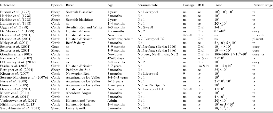
The majority of the studies performed in non-pregnant ruminants aimed to conduct research on the parasite cycle or the possibility of post-natal infection (Table 3). Cyclical oral transmission between dog and cattle was proven when dogs were fed meat from calves inoculated with as few as 300 oocysts. New oocysts isolated from faecal samples from one of these dogs were then administered to a new calf. In turn, the meat from this calf, fed to a new dog, transmitted the infection and caused the shedding of oocysts (Gondim et al. Reference Gondim, Gao and McAllister2002). A similar approach was used to investigate and establish the cyclic transmission between dogs and sheep or goats, also orally inoculated with oocysts shed by dogs (Schares et al. Reference Schares, Heydorn, Cuppers, Conraths and Mehlhorn2001).
Published studies based on Neospora caninum infections in non-pregnant ruminants

DOG, day of gestation when infected; ROI, route of inoculation; ns, not stated; iv, intravenous; im, intramuscular; sc, subcutaneous; it, intrauterine; cnj, conjunctival; tz, tachyzoites; tc, tissue cysts; oocy, sporulated oocysts; mlk infc, milk spiked with tachyzoites; smn cnt, semen spiked with tachyzoites; smn infc, semen from infected sires.
The possibility of post-natal transmission of neosporosis has been addressed in newborn calves. Although tachyzoites are susceptible to digestion in the digestive tract, tachyzoites mixed with colostrum or milk have been shown to be infectious when administered by feeding bottle (i.e. through the oral route) to neonatal calves (Uggla et al. Reference Uggla, Stenlund, Holmdahl, Jakubek, Thebo, Kindahl and Bjorkman1998; Davison et al. Reference Davison, Guy, McGarry, Guy, Williams, Kelly and Trees2001). The reduced production of hydrochloric acid and enzymes in newborn animals, together with the presence of trypsin inhibitors in colostrum, facilitate the survival of tachyzoites (Uggla et al. Reference Uggla, Stenlund, Holmdahl, Jakubek, Thebo, Kindahl and Bjorkman1998). This mechanism opens the possibility of vertical transmission from infected dams to calves, as N. caninum DNA has been identified in colostrum from naturally infected cows (Moskwa et al. Reference Moskwa, Pastusiak, Bien and Cabaj2007). However, it is still unknown whether viable parasites capable of infection are present in colostrum. Attempts to transmit the infection to calves by feeding them milk from experimentally inoculated dams have been unsuccessful (Davison et al. Reference Davison, Guy, McGarry, Guy, Williams, Kelly and Trees2001). A placenta homogenate, obtained from seropositive dams aborting or giving birth to congenitally seropositive calves, did not transmit either the infection to calves or adult cows when given orally (Davison et al. Reference Davison, Guy, McGarry, Guy, Williams, Kelly and Trees2001), although this has shown to be an effective source of infection for dogs, which shed oocysts in the faeces after being fed cotyledons from Neospora-infected cows (Dijkstra et al. Reference Dijkstra, Barkema, Eysker and Wouda2001a , Reference Dijkstra, Eysker, Schares, Conraths, Wouda and Barkema b ).
Neospora DNA has been found in semen from naturally infected bulls (Ortega-Mora et al. Reference Ortega-Mora, Ferre, del-Pozo, Caetano-da-Silva, Collantes-Fernandez, Regidor-Cerrillo, Ugarte-Garagalza and Aduriz2003), leading to a series of experimental infections to investigate the possibility of venereal transmission of the disease. With this purpose, an experimental model based on the intravenous inoculation of adult bulls with 107–108 of Nc 1 tachyzoites was established to analyse the presence of the parasite in semen. These studies have shown that there is intermittent shedding of low numbers of parasites, less than 20 parasites mL−1 estimated by quantitative PCR. This burden was estimated by quantitative PCR, so it remains unclear whether there were actual infectious stages (i.e. tachyzoites) or just remains of parasite nucleic acids. The number of parasites found in the semen was below the threshold for venereal transmission, estimated in 5×10³ tachyzoites (Serrano-Martinez et al. Reference Serrano-Martinez, Ferre, Osoro, Aduriz, Mota, Martinez, Del-Pozo, Hidalgo and Ortega-Mora2007b ; Ferre et al. Reference Ferre, Serrano-Martinez, Martinez, Osoro, Mateos-Sanz, Del-Pozo, Aduriz, Tamargo, Hidalgo and Ortega-Mora2008). This experimental model has been recently transferred to sheep, where a dose titration showed that the frequency and quantity of parasite present in semen is in direct relation with the dose inoculated into the ram. An intravenous inoculum of 107 tachyzoites of Nc NZ1 induced one ram to shed as much as 723 tachyzoites mL−1, estimated using a quantitative PCR technique. Despite the presence of Neospora in semen, breeding with this ram did not transmit the infection to sheep (Syed-Hussain et al. Reference Syed-Hussain, Howe, Pomroy, West, Smith and Williamson2013).
Numerous experimental studies have been carried out to characterize the immune responses occurring after N. caninum infection (Table 3). It was shown that when calves were inoculated orally with sporulated oocysts, or subcutaneously with N. caninum tachyzoites, a cellular immune response was triggered and antigen-specific cell proliferation could be detected (Marks et al. Reference Marks, Lunden, Harkins and Innes1998; De Marez et al. Reference De Marez, Liddell, Dubey, Jenkins and Gasbarre1999). This proliferation was evident from days 4–6 post-inoculation and is associated with the production of IFNγ induced by Neospora antigens (Lunden et al. Reference Lunden, Marks, Maley and Innes1998; Tuo et al. Reference Tuo, Fetterer, Jenkins and Dubey2005). IFNγ has been shown to control the intracellular multiplication of the parasite in cell culture (Innes et al. Reference Innes, Panton, Marks, Trees, Holmdahl and Buxton1995) and is mainly produced by CD4+ after infection. CD8+ and NK cells also play a pivotal role in controlling the parasite during the early stages of infection (Klevar et al. Reference Klevar, Kulberg, Boysen, Storset, Moldal, Bjorkman and Olsen2007). CD4+ cytotoxic T lymphocytes have also been shown to play a relevant role in controlling early N. caninum dissemination in the host through the specific elimination of parasite-infected cells (Staska et al. Reference Staska, McGuire, Davies, Lewin and Baszler2003). A specific serological response could be detected from 2 weeks after inoculation of calves with Neospora parasites and fluctuation in the levels of antibodies has been shown to occur, both in non-pregnant and pregnant cattle (Conrad et al. Reference Conrad, Sverlow, Anderson, Rowe, BonDurant, Tuter, Breitmeyer, Palmer, Thurmond and Ardans1993; Maley et al. Reference Maley, Buxton, Thomson, Schriefer and Innes2001). Interestingly, the antibody titre was not influenced by the dose of inoculation, as 106 and 108 produced similar serological titres (Maley et al. Reference Maley, Buxton, Thomson, Schriefer and Innes2001).
Ruminant models to evaluate pharmacological treatments
Experimental infection of non-pregnant ruminants has also been used to evaluate the efficacy of several drugs. Efficacy is difficult to assess because exogenous infection, or recrudescence of a persistent infection in dams, does not produce evident clinical signs until the occurrence of abortion, when it is too late for treatment. It is also difficult to find clinical evidence of infection if the parasitaemia occurs from mid pregnancy onwards. Field observations have suggested that cows receiving monensin were less likely to abort due to neosporosis (VanLeeuwen et al. Reference VanLeeuwen, Haddad, Dohoo, Keefe, Tiwari and Scott2010). Treatment with this ionophore was assessed in an experimental model, where adult cattle were subcutaneously challenged with 5×10⁶ Nc 1 tachyzoites while being treated with a low-release monensin bolus. Although treated animals showed a lower humoral response at certain days, this result was not significant when adjusting for repeated measures on time (VanLeeuwen et al. Reference VanLeeuwen, Greenwood, Clark, Acorn, Markham, McCarron and O'Handley2011). In a different experiment, designed as a pilot study, calves inoculated subcutaneously and intravenously with a total of 2×10⁸ Nc 1 tachyzoites responded to toltrazuril-sulfone treatment, administered for 6 days after challenge, with lower body temperature, lower specific serological antibodies and no parasite detectable in muscle samples when compared with non-treated animals (Kritzner et al. Reference Kritzner, Sager, Blum, Krebber, Greif and Gottstein2002).
Ruminant models to evaluate vaccines
Controlling ruminant neosporosis, reducing its incidence and vertical transmission are of great global interest. The main efforts in controlling neosporosis are being focused on developing effective vaccines, as these will provide a sustainable prevention and control strategy, reduce the use of pharmacological treatments and minimize the impact on the environment (reviewed by Innes et al. Reference Innes, Bartley, Rocchi, Benavidas-Silvan, Burrells, Hotchkiss, Chianini, Canton and Katzer2011). Field observations suggested that a previously acquired infection by N. caninum would protect against abortion in subsequent infections during pregnancy (McAllister et al. Reference McAllister, Bjorkman, Anderson-Sprecher and Rogers2000). Considering these observations, the role of previous and persistent infections as a source of protection has been investigated. Persistently infected seropositive cattle challenged during the first trimester of gestation through intravenous inoculation of 107 tachyzoites of Nc Liverpool did not suffer any abortions, while seronegative cattle suffered 100% abortions. However, persistent infection did not protect against transplacental transmission of the parasite after challenge (Williams et al. Reference Williams, Guy, Smith, Guy, McGarry, McKay and Trees2003). Furthermore, a recent study with the same experimental approach found foetal lesions consistent with Neospora infection, although no parasite antigen, in persistently infected seropositive cows inoculated at early gestation with 5×10⁷ tachyzoites of the Nc 6 Argentina isolate (Bacigalupe et al. Reference Bacigalupe, Basso, Caspe, More, Lischinsky, Gos, Leunda, Campero, Moore, Schares, Campero and Venturini2013). However, comparisons between these two studies are difficult due to the substantial differences in the experimental designs; thus, the protective effect of persistent infection warrants further investigation.
Several studies have been conducted to assess the protection conferred by inoculation of animals with different strains of N. caninum prior to challenge. In an experimental model in sheep, it was shown that infection through subcutaneous inoculation of 1×10⁷ tachyzoites of Nc 1 prior to mating reduced the abortion rate, although the protection was not enough to avoid vertical transmission, after challenging animals with the same N. caninum strain, dose and route at mid gestation (Buxton et al. Reference Buxton, Wright, Maley, Rae, Lunden and Innes2001). A subsequent study with a similar model applied to cattle showed that cows inoculated with infectious N. caninum prior to mating were protected against vertical transmission when challenged at mid gestation (Innes et al. Reference Innes, Wright, Maley, Rae, Schock, Kirvar, Bartley, Hamilton, Carey and Buxton2001). The protection conferred by the previous infection is not specific for the isolate, as further investigations have shown that both virulent, i.e. Nc 6 Argentina, and low virulent isolates, i.e. Nc Nowra (Miller et al. Reference Miller, Quinn, Windsor and Ellis2002) or Nc Spain 1H (Rojo-Montejo et al. Reference Rojo-Montejo, Collantes-Fernandez, Blanco-Murcia, Rodriguez-Bertos, Risco-Castillo and Ortega-Mora2009), used as the vaccination strain before gestation induce variable degrees of protection against abortion and vertical transmission after inoculation of virulent isolates, i.e. Nc Liverpool or Nc 1, during gestation (Williams et al. Reference Williams, Guy, Smith, Ellis, Bjorkman, Reichel and Trees2007; Hecker et al. Reference Hecker, Moore, Quattrocchi, Regidor-Cerrillo, Verna, Leunda, Morrell, Ortega-Mora, Zamorano, Venturini and Campero2013; Rojo-Montejo et al. Reference Rojo-Montejo, Collantes-Fernandez, Perez-Zaballos, Rodriguez-Marcos, Blanco-Murcia, Rodriguez-Bertos, Prenafeta and Ortega-Mora2013; Weber et al. Reference Weber, Jackson, Sobecki, Choromanski, Olsen, Meinert, Frank, Reichel and Ellis2013). Although these are promising results for vaccine development, there is a risk when using live strains as vaccines, as they may evolve to become more virulent (Miller et al. Reference Miller, Quinn, Windsor and Ellis2002). Other issues for the live vaccines based on attenuated strains are the short shelf-life and the need for a cold chain to deliver the vaccines, although the current existence of commercially available live vaccines shown that these issues may be overcome. A major concern when using live vaccines to protect against bovine neosporosis is that the vaccine should ideally not persist in the host due to the danger of the live vaccine being vertically transmitted to the foetus in utero (reviewed by Innes et al. Reference Innes, Bartley, Rocchi, Benavidas-Silvan, Burrells, Hotchkiss, Chianini, Canton and Katzer2011).
Experimental models have also been used to evaluate the efficacy of vaccines based on killed tachyzoites. While these formulations, inoculated before mating, partially protected sheep against abortion after challenge at mid-late gestation (subcutaneous inoculation of 5×10⁶ tachyzoites of Nc Illinois), they did not prevent vertical transmission of the infection (O‘Handley et al. Reference O'Handley, Morgan, Parker, Jenkins and Dubey2003; Jenkins et al. Reference Jenkins, Tuo and Dubey2004). Furthermore, a similar formulation of adjuvanted killed tachyzoites, administered at early gestation, did not protect pregnant cattle against abortion or vertical transmission when challenged with 4×10⁷ tachyzoites of Nc BPA1 by intramuscular and intravenous routes (Andrianarivo et al. Reference Andrianarivo, Rowe, Barr, Anderson, Packham, Sverlow, Choromanski, Loui, Grace and Conrad2000). Several studies, where relative protection against challenge was achieved through vaccination with live tachyzoites, failed to produced similar protection when vaccinating with killed tachyzoites or antigen formulations from the same isolate used in the challenge (Williams et al. Reference Williams, Guy, Smith, Ellis, Bjorkman, Reichel and Trees2007; Hecker et al. Reference Hecker, Moore, Quattrocchi, Regidor-Cerrillo, Verna, Leunda, Morrell, Ortega-Mora, Zamorano, Venturini and Campero2013; Weber et al. Reference Weber, Jackson, Sobecki, Choromanski, Olsen, Meinert, Frank, Reichel and Ellis2013). This suggests that the lack of protection when using antigen or whole tachyzoite formulations was most probably related to the absence of parasite intracellular multiplication, and hence lack of induction of protective cell-mediated immune responses rather than the lack of cross-recognition of the immunogenic antigens present on the vaccination and challenge isolates. An experimental model of vaccination during gestation, without challenge, has shown that vaccines based on inactivated adjuvanted antigens induced a similar immune response, in terms of serological antibodies, lymphocyte proliferation and IFNγ production, to that found in persistently infected dams during gestation. However, vaccinated and naturally infected animals differed in the predominant IgG isotype (Moore et al. Reference Moore, Leunda, Zamorano, Odeon, Romera, Cano, de Yaniz, Venturini and Campero2005).
Regarding vaccination, experimental models in non-pregnant ruminants have been employed to assess the immunogenicity of several vaccine candidates, based on the evaluation of the desirable CD4+ T cell activation and IFNγ secretion achieved by the immunization products. A preparation of killed whole N. caninum tachyzoites with a variety of adjuvants was subcutaneously inoculated in heifers three times over 3 months. None of the formulations induced the same levels of serological antibodies, cell proliferation or IFNγ compared with natural infection, but among all the adjuvants tested, Polygen was the one that induced the highest humoral response and IFNγ production. This is a low molecular weight, non-particulate polymer that in solution can form a high molecular weight gel (Andrianarivo et al. Reference Andrianarivo, Choromanski, McDonough, Packham and Conrad1999). A DNA subunit vaccine based on NcSRS2, a surface antigen of N. caninum, was effective, after a boost with adjuvanted antigens, in inducing a strong cell-mediated response, measured by T cell activation and IFNγ, similar to that described after infection with live parasites (Baszler et al. Reference Baszler, Shkap, Mwangi, Davies, Mathison, Mazuz, Resnikov, Fish, Leibovitch, Staska and Savitsky2008). Another study showed that the addition of immune stimulation complexes (ISCOMs) to native antigens of N. caninum in a vaccine formulation for calves induced higher titres of serological antibodies and similar production of IFNγ to that observed in calves intravenously infected with live 108 Nc 1 tachyzoites (Moore et al. Reference Moore, Echaide, Verna, Leunda, Cano, Pereyra, Zamorano, Odeon and Campero2011).
FUTURE NEEDS
Different research groups have developed ruminant models of N. caninum infection independently from others, resulting in a high degree of variability. However, a better definition of such models will enable a more robust comparison of available data. Looking ahead, it is important to accurately define and standardize the animal model depending on the purpose of the study, keeping in mind those variables which play an essential role and have been reviewed in this article (ruminant species, breed, term of gestation, route of inoculation, parasite stage, parasite strain/isolate, doses). In addition, several parameters that may influence the final outcome must also be agreed: inoculum preparation, method of quantifying inoculums, storage of challenge inoculum, selection of animals and statistical procedures. Finally, validation and standardization of analytical techniques used to monitor the infection (clinical data, lesion scores, PCR techniques, immunological techniques) also need to be harmonized in order to be able to compare the data obtained.
Most of the animal models currently available are primo-infection models suitable to evaluate exogenous transplacental transmission. However, under field conditions, endogenous transplacental transmission (following a recrudescence of infection in persistently infected cows during pregnancy) is the major transmission route and thus mainly responsible for the maintenance of the infection and occurrence of abortions associated with neosporosis in cattle (Trees and Williams, Reference Trees and Williams2005; reviewed by Dubey et al. Reference Dubey, Schares and Ortega-Mora2007). Therefore, the development of a standardized ruminant model for endogenous neosporosis, in order to study the recrudescence processes of N. caninum infection during pregnancy, is needed to further increase our understanding of the host-parasite relationship and for testing control strategies. Additionally, an abortion model based on challenge at 70 days of gestation may not be the most appropriate model to mimic the natural infection. Neospora caninum-associated abortion in cattle may occur from 3 months of gestation to term, but field data show that the majority of N. caninum abortions are observed between 5–7 months of gestation (reviewed by Dubey et al. Reference Dubey, Schares and Ortega-Mora2007 and Almeria and Lopez-Gatius, Reference Almeria and Lopez-Gatius2013). In addition, the primo-infection at 70 days of gestation by inoculating the cattle intravenously with high parasite doses could be too aggressive for testing the efficacy of vaccines; as a result, the vaccine efficacy could potentially be underestimated.
In summary, experimental ruminant model standardization is urgently required in order to gain knowledge on these relevant protozoan diseases. This is especially relevant when testing vaccine formulations, since the success of developing a vaccine rests not only on identifying the optimal candidate and immunization regimen, but also on devising the best strategy to test vaccine efficacy.
ACKNOWLEDGEMENTS
We gratefully thank members of the SALUVET group, especially Drs Silvia Rojo, Javier Regidor, Mercedes Gómez y Gema Alvarez, and Dr Mark Dagleish (Moredun Research Institute) for the suggestions and constructive criticism of the manuscript.
FINANCIAL SUPPORT
This work was partially supported by research grants from the Spanish Ministry of Economy and Competitiveness (AGL 2010-22191), Junta de Castilla y Leon (LE253U13), Brazilian CNPq (no. 400405/2012-1) and CYTED (Thematic Network 113RT0469-Protozoovac).