Introduction
The Hymenoptera is the third largest insect order, comprising more than 115,000 described species of sawflies, predatory, parasitoid or gall-making wasps, bees, and ants (Sharkey, Reference Sharkey2007). Arrhenotokous haplodiploidy (arrhenotoky), in which haploid males arise from unfertilized eggs through parthenogenesis, whereas diploid females arise from fertilized eggs, is inherent and common, but thelytokous reproduction (thelytoky), where diploid females arise from unfertilized eggs through parthenogenesis, is also widespread in the order (Quicke, Reference Quicke1997).
The cytogenetic mechanism of thelytokous parthenogenesis involves automixis and apomixis. The former (automixis) fulfils the normal chiasmatic meiosis followed by central fusion, terminal fusion, or gamete duplication to restore diploidy, but the latter (apomixis) lacks such a process of chiasmatic meiosis (White, Reference White1984; Suomalainen et al., Reference Suomalainen, Saura and Lokki1987; Rabeling & Kronauer, Reference Rabeling and Kronauer2013). Under automixis, diploidy is restored by fusion or duplication of nuclei after meiosis, thus, the heterozygosity of females decreases more or less at each generation by chromosome recombination (Pearcy et al., Reference Pearcy, Hardy and Aron2006). By contrast, under apomixis, asexual diploid daughters are true clones of the mother due to a lack of genetic recombination. Therefore, genetic effects and evolutionary consequences will differ among the modes of thelytoky.
Behavioural and physiological characteristics ought to be compared between conspecific thelytokous and arrhenotokous populations of parasitoids for biological control (e.g., Ardeh et al., Reference Ardeh, de Jong and van Lenteren2005; Ramirez-Romero et al., Reference Ramirez-Romero, Sivinski, Copeland and Aluja2012). In thelytoky, moreover, to what extent genetic variation can be maintained is different between automixis and apomixis (Beukeboom & Zwaan, Reference Beukeboom, Zwaan and Jervis2007), and thus it is important to understand the modes of thelytoky for management of biocontrol agents.
According to Haccou & Schneider (Reference Haccou and Schneider2004), mutational load and, thus, expected viability of females depend on the reproductive mode. Assuming that the effects of deleterious mutations are multiplicative with a selection coefficient that depends on the ploidy of the individual, they showed that the expected viability of diploid females is much less in apomixis (without chromosome combination) than in automixis (with chromosome combination that makes recessive deleterious mutations homozygous), while it is the highest in arrhenotokous haplodiploidy having haploid males, in which recessive deleterious mutations will be constantly removed. Moreover, they predicted that in Hymenoptera, the evolutionary transition from haplodiploidy to apomixis would be less likely than to automixis.
In fact, automixis is common throughout the four main groups of Hymenoptera, i.e., Tenthredinoidea (sawflies), Aculeata (bees, ants), Ichneumonoidea (parasitoids), and Proctotrupomorpha (parasitoids, gall-makers) (e.g., Suomalainen et al., Reference Suomalainen, Saura and Lokki1987; Mateo Leach et al., Reference Mateo Leach, Pannebakker, Schneider, Driessen, Van de Zande, Beukeboom, Schön, Martens and Van Dijk2009; Tagami & Miura, Reference Tagami and Miura2007; Rabeling & Kronauer, Reference Rabeling and Kronauer2013; see Sharkey et al. (Reference Sharkey, Carpenter, Vilhelmsen, Heraty, Liljeblad, Dowling, Schulmeister, Murray, Deans, Ronquist, Krogmann and Wheeler2012) for the phylogeny of these taxa); especially in the Proctotrupomorpha, automictic gamete duplication induced by bacterial endosymbionts is prevalent (Stouthamer & Kazmer, Reference Stouthamer and Kazmer1994; Pannebakker et al., Reference Pannebakker, Pijnacker, Zwaan and Beukeboom2004). On the contrary, although apomixis is the most common form of parthenogenesis in diplodiploid arthropods (Suomalainen et al., Reference Suomalainen, Saura and Lokki1987), it is uncommon in the Hymenoptera (Van Wilgenburg et al., Reference Van Wilgenburg, Driessen and Beukeboom2006; Rabeling & Kronauer, Reference Rabeling and Kronauer2013); a few cases are known only from Tenthredinoidea (sawflies) and Proctotrupomorpha (parasitoids, gall-makers) (Suomalainen et al., Reference Suomalainen, Saura and Lokki1987; Vavre et al., Reference Vavre, de Jong and Stouthamer2004; Naito & Inomata, Reference Naito, Inomata, Blank, Schmidt and Taeger2006; Adachi-Hagimori et al., Reference Adachi-Hagimori, Miura and Stouthamer2008). Such rarity of apomixis in the haplodiploid order Hymenoptera supports the above theoretical prediction by Haccou & Schneider (Reference Haccou and Schneider2004). Nevertheless, we found a case of spontaneous apomixis in a parasitoid wasp of Ichneumonoidea, seemingly contradicting their prediction.
The ichneumonoid wasp Meteorus pulchricornis (Wesmael) (Hymenoptera, Braconidae, Euphorinae) is a koinobiont parasitoid of lepidopteran larvae (Huddleston, Reference Huddleston1980; Maeto, Reference Maeto1989; Berry & Walker, Reference Berry and Walker2004; Stigenberg & Ronquist, Reference Stigenberg and Ronquist2011), having both arrhenotokous (biparental) and thelytokous (uniparental) strains (Fuester et al., Reference Fuester, Taylor, Peng and Swan1993), which are occasionally sympatric in East Asia (our unpublished data). The thelytokous strains show a marked degree of genetic variations among them (Abe et al., Reference Abe, Nishimura and Maeto2013) and are promising biocontrol agents against noctuid and lymantrid pests (e.g., Marsh, Reference Marsh1979; Takashino et al., Reference Takashino, Kobayashi and Okada1998; Liu & Li, Reference Liu and Li2006), but little is known of their genetic system. Similar intraspecific variation of reproductive modes, arrhenotoky and thelytoky, is also known in another congeneric species of potential use for biological control of tortricid pests (Bürgi & Mills, Reference Bürgi and Mills2013), and thus it is imperative to understand the mechanism of thelytoky in this group of wasps.
Here, we describe the cytological mechanism of thelytoky, its genetic consequences, and involvement with bacterial symbionts in the thelytokous strains of M. pulchricornis, with discussion on the conditions that enable rare cases of apomictic thelytoky in the Hymenoptera.
Materials and methods
Insect strains and rearing
We used four thelytokous strains (KAGAWA_01_U, collected at Zentsuji, Kagawa Prefecture, Shikoku in 2001; KAGAWA_11_U, at Takamatsu, Kagawa Prefecture in 2011; and OSAKA_11#1_U and OSAKA_11#2_U, at Takatsuki, Osaka Prefecture, Honshu in 2011) and one arrhenotokous strain (KAGAWA_09_B, at Takamatsu in 2009) of M. pulchricornis from Japan. Each thelytokous strain was originated from a single female; and the arrhenotokous strain was originated from a single female, although its offspring were mated with males of the same population. The thelytokous strain KAGAWA_01_U has been maintained for more than 10 years (50 generations or more) in the laboratory without emergence of any adult male. All strains were cultured on larvae of Spodoptera litura (Fabricius) (Lepidoptera, Noctuidae) as host insects, and adult wasps were fed using honey solution. Wasps and host larvae were reared at 20–25 °C under a 16/8-h light–dark cycle.
Cytological technique
To clarify the chromosome complement of this species, ovaries were extracted from the pupae (on the 7th day after cocoon spinning) of the thelytokous strain KAGAWA_01_U in 1% sodium citrate solution and transferred onto a pre-cleaned slide on which the fixation and stain of chromosomes were made following the air-drying technique of Imai et al. (Reference Imai, Crozier and Taylor1977). Chromosomes of oogonia at the mitotic metaphase were examined under a microscope (Microphot FXA, Nikon) and photographed using a CCD colour camera (DS-5M-L1, Nikon).
To observe meiotic and mitotic stages in deposited eggs of the arrhenotokous (KAGAWA_09_B) and thelytokous (KAGAWA_01_U) strains, host larvae were dissected approximately every 5–10 min to extract the eggs until 3 h after oviposition, by which mitotic cell division started. Eggs were fixed on a microscopic slide by using the following fixatives in order: (1) distilled water:absolute ethanol:glacial acetic acid at 4:3:3, (2) absolute ethanol:glacial acetic acid at 1:1, and (3) glacial acetic acid. After drying, fixed eggs were stained using Vectashield Hard Set Mounting Medium with 4′,6-diamidino-2-phenylindole (DAPI; Vector Laboratories) and covered using a cover glass. The following day, the slides were examined under a fluorescence microscope (BX50, Olympus), and the selected images were photographed using a CCD camera (DP70, Olympus).
Microsatellite markers and genotyping
For the development of microsatellite markers, genomic DNA was extracted from adult wasps of the thelytokous strain KAGAWA_01_U using the Qiaquick DNeasy tissue kit (Qiagen). Microsatellite loci were enriched and isolated by the combined procedures of Fischer & Bachmann (Reference Fischer and Bachmann1998) and Hale et al. (Reference Hale, Bevan and Wolff2001), as precisely reported by Hamaguchi et al. (Reference Hamaguchi, Matsumoto, Maruyama, Hashimoto, Yamane and Itioka2007, Reference Hamaguchi, Kato, Esaki and Kamata2011). Three sets of microsatellite primers (table 1) were established to study allele segregation.
Primer sequences used for genotyping three microsatellite loci.

DNA was extracted from each initial female and her progeny (in two or three generations) of the four thelytokous strains, and all individuals were genotyped using these three microsatellite markers. For DNA extraction, a middle leg removed from an adult wasp or half a mature larva was incubated using 20 μl of 50 mM NaOH in a 0.2-ml tube at 95 °C for 15 min. The solution, mixed using 20 μl of 0.2 mM Tris–HCl (pH 8.0), was used as a template. Polymerase chain reaction (PCR) amplifications and fluorescent labelling of the amplified products were according to the method of Schuelke (Reference Schuelke2000) using AmpliTaq Gold 360 Master Mix (Applied Biosystems) in 10 μl volumes. The PCR products were electrophoresed on an automated sequencer (ABI PRISM 310 Genetic Analyser) and analysed using GeneScan software (Applied Biosystems).
The rate of transition to homozygosity of a heterozygous locus during parthenogenesis is expected to be 0 in apomixis and automixis after premeiotic doubling, 0–1/3 in automixis with central fusion, 1/3–1 with terminal fusion, and 1 with gamete duplication (Pearcy et al., Reference Pearcy, Hardy and Aron2006, Reference Pearcy, Hardy and Aron2011). A 95% confidence interval (CI) of the observed rate of transition for each microsatellite locus was estimated by the adjusted Wald method (Agresti & Coull, Reference Agresti and Coull1998).
Antibiotic treatment
The effects of antibiotic treatments on the production of male offspring were determined using five female wasps of the thelytokous strain KAGAWA_01_U. The wasps and their female offspring were provided a cotton ball containing honey solution with 5% tetracycline hydrochloride for 2 days before oviposition. The number of wasps that emerged and their gender were recorded for five generations. The mortality of wasps increased markedly when they were fed a honey solution with over 5% tetracycline hydrochloride in a preliminary experiment; thus, we fixed the concentration of tetracycline hydrochloride for this experiment.
Diagnostic PCR for checking bacterial infection
To check bacterial infection in the thelytokous strain KAGAWA_01_U, e.g., Wolbachia, Spiroplasma, Richettsia, and Cardinium, PCR amplifications were conducted using the bacterial general primers for the 16S rRNA gene, which amplify this gene across all known bacteria (Weisburg et al., Reference Weisburg, Barns, Pelletier and Lane1991).
DNA was extracted from individual specimens (n=12) by crushing them using a clean plastic rod in 30 μl of 5% Chelex, and incubating with 2 μl of 0.5 mg ml−1 proteinase K at 56 °C for 12 h. Homogenates were boiled at 99.9 °C for 3 min to inactivate the proteinase K, and then, they were used as templates for PCR. PCR amplifications were conducted under the following conditions: 16.5 μl of AmpliTaq Gold PCR Master Mix (PE Applied Biosystems, Tokyo, Japan), 1.3 μl forward and reverse primers (10 pmol μl−1), and 13.9 μl of sterile water, to obtain a total PCR reaction volume of 33 μl. All reactions were performed using an ABI thermal cycler (PE Applied Biosystems PCR System 9700) with the following programme: initial denaturing at 96 °C for 10 min; 40 cycles at 95 °C for 1 min; annealing at 55 °C for 1 min and 72 °C for 1 min 30 s; and final extension of 72 °C for 1 min 30 s. Primers 27f (5′-GAG AGT TTG ATC CTG GCT CAG-3′) and 1495r (5′-CTACGGCTA CCT TGT TAC GA-3′) were used (Weisburg et al., Reference Weisburg, Barns, Pelletier and Lane1991). The PCR included a negative control (sterile water instead of DNA) and a positive control (Wolbachia of Ephestia kueniella) to detect contamination or a mistake in the PCR method. The PCR products were resolved on a 1.5% agarose gel, stained using ethidium bromide and visualized under a UV transilluminator. To confirm that DNA was properly extracted, the mitochondrial cytochrome oxidase-I (COI) gene primers, COI 1 (5′-CTT TAT CAA CAT TTA TTT TGA TTT TTT-3′) and COI 2 (5′-TAC TCC AAT AAA TAT TAT AAT AAA TTG-3′) (an annealing step at 53 °C), were used to amplify mitochondrial DNA as a positive control to check the template DNA quality (Hoshizaki & Shimada, Reference Hoshizaki and Shimada1995).
Results
Cytological observations of arrhenotokous and thelytokous strains
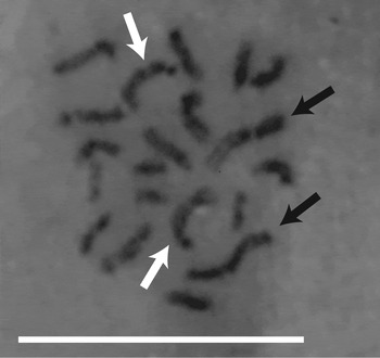
We counted 20 chromosomes at the mitotic metaphase in the pupal oogonia of the thelytokous strain (fig. 1), composed of the two longest (indicated using black arrows), two second longest (with white arrows), and 16 other chromosomes in eight cases. This showed that the thelytokous strain is diploid with a haploid set of ten chromosomes.
Metaphase of mitosis in a pupal oogonium of the thelytokous strain. The pairs of two longest and two second longest chromosomes are indicated using black arrows and white arrows, respectively. Scale bar represents 10 μm.

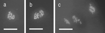
In the arrhenotokous strain, ten bivalent chromosomes were observed in the first metaphase of meiosis just after oviposition (fig. 2a). Within 1 h of oviposition, a reduction division of chromosomes (fig. 2b) and subsequent second phase of meiosis proceeded resulting in four sets of chromosomes (fig. 2c), presumably pronucleus and polar bodies. Mitotic cell division was observed to start about 2 h after oviposition.
Chromosomes in the deposited eggs of the arrhenotokous strain. (a) First metaphase of meiosis after oviposition, showing ten bivalents, (b) first anaphase (20 min after oviposition), and (c) second meiosis (30 min after oviposition). Scale bar represents 10 μm.

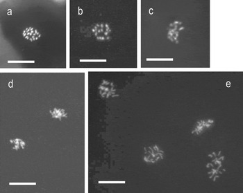
In the thelytokous strain, we counted 20 univalent chromosomes in deposited eggs just after oviposition (fig. 3a). This complement of 20 chromosomes remained unchanged for at least 1 h (fig. 3b, c). We examined more than 100 cases but never observed nuclear division and chromosomal complements in the haploid number (n=10), nor polar bodies, until mitotic cell division started approximately 1.5 h after oviposition (fig. 3d, e). These observations did not show meiosis in the egg maturation stages after oviposition in the thelytokous strain.
Chromosomes in the deposited eggs of the thelytokous strain. (a) Metaphase just after oviposition, showing 20 univalents, (b) metaphase (45 min after oviposition), (c) metaphase (1 h and 25 min after oviposition), (d) first mitotic division (1 h and 50 min after oviposition), and (e) subsequent mitotic division (2 h and 45 min). Scale bar represents 10 μm.

Allele segregation in thelytokous strains
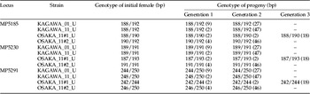
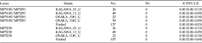
All three microsatellite loci examined were heterozygous in the four thelytokous strains examined, except for MP5230 in the strain OSAKA_11#2_U, and no male or homozygous female progeny were produced from heterozygous females (table 2). The upper 95% CI of the rate of transition to homozygosity of every heterozygous locus was lower than 0.05 on pooled data of all thelytokous strains (table 3), indicating that the cytogenetic mechanism of this thelytokous diploidization is apomictic or less probably automixis with central fusion.
Genotype of the initial females and their parthenogenetically produced progeny in the four thelytokous strains for three microsatellite loci. Numbers in parenthesis indicate the number of progeny with that genotype.

Observed rates of transition to homozygosity of heterozygous loci during parthenogenesis. No, number of progeny; Nt, number of transitions to homozygosity; R, observed rate of transition; CI, confidence interval.

Effect of antibiotic treatment on a thelytokous strain
We found no males in the 99 wasps produced by females treated using tetracycline hydrochloride for up to five generations (see the electronic supplementary material, fig. S1), indicating that the thelytoky of this species is not caused by the infection of any bacterial endosymbionts.
Bacterial infection in a thelytokous strain
Diagnostic PCR using the bacterial general primers showed no bacterial infection in the thelytokous wasps. This is consistent with the results of the antibiotic treatment described above and supports its indication that no bacterial endosymbionts cause thelytoky in this species.
Discussion
Our results reveal that the thelytoky of M. pulchricornis is apomixis without meiosis, thus, making no genetic recombination as a result to maintain heterozygosity in subsequent generations. While no observation of the transition to homozygosity of heterozygous loci in the thelytokous strains cannot exclude automixis with central fusion (Pearcy et al., Reference Pearcy, Hardy and Aron2006; Rey et al., Reference Rey, Loiseau, Facon, Foucaud, Orivel, Cornuet, Robert, Dobigny, Delabie, Mariano and Estoup2011), the diploid number of univalent chromosomes in the metaphase just after oviposition refutes it. Although automixis after premeiotic doubling may show the same number of bivalent chromosomes in the metaphase, the fact that no nuclear division and polar bodies were observed within about 1 h after oviposition, in which meiosis occurred in the arrhenotokous strain, indicate the absence of meiosis in the thelytokous strain.
In the Hymenoptera (Proctotrupomorpha), a case of apomictic thelytoky is known to be induced by endosymbiotic bacteria Rickettsia (Hagimori et al., Reference Hagimori, Abe, Date and Miura2006; Adachi-Hagimori et al., Reference Adachi-Hagimori, Miura and Stouthamer2008); this is in addition to the many cases of automictic thelytoky (gamete duplication) induced by Wolbachia (Stouthamer & Kazmer, Reference Stouthamer and Kazmer1994; Tagami & Miura, Reference Tagami and Miura2007). Recently, Wolbachia-induced thelytoky was observed in a braconid parasitoid as its first case in Ichneumonoidea (Kremer et al., Reference Kremer, Charif, Henri, Bataille, Pre´vost, Kraaijeveld and Vavre2009), although the cytogenetic mechanism is unknown. No positive evidence, however, was given for the induction of thelytoky by any bacterial symbionts in M. pulchricornis, indicating that the apomictic thelytoky of this species is spontaneous, i.e., genetically determined by the wasp itself. Nevertheless, the possible cause of thelytokous reproduction by horizontal gene transfer from a bacterial symbiont once existed to the nucleus of wasp cannot be excluded (Dunning Hotopp, Reference Dunning Hotopp2011).
Although automictic thelytoky is widely distributed in the Hymenoptera, apomictic thelytoky is known for only two species of Tenthredinoidea and three species of Proctotrupomorpha. Pachyprotasis youngiae (Tenthredinidae) is the only known triploid species in the Hymenoptera (Naito & Inomata, Reference Naito, Inomata, Blank, Schmidt and Taeger2006), and Strongylogaster (=Thrinax) macula (Tenthredinidae) shows achiasmatic meiosis with the expulsion of a single polar body, although with the occasional emergence of haploid males (Peacock & Sanderson, Reference Peacock and Sanderson1939). Recently, a similar apomictic cloning mechanism of achiasmatic meiosis with the expulsion of a single polar body has been found in two parasitoid wasps of Proctotrupomorpha, which likely originated from interspecific hybridization in Trichogramma cacoeciae (Trichogrammatidae) (Vavre et al., Reference Vavre, de Jong and Stouthamer2004) or was obviously induced by the endosymbiotic bacteria Rickettsia in Neochrysocharis formosa (Eulophidae) (Adachi-Hagimori et al., Reference Adachi-Hagimori, Miura and Stouthamer2008). The female-producing eggs of the unisexual spring generation in a gall-maker Neuroterus quercusbaccarum (=N. baccarum) (Cynipidae) of Proctotrupomorpha, which undergoes seasonal cyclic parthenogenesis, are also regarded to be produced through apomixis (Doncaster, Reference Doncaster1916; Dodds, Reference Dodds1939; Suomalainen et al., Reference Suomalainen, Saura and Lokki1987). Nevertheless, the spontaneous and non-cyclic apomixis of diploid thelytoky, without expulsion of polar bodies, in the present species M. pulchricornis is new to the Hymenoptera.
Apomixis definitely has the advantage of maintaining favourable gene combinations in a fixed manner, with no recombination in meiosis. Although apomictic thelytoky is the most common type of parthenogenesis found in arthropods (Suomalainen et al., Reference Suomalainen, Saura and Lokki1987), it is rare within the haplodiploid order Hymenoptera, while automictic thelytoky is rather frequent, as already pointed out by Mateo Leach et al. (Reference Mateo Leach, Pannebakker, Schneider, Driessen, Van de Zande, Beukeboom, Schön, Martens and Van Dijk2009) and by Rabeling & Kronauer (Reference Rabeling and Kronauer2013). In haplodiploidy, deleterious mutations can be effectively eliminated from haploid males, and thus, the estimated expected viability is high for diploid females. The expected viability of females is the highest in haplodiploidy (arrhenotoky), low in automictic thelytoky, and definitely the least in apomictic thelytoky as well as in diplodiploidy (amphimixis) (Haccou & Schneider, Reference Haccou and Schneider2004). Therefore, the transition from haplodiploidy to apomictic thelytoky will be more difficult than that to automictic thelytoky. Then, what are the conditions that cause rare cases of apomictic thelytoky in the Hymenoptera?
It is most likely that fixed heterosis, caused by triploidization in P. youngiae (Naito & Inomata, Reference Naito, Inomata, Blank, Schmidt and Taeger2006) or by hybridization in T. cacoeciae (Vavre et al., Reference Vavre, de Jong and Stouthamer2004), has overwhelmed the fast accumulation of deleterious mutations in diploidy. On the other hand, apomictic thelytoky of N. formosa is caused and maintained by the infection of bacterial symbionts (Adachi-Hagimori et al., Reference Adachi-Hagimori, Miura and Stouthamer2008), where horizontal transmission is involved. In those cases, males are seldom or never known, or if present, they are infertile (Adachi-Hagimori et al., Reference Adachi-Hagimori, Miura and Stouthamer2008).
The cyclic parthenogenesis of N. quercusbaccarum involves a generation of haploid males (Doncaster, Reference Doncaster1916; Dodds, Reference Dodds1939); thus, deleterious mutations can be eliminated from the males. In addition, in the case of probably spontaneous apomixis of S. macula, occasionally appearing haploid males indicate facultative thelytoky or deuterotoky (Peacock & Sanderson, Reference Peacock and Sanderson1939), in which unmated females produce both diploid females and haploid males. Such occasional sexual reproduction (D'Souza & Michiels, Reference D'Souza and Michiels2010) may cover the cost of apomictic parthenogenesis.
In the present species M. pulchricornis, apomixis is unlikely to be caused by polyploidization, specific hybridization, or bacterial symbiosis. Thelytokous strains usually do not produce males for many generations in the laboratory, but both arrhenotokous and thelytokous strains are sometimes sympatric in the fields (our unpublished data). Thus, it could be suggested that apomictic thelytoky of this species is primarily facultative in combination with occasional arrhenotoky, that is nothing but deuterotoky in nature, and deleterious mutations would be regularly eliminated from occasional haploid males. The examined strains of obligate thelytoky may be dead ends recently derived from a spectrum of deuterotokous strains. Further investigation of nuclear gene flow among sympatric strains of so-called arrhenotoky and thelytoky will test this suggestion.
Our finding suggests the possibility of operational shifting between sexual and asexual reproductions in parasitoid wasps, and thus, it will encourage further genetic studies to use asexual lines of wasps more wisely for biological control.
Supplementary Material
The supplementary material for this article can be found at http://www.journals.cambridge.org/BER
Acknowledgements
We thank Yasuhisa Kunimi, Madoka Nakai, Sho Furue, Shunpei Fujie, Hironobu Umemoto, and Naotaka Kumagawa for providing us with the wasp strains that we examined in this study, Makio Takeda and Katsuhiko Sakamoto for accommodating us in the laboratory of molecular biology, and Takuma Nishimura and Yosuke Abe for their help in rearing insects. This research was in part supported by JSPS KAKENHI (Grant numbers 19380036 and 25292034).