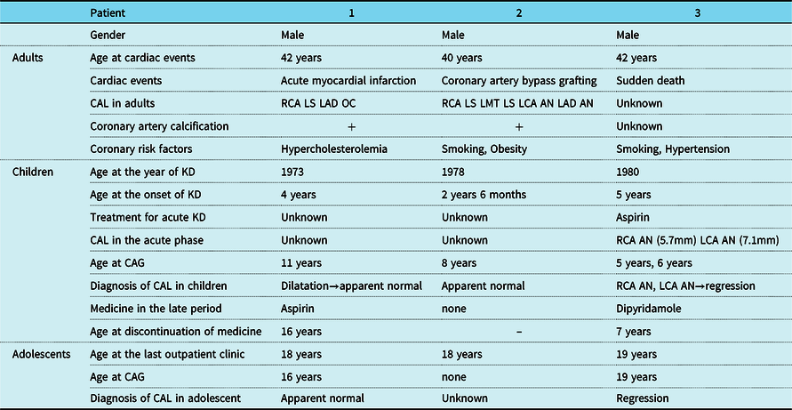
Fifty years have passed since the first description of Kawasaki disease. However, its long-term prognosis remains unclear. Especially, there is no information about the prognosis in patients with regressed coronary aneurysms caused by Kawasaki disease, because most patients are not followed-up as adults. Reference Tsuda, Tsujii and Hayama1 We reported three male patients with cardiac events in their forties; however, they had been confirmed as having almost normal coronary arteries during the coronary angiograms when they were children and adolescents.
Patient 1
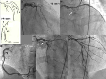
A 42-year-old man visited an emergency hospital, because he had chest pain and respiratory distress during jogging at night. His body length and body weight were 170 cm and 51 kg, respectively. A 12-lead electrocardiogram revealed ST-T elevation in leads V2–V5. He was diagnosed with an acute anterior myocardial infarction. Coronary angiograms revealed an occlusion of the left anterior descending artery (Fig 1). Emergency percutaneous coronary intervention was performed. After thrombotic aspiration, a bare metal stent was inserted at segment 6 (3.5/24 mm). Coronary angiograms revealed 25% stenosis with calcifications at segment 1 of the right coronary artery. Intra-aortic balloon pumping and catecholamines were used for cardiogenic shock. A beta-blocker, aspirin, and statin were prescribed. The total cholesterol was 228 mg/dl. After 6 months, 25% stenosis in the stent was detected in the follow-up angiograms. Hypokinesis at the apex of the left ventricle was also found in his left ventriculogram. In the two-dimensional echocardiography, the left ventricular end-diastolic dimension and ejection fraction were 57.6 mm and 49%, respectively. He was referred to our hospital at 45 years. Further, he had a cerebral infarction during exercise at 48 years. He underwent an immediate thrombolysis for the left mid-cerebral artery, and he had no disturbance due to the cerebral infarction after rehabilitation. Coumadin was added.
Coronary angiograms at the age of 16 and 42 years (Patient 1). Coronary angiograms at 16 years revealed almost angiographic normal coronary arteries. (Coronary angiograms at the age of 16 were discarded.) ( A and B ). At 42 years, an occlusion of the left anterior descending artery and mild localised stenosis with calcification at segment 1 were detected. ( C , D and E ) Percutaneous coronary intervention was successful for the occlusion of the left anterior descending artery.

He was admitted for 20 days because of incomplete Kawasaki disease at the age of 4 in 1973 (Table 1). The detail of the acute treatment was unknown. After the school examination at the age of 11, he was referred to our hospital because of a history of Kawasaki disease. Coronary angiograms revealed coronary artery dilatation of the right coronary artery and left anterior descending artery; however, the coronary aneurysms were not detected by the two-dimensional echocardiography. Aspirin was prescribed. The coronary angiogram findings at the age of 16 were almost the same as the previous coronary angiograms (Fig 1). He was diagnosed as “normal”, and aspirin was stopped. Follow-up was recommended once a year. He no longer visited our hospital after the age of 18. He has the habit of jogging every day as an adult. Hypercholesterolemia was pointed out once during an examination at the age of 41. However, he had no symptoms until an episode when he was 42 years old.
Patient characteristics

Note: CAL, coronary artery lesion, RCA, right coronary artery, LCA left coronary artery, LAD, left anterior descending, LMT, left main trunks, KD, Kawasaki disease, AN aneurysm, LS localised stenosis, OC occlusion.
Patient 2
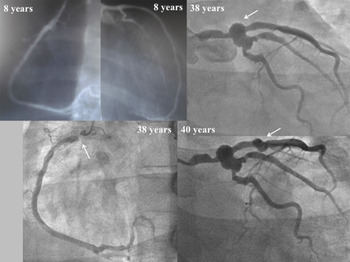
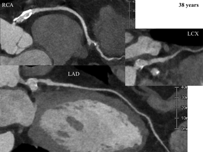
A 20-year-old man visited a hospital because of chest pain. However, no cardiac abnormalities were detected at that time. At 26 years, he had palpitation during the night. He was transferred to an emergency hospital, and was diagnosed with paroxysmal atrial fibrillation. He was restored to normal sinus rhythm by direct cardioversion. Although he had several episodes of paroxysmal atrial fibrillation after that episode, it stopped spontaneously. He underwent a chest computed tomography at the age of 37 years, because he was aware of having a history of Kawasaki disease. Coronary artery calcifications were detected in his computed tomography (Fig 2). A 12-lead electrocardiogram revealed an abnormal Q wave in lead III. His coronary angiograms revealed severe localised stenosis of the right coronary artery and a new appearing coronary artery aneurysm at the bifurcation of the left coronary artery (Fig 3). He was transferred to our hospital at the age of 38, because hypoperfusion was detected in the inferior wall of the left ventricle by 99 m tetorofosmin myocardial perfusion imaging. His height and weight were 176 cm and 88 kg, respectively. His low-density-lipoprotein cholesterol was 114 mg/dl. The values of fractional flow reserve in the left anterior descending artery and left circumflex at 40 years were 0.70 and 0.74, respectively. Further, coronary angiograms revealed a new aneurysm of the left anterior descending artery. Although the values of myocardial blood flow ratio in both areas of the right coronary artery and left anterior descending artery were reduced, the value of the whole myocardial blood flow was 2.16 and within normal limits by radioisotope myocardial perfusion imaging. He had chest pain with inversion of T wave in leads II, III, and aVF in the treadmill test. Coronary artery bypass grafting to the right coronary artery and left anterior descending artery were performed, and it was confirmed that the grafts were patent in computed tomography angiograms at the age of 41.
Coronary angiograms at the age of 8, 38, 40 years (Patient 2). Coronary angiograms at 8 years revealed almost angiographic normal coronary arteries. At 38 years, severe localised stenosis of segment 1 and a new appearing coronary aneurysm at the bifurcation of the left coronary artery were detected. Further, a new coronary artery aneurysm was detected in the left anterior descending artery in the coronary angiogram at 40 years.

Computed tomography angiograms at the age of 38 (Patient 2). Coronary artery aneurysms with calcification at the proximal portion in the right coronary artery and coronary aneurysm at bifurcation of the left coronary artery were detected.

He had acute Kawasaki disease at the age of 2 years and 6 months in 1978. The detail of the acute treatment was unknown. In his elementary school cardiac examination, it was recommended to visit the hospital, because he had a history of Kawasaki disease. He was diagnosed with “normal coronary arteries” in his coronary angiograms at the age of 8. He was followed with master double electrocardiograms each 2 years until 18 years. The follow-up was stopped at the age of 18. He had been smoking for 20 years after that.
Patient 3
The 3rd patient had an unexpected and unknown sudden death at the age of 42. He called his office to ask for the day off the previous morning before his death, because of his poor condition. He did not go to his office the next morning. His superior officials visited his home, and found that he had died in bed. The cause of the sudden death was unknown because an autopsy was not performed. He had a medical examination in the office 1 month before his death. His height and weight were 158 cm and 49 kg, respectively. His blood pressure was 167/100 mmHg. There were no abnormalities in his urinalysis. He had been a smoker. Although the examination for his hypertension had been recommended, he had not visited the hospital.
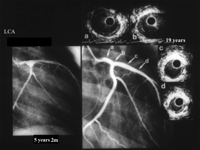
He had Kawasaki disease at the age of 5 in 1980, and he had an aspirin therapy. He had bilateral coronary artery aneurysms after Kawasaki disease. He was referred to our hospital. His coronary angiograms 1 month after the onset of Kawasaki disease revealed coronary aneurysms in the right coronary artery and left coronary artery (Fig 4). The maximal diameter of the right coronary artery and bifurcation of the left coronary artery were 5.7 and 7.1 mm, respectively. Dipyridamole was prescribed. The bilateral coronary aneurysms had regressed 1 year after Kawasaki disease, and the medication was stopped. Further, intimal thickening at the previous right and left coronary aneurysms observed by intravascular ultrasound was mild at the age of 19 years. Follow-up once a year was recommended by his doctor. However, he had never visited our hospital since that time.
Left coronary angiograms at the age of 5 and 19 years, and the findings by intravascular ultrasound (Patient 3). Coronary angiogram at 5 years revealed coronary artery aneurysm at the bifurcation of the left coronary artery. The maximal diameter of coronary aneurysm at bifurcation of the left coronary artery was 7.1 mm. Coronary angiogram at 19 years revealed regression of the coronary artery aneurysm. The intimal thickening in the coronary artery wall at a previous coronary aneurysm was mild.

Discussion
Coronary artery lesions in the acute phase of Kawasaki disease were not evaluated in the former two patients, because they had acute Kawasaki disease in the 1970’s, and their coronary arteries were diagnosed as almost normal in the coronary angiograms in their school-ages. In the 3rd patient, medium-sized coronary aneurysms in the acute phase and regression of the coronary aneurysms at 1 year after Kawasaki disease were confirmed in the coronary angiograms. All three patients had no apparent aneurysms or stenosis in their adolescents after acute Kawasaki disease. However, it was suspected that their findings in the late period were slight dilatation in some portions. In the former two patients, the portions with slight dilatation became the culprit lesions of the cardiac events. In their forties, coronary artery calcifications were found in those portions.
Almost all the small aneurysms (<4 mm) in the acute phase regressed. Reference Tsuda, Tsujii and Hayama1,Reference Tsuda, Kamiya, Ono, Kurosaki and Echigo2 The medium aneurysms (≧4 mm and <8 mm) in the acute phase regressed or persisted as dilatation. Most giant aneurysms (≧8 mm) persisted as aneurysms. Reference Tsuda, Kamiya, Ono, Kurosaki and Echigo2,Reference Tsuda, Hamaoka and Suzuki3 Those regressed aneurysms could persist as slight dilatation or apparently normal coronary arteries for a long period of more than 10 years. Reference Suzuki, Kamiya, Tsuda and Tsukano4 However, it is considered that intimal thickening continues in such coronary artery walls and the remodelling of the coronary artery wall occurred gradually many years after acute Kawasaki disease. Reference Suzuki, Yamagishi and Kimura5–Reference Suzuki, Miyagawa and Komatsu7 Further, it is known that coronary artery calcification is the marker of such coronary arterial involvement caused by Kawasaki disease. Reference Tsuda, Matsuo and Kurosaki8 In atherosclerosis, coronary artery calcification usually increases with aging. Coronary artery calcium scoring may be a useful tool to screen patients with a remote history of Kawasaki disease or unknown coronary artery status such as probable Kawasaki disease. Reference Kahn, Budoff and Duniels9 We reported that the larger coronary artery diameter caused by Kawasaki disease, resulted in an earlier appearance of coronary artery calcifications in the coronary artery lesions. Reference Tsujii, Tsuda, Kanzaki, Ishiduka, Nakashima and Kurosaki10 The timing of the appearance of coronary artery calcifications between the giant aneurysms and medium aneurysms differed. Regressed coronary aneurysms need to be re-evaluated more than 20 years after acute Kawasaki disease.
There is also a difference in the time of cardiac events between the patients with giant aneurysms and those with medium-sized aneurysms. The cardiac events free rate at 20 years in patients with giant aneurysms was significantly lower than that in patients with medium aneurysms. During the 1970’s and 1980’s, the infants with giant aneurysms often had acute myocardial infarctions within a year of the initial episode of Kawasaki disease. Reference Tsuda, Arakaki and Shimizu11 Further, most of them had coronary events such as coronary revascularisation in children or adolescents. Reference Tsuda, Kamiya, Ono, Kurosaki and Echigo2,Reference Tsuda, Hamaoka and Suzuki3,Reference Suda, Iemura and Nishiono12 On the other hand, cardiac event in patients with regression of coronary aneurysms is very rare until 20 years. Reference Tsuda, Tsujii and Hayama13 However, some patients with acute coronary syndrome in their twenties and thirties have been reported, Reference Gordon, Daniels and Kahn14–Reference Kawai, Takakura and Naruse17 and we encountered three patients in their forties.
Involvement of the coronary arterial wall has continued since the appearance of coronary artery aneurysms due to acute Kawasaki disease vasculitis. It is very interesting for their cardiac events to have occurred in adulthoods, although they were asymptomatic for a long time after the time of the regression of the coronary aneurysms after Kawasaki disease. The symptoms caused by Kawasaki disease are rare and evidence of ischemia is often not present until actual cardiac event occurs. Various coronary risk factors would be added with aging. During their follow-up, the medication was given to two patients in their childhood (Patient 1 and 3). However, the medication was stopped, because their coronary arteries were diagnosed as “angiographic normal coronary arteries”. However, it was suspected that they had cardiac events in their forties due to coronary artery lesions caused by Kawasaki disease. To prevent serious cardiac events in such patients, it may be needed to re-start the anti-thrombotic therapy again in their adulthoods. It is probable that more effective antithrombotic therapy including the use of antiplatelets has decreased the prevalence of acute myocardial infarctions. In addition, treatment for coronary risk factors such as lowering of low-density lipoprotein cholesterol would be needed. The usefulness of long-term aspirin and a statin to promote endothelial cell health should be investigated in the future.
There is a patient population who had acute Kawasaki disease in the 1970’s and 1980’s when appropriate examinations of coronary artery lesions in the acute phase were not established. During that period many patients probably died, because a precise means of diagnosis for cardiac sequelae were not determined. Reference Tsuda, Arakaki and Shimizu11 Even if cardiac events in the 1970’s and 1980’s did not occur in patients who were not diagnosed with acute Kawasaki disease and coronary artery disease caused by Kawasaki disease, they might have had cardiac events over 40 years. Nowadays, it is not unusual to discover patients, not previously diagnosed as having had Kawasaki disease, with coronary artery lesions suggestive of previous Kawasaki disease such as calcified giant aneurysms and segmental stenosis. Unfortunately, they are often found because of acute myocardial infarction or sudden death. However, it might be difficult to suspect previous Kawasaki disease in such cases with regression of coronary aneurysms, because they do not always have coronary artery lesions suggestive of previous Kawasaki disease. One of the causes of cardiac events is progressive stenosis and another is thrombus formation without coronary aneurysms. Furthermore, new appearing coronary aneurysms after regression rarely occur in the late period. Reference Tsuda, Kamiya, Ono, Kimura and Echigo18,Reference Ozawa, Suzuki and Hasegawa19
The cardiac events in this report were completely unexpected. They were born in the 1970’s and suffered from acute Kawasaki disease, and had been asymptomatic for many years prior to their cardiac events. The possibility of cardiac events in patients with regression of coronary aneurysms has been considered to be very low. However, this may be incorrect. Actually, the prevalence of cardiac events in this population remains unknown, and it must be clarified in this future. There are probably many such patients, now in their forties and fifties, at risk, and the cause of the cardiac events in this group and its prevention is an important issue. Patients with regression of coronary aneurysms and families of patients at a potential risk of cardiac complications should be educated about the need for re-evaluation in young adults. Computed tomography angiography or magnetic resonance angiography can then be non-invasive and useful to delineate the coronary artery lesions. All patients who had had coronary artery aneurysms should be followed for life. It is recommended to be followed-up every 3–5 years in such patients, even if they are asymptomatic.
Conclusion
We encountered three male patients with a history of Kawasaki disease, who had their 1st cardiac events in their forties. Re-evaluation of coronary artery lesions after regressed coronary artery aneurysms is needed to prevent fatal cardiac events in such patients. Coronary artery calcification in the proximal portion of the major coronary arteries can be a predictive marker after the regression of coronary aneurysm caused by Kawasaki disease.
Acknowledgements
We thank John Martin for his kind English language consultation.
Financial support
This research received no specific grant from any funding agency, commercial, or not-for-profit sectors.
Conflicts of interest
The authors state that they have no conflict of interest.
Ethical standards
All procedures performed in studies involving human participants were in accordance with the ethical standards of the institutional committee with 1964 Helsinki declaration and its later amendments or comparable ethical standards.