


Introduction
Cryptosporidium is an important etiologic agent of human and animal disease
Cryptosporidium species are the second most common cause of diarrhoea in children and are also linked to higher morbidity. In fact, across all age categories, Cryptosporidium caused 57,200 deaths, with children under 5 accounting for over 80% of Cryptosporidium infection-related mortality (Khalil et al., Reference Khalil, Troeger, Rao, Blacker, Brown, Brewer, Colombara, De Hostos, Engmann, Guerrant, Haque, Houpt, Kang, Korpe, Kotloff, Lima, Petri, Platts-Mills, Shoultz, Forouzanfar, Hay, Reiner and Mokdad2018). Numerous outbreaks of human cryptosporidiosis have been linked to contaminated drinking water, recreational water and food (Karanis et al., Reference Karanis, Kourenti and Smith2007; Baldursson and Karanis, Reference Baldursson and Karanis2011; Efstratiou et al., Reference Efstratiou, Ongerth and Karanis2017; Bourli et al., Reference Bourli, Eslahi, Tzoraki and Karanis2023). Furthermore, the genus Cryptosporidium includes parasitic protozoa that can infect the epithelial cells lining the luminal surfaces of the digestive and respiratory tracts. This infection affects a wide range of mammals, birds, reptiles, amphibians and fish. Notably, cryptosporidial oocysts are environmentally resilient (Golomazou et al., Reference Golomazou, Mamedova, Eslahi and Karanis2024), exhibiting remarkable resistance to chemical disinfection (e.g. drinking water chlorination) but remaining susceptible to extreme temperatures, including freezing and pasteurization.
Cryptosporidium life cycle
Cryptosporidium completes its complex life cycle within a single host, primarily targeting the microvillar border of the intestinal epithelium. During its development, the parasite occupies a unique intracellular, extracytoplasmic niche, often referred to as ‘epicellular’ localization (Barta and Thompson, Reference Barta and Thompson2006; Aldeyarbi and Karanis, Reference Aldeyarbi and Karanis2016a,Reference Aldeyarbi and Karanisb; Karanis, Reference Karanis2018). Transmission occurs predominantly via the fecal–oral route following the ingestion of sporulated oocysts. Upon ingestion, oocysts undergo excystation in the gastrointestinal tract, releasing 4 motile sporozoites into the intestinal lumen. These sporozoites invade the apical surface of epithelial cells, where they differentiate into trophozoites within a specialized parasitophorous vacuole. This unique, closed epicellular compartment is derived from membranes contributed by both the host cell and the parasite, keeping the organism topologically outside the host cytoplasm (Bones et al., Reference Bones, Jossé, More, Miller, Michaelis and Tsaousis2019).
According to the updated lifecycle paradigm, the intracellular developmental phase progresses through a highly regulated timeline rather than distinct, morphologically diverse meront classes. Trophozoites undergo merogony (schizogony) to form meronts, which consistently yield 8 merozoites (Karanis et al., Reference Karanis, Kimura, Nagasawa, Igarashi and Suzuki2008; English et al., Reference English, Guérin, Tandel and Striepen2022). Upon release, these merozoites reinvade adjacent epithelial cells to propagate the asexual phase. The parasite undergoes exactly 3 sequential rounds of this asexual replication (trophozoites, meronts and merozoites) before automatically initiating sexual development (gametogony). Following the third round of merogony, the released merozoites enter host cells and differentiate into either unicellular macrogamonts (female forms) or multinucleated microgamonts (male forms). During fertilization, mature microgametes are released from the microgamonts and fuse with macrogamonts to form a diploid zygote. The zygote subsequently undergoes sporogony (sporulation), yielding 4 motile sporozoites within a protective oocyst wall. These developing oocysts follow 2 distinct pathways: thin-walled oocysts undergo autoinfection within the host gastrointestinal tract, whereas thick-walled oocysts are shed in feces into the environment, thereby initiating transmission to new hosts (Fayer, Reference Fayer, Fayer and Xiao2008; English et al., Reference English, Guérin, Tandel and Striepen2022).
Concurrently, alternative developmental pathways have been proposed to complement this intracellular lifecycle. Independent microscopic evaluations have documented the presence of free-living, extracellular gamont-like stages that exhibit syzygy (the pairing of gamonts without immediate fusion) (Aldeyarbi and Karanis, Reference Aldeyarbi and Karanis2016c). While these extracellular forms remain a subject of ongoing characterization within the research community, they underscore the parasite’s unexpected developmental plasticity.
Due to its unique biological features, such as the epicellular position and potential for extracellular stages, Cryptosporidium has been reclassified from classical coccidia to the subclass Cryptogregarina (Ryan et al., Reference Ryan, Paparini, Monis and Hijjawi2016). This taxonomic distinction is important for in vitro cultivation, as understanding its gregarine-like metabolism and extracellular survival informs the development of host-free media and optimized culture conditions. Although the intestine is the primary site of infection, the parasite can also colonize extraintestinal epithelial tissues, such as the biliary tree and respiratory tract, particularly in immunocompromised hosts. This systemic plasticity underscores the need for tissue-specific culture models (Sponseller et al., Reference Sponseller, Griffiths and Tzipori2014).
Classification challenges in Cryptosporidium
The genus Cryptosporidium belongs to the phylum Apicomplexa. Initially classified as a coccidian due to shared life-cycle features (Levine, Reference Levine1988), later molecular and morphological research showed that Cryptosporidium is more closely related to gregarines than to coccidia (Carreno et al., Reference Carreno, Martin and Barta1999). This phylogenetic shift led to a formal taxonomic reclassification by Cavalier-Smith (Reference Cavalier-Smith2016), who moved Cryptosporidium from the class Coccidiomorphea to the class Gregarinomorphea and erected the subclass Cryptogregaria (order Cryptregarida) to accommodate the genus.
As synthesized by Ryan et al. (Reference Ryan, Paparini, Monis and Hijjawi2016), this reclassification into Cryptogregaria is supported by several fundamental biological traits that diverge sharply from classical coccidia and mirror gregarine behaviour. These include the ability to undergo extracellular, host-free environmental life-cycle stages (axenic development), the presence of sizable extracellular gamont-like stages, syzygy-like pairings and a notorious biological resistance to all standard anti-coccidial therapies (Ryan et al., Reference Ryan, Paparini, Monis and Hijjawi2016). However, while the gregarine affinity is strongly supported by 18S rRNA sequencing and ultrastructural evidence, this systematic shift remains a subject of ongoing debate. A faction of the parasitology community continues to question a definitive gregarine classification, arguing that certain biological parameters or alternative evolutionary trees place Cryptosporidium either as a deeply branching basal apicomplexan lineage or closer to ancestral coccidians, highlighting a persistent lack of universal taxonomic consensus.
The complete life cycles of C. andersoni and C. parvum were described, with extracellular gamont-like stages and syzygy observed in some of these stages (Hijjawi et al., Reference Hijjawi, Meloni, Ryan, Olson and Thompson2002, Reference Hijjawi, Meloni, Ng’anzo, Ryan, Olson, Cox, Monis and Thompson2004; Aldeyarbi and Karanis, Reference Aldeyarbi and Karanis2016c). Identifying novel extracellular stages in the Cryptosporidium life cycle offers strong biological evidence for its formal taxonomic reclassification from classical coccidia. Detailed description of the early development of G. steini and C. muris and searched for explicit characteristics that may address the nature of the relationship between gregarines and cryptosporidians (Valigurová et al., Reference Valigurová, Hofmannová, Koudela and Vávra2007). Different aspects of the evolutionary biology and invasion mechanisms of Cryptosporidium need to be examined in light of knowledge of gregarines (Aldeyarbi and Karanis, Reference Aldeyarbi and Karanis2016c). The major factor limiting understanding of biological details and classification is the lack of a practical, reproducible in vitro cultivation system for this intestinal parasite (Karanis and Aldeyarbi, Reference Karanis and Aldeyarbi2011).
This present review provides a state-of-the-art overview of in vitro efforts to cultivate Cryptosporidium, discusses recent developments, highlights the advantages and drawbacks of these emerging systems, and pinpoints research work published exclusively between 2018 and 2025. It is an update of the invited review ‘The truth about in vitro culture of Cryptosporidium species’ (Karanis, Reference Karanis2018).
Materials and methods
Study design
This systematic review was conducted in accordance with PRISMA guidelines and included peer-reviewed articles published between 2018 and 2025 that reported Cryptosporidium cultivation methods.
Search strategy and criteria
A comprehensive PubMed search was conducted to identify relevant studies published in English between 2018 and 2025. The search used the keywords ‘Cryptosporidium’, ‘cultivation’, ‘plant compounds’ and ‘drugs’. Reference lists of included studies were also checked for related articles. Articles with titles indicating Cryptosporidium or cultivation were screened and selected for inclusion.
Inclusion and exclusion criteria
Articles were included if they met the following objective criteria: (i) published after 2018 and addressed Cryptosporidium cultivation methods; (ii) described quantifiable in vitro cultivation systems. These systems were evaluated based on their ability to support specific parasite developmental milestones, such as asexual replication, sexual differentiation or complete life cycle progression, across established host systems, including standard cell lines like HCT-8 or advanced platforms like three-dimensional (3D) organoids, tissue-engineered monolayers and microfluidic models; and (iii) were written in English. Studies were excluded if they were review articles, case reports, unrelated to the core objective of in vitro propagation, lacked accessible full-text PDFs or were published in languages other than English.
Data extraction
For the full-text review, a standardized data extraction approach captured key biological and technical variables from each eligible study. Extracted parameters included the year of publication, the specific Cryptosporidium species and strain evaluated (e.g. C. parvum), and the cultivation models used. When necessary, these models and parasite species were inferred from the text.
The included studies were grouped by cultivation model and categorized into conventional two-dimensional (2D) cell lines, 3D organoids/enteroids, air–liquid interface (ALI) systems and microfluidic gut-on-chip devices, with animal models serving as primary sources and foundational baselines for in vitro platforms. The number of studies in each category was determined, the parasite species used within those models were mapped and the corresponding references were compiled. This provided a structured, taxonomically clear overview of the available evidence.
Results
Literature search and eligible studies
The systematic search initially identified 72 records. The review was conducted in accordance with the PRISMA Statement (Page et al., Reference Page, McKenzie, Bossuyt, Boutron, Hoffmann, Mulrow, Shamseer, Tetzlaff, Akl, Brennan, Chou, Glanville, Grimshaw, Hróbjartsson, Lalu, Li, Loder, Mayo-Wilson, McDonald, McGuinness, Stewart, Thomas, Tricco, Welch, Whiting and Moher2021), applying predefined inclusion and exclusion criteria and a risk-of-bias assessment, as outlined below (Figure 1).
Flow diagram of the study design process showing the total number of retrieved reports, number of records after duplicates were removed, number of records screened and excluded, number of full-text articles assessed for eligibility, number of selected articles for analysis and number of studies included in the review.

Figure 1 Long description
The study design process flowchart. Under Identification, records identified from databases and PubMed total 72, with 0 removed before screening. In Screening, 72 records are screened and 52 are excluded for reasons like review articles (7), case reports (1), unrelated objectives (35), non-English publications (1) and unavailable full text (8). Reports sought for retrieval are 20, with 11 not retrieved. Nine reports are assessed for eligibility. In Included, 20 new studies are from databases and 7 additional studies are from other methods, totaling 27 studies included in the review.
Of the studies reviewed, 20 were considered eligible and underwent full-text evaluation, while 52 were excluded. The reasons for exclusion were review articles (n = 7), case reports (n = 1), studies unrelated to the research objective (n = 35), non-English publications (n = 1) and lack of accessible full-text PDF versions (n = 8).
Following full-text evaluation, 9 reports were retrieved and assessed in greater detail. Of these, 7 additional studies met the predefined inclusion criteria and were subsequently incorporated into the review. In total, 27 studies evaluating Cryptosporidium cultivation systems, including those using cell lines and recent experimental models, were included in the final analysis.
Characteristics of the included studies
The studies included in the present review were published between 2018 and 2025. No eligible studies investigating cell-free methods (axenic) for culturing Cryptosporidium were identified. Twenty-seven studies were analysed, focusing exclusively on Cryptosporidium cultivation systems, including culture conditions, durations and factors influencing growth. The analysis primarily focused on the cultivation systems employed rather than on the specific ingredients in the media and their effects.
Cultivation systems applied during the period 2018–2025
Of the 27 included studies, 24 evaluated a single primary cultivation model, while 3 studies assessed 2 distinct models within the same publication. These often used standard HCT-8 cell cultures alongside newer platforms for comparative analysis. Among single-model studies, organoid-based systems were most common (n = 5), followed by HCT-8 (n = 4), ALI systems (n = 4), hollow-fibre technology (n = 3), gut-on-a-chip platforms (n = 3), in vivo animal models (n = 3) and COLO-680N (n = 2). In the 3 studies using multiple methodologies, platforms were evaluated side by side: HCT-8 was compared with COLO-680N (n = 1), organoid-based systems (n = 1) and ALI (n = 1) (Figure 2).
Number of publications included in the review by models.

Among these, the conventional HCT-8 cell line remains a cornerstone in Cryptosporidium research. It serves not only as a standard platform for drug screening and developmental investigations but also as a fundamental baseline model and integral biological component upon which many advanced, novel culture systems are established. Recent methodological advances have increasingly focused on 3D culture systems, including hollow-fibre technology, organoid-based platforms, ALI models and organ-on-a-chip technologies, which aim to more accurately replicate the physiological intestinal environment.
Frequency of cultivation systems applied during 2018–2025
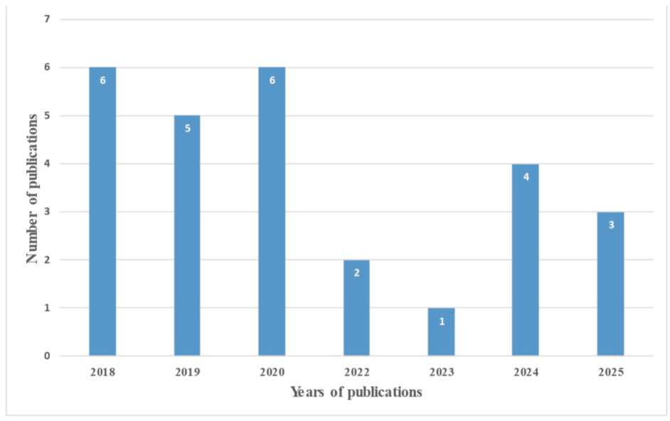
Figure 3 illustrates the distribution of publications included in the review by year (2018–2025). The number of studies increased between 2018 and 2020. However, a decline in publications was noted during 2022–2023 compared with previous years. A relative resurgence in research activity was observed in 2024–2025 (Figure 3).
Number of publications included in the submission: Cryptosporidium reviews by years (2018–2025).

Discussion
Advanced in vitro cultivation systems are essential for reducing reliance on animal models in Cryptosporidium research (Karanis, Reference Karanis2018; Feix et al., Reference Feix, Cruz-Bustos, Ruttkowski and Joachim2023). Animal models remain necessary for studying complex host factors such as immune responses, interactions with the gut microbiota and metabolic changes. However, in vitro platforms that closely mimic the physiological gut environment enable detailed investigation of Cryptosporidium biology, invasion kinetics and cellular development (Karanis, Reference Karanis2018; Vélez et al., Reference Vélez, Silva, Kamena, Daugschies, Mazurek, Taubert and Hermosilla2022). Intracellular host-cell culture systems are currently required to study complete parasite propagation, merozoite formation and the full life cycle (Miller et al., Reference Miller, Jossé, Brown, Blakeman, Povey, Yiangou, Price, Cinatl, Xue, Michaelis and Tsaousis2018; Bones et al., Reference Bones, Jossé, More, Miller, Michaelis and Tsaousis2019). In contrast, host-free (cell-free) incubation systems do not support continuous parasite propagation or large-scale oocyst production. Although earlier studies suggested extracellular development in cell-free media (Hijjawi et al., Reference Hijjawi, Meloni, Ng’anzo, Ryan, Olson, Cox, Monis and Thompson2004), current consensus recognizes these systems as specialized tools for short-term functional assays limited to extracellular oocysts and sporozoites (Zhang et al., Reference Zhang, Sheoran and Widmer2009). These systems allow researchers to study baseline oocyst or sporozoite metabolism (Zhang et al., Reference Zhang, Guo and Zhu2015), assess excystation kinetics in response to environmental triggers (King et al., Reference King, Keegan, Robinson and Monis2011) and screen therapeutic compounds targeting extracellular sporozoites before host-cell invasion, without confounding variables from host cells.
Diversification of cultivation systems
Over the past 8 years, research has used a variety of cultivation frameworks from basic cell monolayers (e.g. HCT-8 and COLO-680N) to advanced 3D organoids, ALI systems, hollow-fibre bioreactors and in vivo animal models (Table 1).
Studies included in the review by cultivation models and the number of studies

Table 1 Long description
Columns list cultivation model, short description, full life cycle achieved, duration, and studies included in review. Cell culture using HCT 8 or Caco 2 is described as human intestinal epithelial cell lines in standard static two dimensional monolayers or three dimensional matrices, with full life cycle not achieved and duration of 3 to 4 days; studies listed are Melicherova et al., 2018; Tandel et al., 2019; Funkhouser Jones et al., 2020; English et al., 2022; Velez et al., 2022; Gunasekera et al., 2023; Korwin Mihavics et al., 2025. Cell culture using COLO 680 N is described as a human esophageal squamous cell carcinoma line in standard two dimensional culture, with full life cycle marked contested with an asterisk and duration up to 8 weeks; studies listed are Miller et al., 2018; Josse et al., 2019; Velez et al., 2022. Hollow fiber technology is described as a bioreactor using artificial capillary like fibers to mimic gut dynamics and fluid flow, with full life cycle achieved and duration over 20 months; studies listed are Yarlett and Morada, 2018; Yarlett et al., 2020; Yarlett et al., 2024. Organoid based systems are described as three dimensional mini organs differentiated from primary human or animal intestinal stem cells, with full life cycle noted as system dependent and duration up to 3 to 4 weeks; studies listed are Heo et al., 2018; Wilke et al., 2018; Dutta et al., 2019; Cardenas et al., 2020; Korwin Mihavics et al., 2025; Varegg et al., 2025. Air liquid interface is described as organoid derived cells grown on a porous membrane with luminal air exposure, with full life cycle achieved and duration up to 20 days; studies listed are Wilke et al., 2019; Funkhouser Jones et al., 2020; Wilke et al., 2020; Deng et al., 2024; Greigert et al., 2024. Gut on a chip is described as microfluidic platforms introducing continuous fluid shear stress to host monolayers, with full life cycle noted as system dependent and duration up to 10 days; studies listed are Nikolaev et al., 2020; Gunasekera et al., 2024; Gunasekera et al., 2025. Animal models are described as neonatal, immunocompromised, or gene ablated in vivo hosts including mice, calves, and piglets, with full life cycle achieved and duration listed as variable; studies listed are Graef et al., 2018; Sateriale et al., 2019; Riggs and Schaefer, 2020. A final row labeled Total Included Empirical Studies lists 27 studies with a double asterisk in the studies column, and dashes in the other columns.
Notes: a Note on COLO-680N: While initial investigations reported multi-passage continuous propagation and full life cycle completion (Miller et al., Reference Miller, Jossé, Brown, Blakeman, Povey, Yiangou, Price, Cinatl, Xue, Michaelis and Tsaousis2018), independent replication attempts have widely disputed these findings, reporting a failure to maintain long-term continuous culture (Vélez et al., Reference Vélez, Silva, Kamena, Daugschies, Mazurek, Taubert and Hermosilla2022).
b Dual-model approaches are applied in several studies, including cell culture (HCT-8) combined with organoid-based systems (Korwin-Mihavics et al., Reference Korwin-Mihavics, Dews, Miller, Cameron, Martorelli Di Genova and Huston2025), air–liquid interface (ALI) systems (Funkhouser-Jones et al., Reference Funkhouser-Jones, Ravindran and Sibley2020) and COLO-680N (Vélez et al., Reference Vélez, Silva, Kamena, Daugschies, Mazurek, Taubert and Hermosilla2022).
From 2018 to 2025, studies employing conventional cell monolayer culture systems were progressively optimized, alongside a marked increase in research utilizing advanced 3D technologies and ALI culture systems. Most recently, in 2025, an emerging in vitro cultivation of Cryptosporidium was reported.
The use of monolayer cultures
Progress in the in vitro propagation of C. parvum has been reported for several cell lines, including HCT-8 (human adenocarcinoma), COLO-680N (oesophageal squamous-cell carcinoma), MDCK (Madin-Darby canine kidney), MDBK (Madin-Darby bovine kidney), BFTE (bovine fallopian tube epithelial), Caco-2 (human colonic adenocarcinoma), HT29 (human colon cancer) and BS-C-1 (African green monkey kidney) cell lines (Feix et al., Reference Feix, Cruz-Bustos, Ruttkowski and Joachim2023).
Cell culture (HCT-8 and HT-29)
Traditional cell lines, most notably human ileocecal colorectal adenocarcinoma (HCT-8) cells, remain a foundational tool in Cryptosporidium research. They serve as the baseline for evaluating both classical growth kinetics and advanced engineering platforms. For instance, Gunasekera et al. (Reference Gunasekera, Clode, King, Monis, Thierry, Carr, Chopra, Watson, O’Dea, Hijjawi and Ryan2023) used standard static HCT-8 monolayers to compare the early development of 2 C. parvum isolates and 1 C. hominis isolate by scanning electron microscopy. Their morphological findings showed that asexual phases progressed similarly across species, whereas sexual differentiation occurred more rapidly in C. hominis, with macrogamonts appearing within 48 h (Gunasekera et al., Reference Gunasekera, Clode, King, Monis, Thierry, Carr, Chopra, Watson, O’Dea, Hijjawi and Ryan2023).
For in vitro cultivation of the gastric parasite C. proliferans strain TS03 (Melicherová et al., Reference Melicherová, Hofmannová and Valigurová2018), HCT-8 and HT-29 cell lines have been used. Electron and confocal laser scanning microscopy were performed at 24, 48 and 72 h after inoculation with a mixture of C. proliferans oocysts and sporozoites. Free sporozoites and putative merozoites seemingly searched for infection sites, while intracellular developmental stages – including trophozoites and meronts/gamonts enclosed within parasitophorous sacs, as well as empty sacs – were also detected. Notably, no structures resembling extracellular Cryptosporidium stages were present, nor was there evidence of sexual replication or the production of infective oocysts. Only a few parasites managed to attach and develop throughout the experiment. These findings highlight a comparative point: while HCT-8 and HT-29 cell lines are suitable for cultivating intestinal pathogens such as C. parvum and C. hominis, as well as the human-infecting gastric pathogen C. andersoni, they are not effective for cultivating the gastric pathogen C. proliferans. This confirms that selecting a cell line suitable for the specific Cryptosporidium species is crucial for successful in vitro cultivation (Melicherová et al., Reference Melicherová, Hofmannová and Valigurová2018).
In HCT-8 cultures, Tandel and colleagues mapped the C. parvum life cycle, labelling and isolating developmental stages to identify genes involved in parasite sexual development. Sexual differentiation of Cryptosporidium occurs in HCT-8 cultures; however, gamete fusion is blocked, and new oocyst formation does not occur, resulting in arrested parasite cultures. The cause of this block is not clearly understood, though it seems to relate to the host rather than parasite physiology. While traditional HCT-8 culture enables both asexual reproduction and sexual differentiation, this system does not promote substantial new oocyst production, indicating a barrier to fertilization and zygote development (Tandel et al., Reference Tandel, English, Sateriale, Gullicksrud, Beiting, Sullivan, Pinkston and Striepen2019).
Building on these observations, English et al. (Reference English, Guérin, Tandel and Striepen2022) used the HCT-8 cell line to revise established paradigms of the Cryptosporidium life cycle. By tracking host-cell invasion, intracellular development and egress kinetics over successive generations, the study clarified the timing of life-cycle stages and mapped the developmental fate of distinct parasite populations.
Recent evidence indicates that, within traditional static HCT-8 cell cultures, fertilization events, subsequent oocyst production and autoinfection cycles occur only under rare or highly restricted conditions (Korwin-Mihavics et al., Reference Korwin-Mihavics, Dews, Miller, Cameron, Martorelli Di Genova and Huston2025). Critically, even when rare oocyst formation is detected microscopically or molecularly, collected supernatants from these standard HCT-8 systems have repeatedly failed to establish productive in vivo infections in mice. This functional limitation strongly indicates that the quantity of newly generated oocysts is either far below the threshold required to initiate a productive in vivo infection or that the in vitro-derived oocysts are developmentally defective and therefore functionally inviable.
Among in vitro cell lines, HCT-8 (human ileocecal adenocarcinoma) cells are the most widely used and effective for screening natural products, plant extracts and drugs against Cryptosporidium species. These cells provide a robust infection model well-suited for high-throughput screening and in-depth study of infection (Gaur et al., Reference Gaur, Kuhlenschmidt, Kuhlenschmidt and Andrade2018; Woolsey et al., Reference Woolsey, Valente, Williams, Thamsborg, Simonsen and Enemark2019, Reference Woolsey, Zeller, Blomstrand, Øines and Enemark2022; Shahiduzzaman et al., Reference Shahiduzzaman, Ras and Widmer2020; Abenoja et al., Reference Abenoja, Cotto-Rosario and O’Connor2021; Blomstrand et al., Reference Blomstrand, Enemark, Øines, Steinshamn, Aasen, Mahnert, Sørheim, Athanasiadou, Thamsborg and Woolsey2021; Kabir et al., Reference Kabir, Recuenco, Mohd Zin, Watanabe, Fukuda, Bando, Watanabe, Bochimoto, Xuan and Kato2022; Ranasinghe et al., Reference Ranasinghe, Armson, Lymbery, Zahedi and Ash2023; Ampama et al., Reference Ampama, Hanke, Velásquez, Wäber, Hermosilla, Taubert and Mazurek2025; Liu et al., Reference Liu, Wang, Zheng, Zhang, Wang, Wang, Yin, Duan and Zhu2026).
Cell culture (COLO-680N)
An alternative 2D host system using the human oesophageal squamous cell carcinoma line (COLO-680N) was proposed to support the complete life cycle of C. parvum, reportedly enabling continuous sexual reproduction and the production of new, infectious oocysts (Miller et al., Reference Miller, Jossé, Brown, Blakeman, Povey, Yiangou, Price, Cinatl, Xue, Michaelis and Tsaousis2018). In that foundational study, inoculating COLO-680N monolayers yielded sufficient viable oocysts to enable serial propagation over multiple passages. Comparative proteomic analysis also showed that infected COLO-680N cultures exhibited more pronounced proteomic changes than infected HCT-8 cells, indicating either a higher parasite burden, as evidenced by greater parasite protein abundance, or a more pronounced disruption of the host cell proteome (Miller et al., Reference Miller, Jossé, Brown, Blakeman, Povey, Yiangou, Price, Cinatl, Xue, Michaelis and Tsaousis2018).
However, reproducing this continuous cultivation protocol has proven challenging for the broader research community. Although initially considered a successful system (Miller et al., Reference Miller, Jossé, Brown, Blakeman, Povey, Yiangou, Price, Cinatl, Xue, Michaelis and Tsaousis2018; Jossé et al., Reference Jossé, Bones, Purton, Michaelis and Tsaousis2019), COLO-680N cells did not support sustained C. parvum development in later independent studies. For example, Vélez et al. (Reference Vélez, Silva, Kamena, Daugschies, Mazurek, Taubert and Hermosilla2022) were unable to replicate the original results and reported that COLO-680N cells do not support multi-passage amplification or produce sufficient oocysts for long-term maintenance due to a consistent developmental block during gamogony.
Consequently, while COLO-680N remains an intriguing model for acute infection kinetics, it has not yet achieved widespread adoption as a reliable platform for continuous in vitro propagation.
Alternative intestinal cell lines (HT-29 and Caco-2)
While HCT-8 remains the preferred standard, investigators have concurrently explored other human colorectal adenocarcinoma variants to evaluate their suitability to Cryptosporidium development. For instance, Bayoumy et al. (Reference Bayoumy, Zaalouk, Faramawy and Elagmy2020) utilized HT-29 monolayers to monitor the chronological appearance of early life-cycle phases. At 6 h post-infection, high parasite counts were noted, though these consisted entirely of immature trophozoite stages. At 24 and 48 h post-infection, HT-29 monolayers supported the development of C. parvum asexual stages, with many immature meronts and few mature meronts (Bayoumy et al., Reference Bayoumy, Zaalouk, Faramawy and Elagmy2020). However, much like standard static HCT-8 cultures, the HT-29 system has not translated into a functionally complete platform for long-term cultivation, as parasite development stalls past the asexual phase and fails to yield meaningful quantities of viable oocysts.
Three-dimensional technologies
Three-dimensional technologies serve as a critical experimental bridge between traditional 2D monolayer cultures and in vivo animal models (Feix et al., Reference Feix, Cruz-Bustos, Ruttkowski and Joachim2023). In contrast to conventional 2D setups, 3D culture systems for Cryptosporidium employ advanced platforms – such as hollow-fibre technology, intestinal organoids, bioengineered scaffolds and ALI systems – to support parasite development within a more physiologically relevant microenvironment. These systems closely resemble the host’s native intestinal architecture, enabling the study of the parasite’s entire life cycle, which is essential for understanding its aetiology and screening potential therapeutic compounds.
While these 3D culturing systems successfully replicate the structural features of enterocytes, including tight junctions and microvilli brush borders, most static platforms still fail to support continuous, long-term visualization of host–parasite interactions (Gunasekera et al., Reference Gunasekera, Zahedi, O’Dea, King, Monis, Thierry, Carr and Ryan2020). To complement stem-cell-derived models, investigators have also developed bioengineered tissue scaffolds. For instance, Cardenas et al. (Reference Cardenas, Bhalchandra, Lamisere, Chen, Zeng, Ramani, Karandikar, Kaplan, Estes and Ward2020) described a 3D silk scaffold-based model utilizing transformed intestinal epithelial cells (IECs) co-cultured with human intestinal myofibroblasts, which sustained C. parvum infection for up to 2 weeks and successfully supported the completion of its life cycle.
Hollow-fibre technology
One effective method for cultivating challenging-to-grow cells is hollow-fibre technology. In 2016, 3D devices mimicking the gut were used to achieve cell-culture-based oocyst production. This was done using HCT-8 cells and hollow-fibre technology, despite the availability of the COLO-680N platform (Morada et al., Reference Morada, Lee, Gunther-Cummins, Weiss, Widmer, Tzipori and Yarlett2016). Following these advancements, Yarlett and Morada (Reference Yarlett and Morada2018) and Yarlett et al. (Reference Yarlett, Morada, Gobin, Van Voorhis and Arnold2020) provided protocols for long-term in vitro culture of C. parvum.
This methodology supplies oxygen and nutrients to the basal surface of host intestinal cells attached to the exterior of hollow fibres. It provides a way to set up hollow-fibre bioreactors that can replicate in vivo conditions. The apical surface of intestinal cells is formed in a low-redox, high-nutrient environment. This environment promotes high growth rates and long-term maintenance of C. parvum by mimicking the gut lumen in the reactor. This technique yields 108–109 oocysts suitable for molecular and biochemical research.
A major reason for the adoption of these advanced systems is the challenge posed by the multistage sexual and asexual life cycle of C. parvum, which has traditionally been difficult to cultivate in vitro. One approach uses a hollow-fibre bioreactor to grow C. parvum continuously in vitro, producing both sexual and asexual stages. Preliminary pharmacokinetic and pharmacodynamic data can be obtained by evaluating prospective therapeutic substances under conditions that mimic those in the gut (Yarlett et al., Reference Yarlett, Morada, Gobin, Van Voorhis and Arnold2020).
Yarlett and his associates have used this technique to sustain C. parvum in vitro cultures that generate every step of the life cycle for 20 months. In vitro cultures of C. parvum in a hollow-fibre bioreactor for 20 months produced parasites that showed no decrease in virulence, similar clinical scores in a calf model and genomic variation patterns like those observed in animal-cultured parasites (Yarlett et al., Reference Yarlett, Morada, Schaefer, Ackman, Carranza, Baptista, Riggs and Kissinger2024).
Organoid-based systems
Early efforts to move beyond traditional monolayers led to the development of primary intestinal organoid platforms (Wilke et al., Reference Wilke, Ravindran, Funkhouser-Jones, Barks, Wang, VanDussen, Stappenbeck, Kuhlenschmidt, Kuhlenschmidt and Sibley2018). These biomimetic systems address the limitations of neoplastic cell lines by using primary cells from healthy human or murine adult stem cells. As a result, they closely replicate the native 3D structure, polarized cellular orientation, multi-lineage differentiation and diverse cell composition of the in vivo epithelium (Heo et al., Reference Heo, Dutta, Schaefer, Iakobachvili, Artegiani, Sachs, Boonekamp, Bowden, Hendrickx, Willems, Peters, Riggs, O’Connor and Clevers2018; Wilke et al., Reference Wilke, Ravindran, Funkhouser-Jones, Barks, Wang, VanDussen, Stappenbeck, Kuhlenschmidt, Kuhlenschmidt and Sibley2018).
The pioneering investigation utilizing a primary organoid platform to examine Cryptosporidium biology successfully employed human biopsy-derived intestinal and lung tissues to sustain C. parvum proliferation across both the asexual and sexual phases of its life cycle (Heo et al., Reference Heo, Dutta, Schaefer, Iakobachvili, Artegiani, Sachs, Boonekamp, Bowden, Hendrickx, Willems, Peters, Riggs, O’Connor and Clevers2018). Dutta et al. (Reference Dutta, Heo and O’Connor2019) subsequently optimized this approach by establishing standardized protocols for preparing infectious oocysts and sporozoites, specifically tailored for inoculating human intestinal and airway organoids. These systems offer a highly physiological microenvironment; comparative data demonstrate that C. parvum infection is significantly more robust in small intestinal organoids than in lung organoids, with the parasite selectively targeting fully differentiated, mature enterocytes. Because these primary organoids consist entirely of epithelial cells, they enable direct exploration of host–parasite epithelial interactions and the identification of functional genes and potential therapeutic targets without the confounding influence of stromal elements (Heo et al., Reference Heo, Dutta, Schaefer, Iakobachvili, Artegiani, Sachs, Boonekamp, Bowden, Hendrickx, Willems, Peters, Riggs, O’Connor and Clevers2018; Dutta et al., Reference Dutta, Heo and O’Connor2019; Bhalchandra et al., Reference Bhalchandra, Lamisere and Ward2020).
The specific spatial orientation of these advanced platforms dictates how the parasite interacts with the host epithelium. To accommodate different experimental mandates, current organoid-derived models generally fall into 3 structural categories:
Conventional 3D Organoids (Apical-In): When grown embedded in standard extracellular matrix (ECM) hydrogels, organoids naturally develop an ‘apical-in’ polarity, where the mature brush border and microvilli face an internal, enclosed central lumen. Because C. parvum obligately targets enterocytes via their apical surface, infection in these closed systems requires technically demanding microinjection protocols or physical shearing/fragmentation of the organoid architecture to expose the lumen (Heo et al., Reference Heo, Dutta, Schaefer, Iakobachvili, Artegiani, Sachs, Boonekamp, Bowden, Hendrickx, Willems, Peters, Riggs, O’Connor and Clevers2018; Bhalchandra et al., Reference Bhalchandra, Lamisere and Ward2020).
‘Apical-Out’ 3D Systems: To bypass the physical hurdles of microinjection, recent protocols manipulate developmental polarity to generate ‘apical-out’ organoids, reversing the cellular orientation so that the brush border faces outward into the culture medium. For instance, Varegg et al. (Reference Varegg, Robertson, Hovd, Stokstad and Jiménez-Meléndez2025) recently established a bovine apical-out ileal organoid panel. This model demonstrated highly efficient infection and robust asexual reproduction of C. parvum by allowing direct, uncomplicated inoculation into the surrounding culture media.
Organoid-Derived Monolayers (ODMs): Alternatively, 3D organoids can be enzymatically dissociated and seeded onto Matrigel-coated permeable transwell supports to grow as confluent, flat monolayers. While structurally 2D, these ODMs retain the non-transformed genetic profile of the source stem cells and are fully differentiated into multi-lineage epithelial cell types prior to inoculation, ensuring a highly polarized, accessible epithelium (Wilke et al., Reference Wilke, Ravindran, Funkhouser-Jones, Barks, Wang, VanDussen, Stappenbeck, Kuhlenschmidt, Kuhlenschmidt and Sibley2018). This model facilitates direct apical access for the parasite, supports robust infection, promotes parasite mating and successfully accommodates the formation of new, morphologically mature oocysts that are functionally infectious in vivo (Wilke et al., Reference Wilke, Funkhouser-Jones, Wang, Ravindran, Wang, Beatty, Baldridge, VanDussen, Shen, Kuhlenschmidt, Kuhlenschmidt, Witola, Stappenbeck and Sibley2019; Korwin-Mihavics et al., Reference Korwin-Mihavics, Dews, Miller, Cameron, Martorelli Di Genova and Huston2025).
Milestones and current limitations of organoid systems. The integration of organoid-based techniques has opened unprecedented avenues for exploring the complete in vitro life cycle of Cryptosporidium, producing infectious oocysts similar to those found in host animals (Heo et al., Reference Heo, Dutta, Schaefer, Iakobachvili, Artegiani, Sachs, Boonekamp, Bowden, Hendrickx, Willems, Peters, Riggs, O’Connor and Clevers2018; Karanis, Reference Karanis2018). Notably, these platforms have confirmed the in vivo generation of infectious oocysts, validating a functionally viable lifecycle outside a live host (Heo et al., Reference Heo, Dutta, Schaefer, Iakobachvili, Artegiani, Sachs, Boonekamp, Bowden, Hendrickx, Willems, Peters, Riggs, O’Connor and Clevers2018; Korwin-Mihavics et al., Reference Korwin-Mihavics, Dews, Miller, Cameron, Martorelli Di Genova and Huston2025).
Despite these breakthroughs, significant limitations remain that present opportunities for future optimization:
• Population decline: While stem-cell-derived organoids successfully sustain Cryptosporidium cultivation for up to 28 days, the total parasite population gradually declines after peaking at 48–72 h post-infection, and the quantitative yield of offspring oocysts remains far lower than what is naturally shed by host animals (Heo et al., Reference Heo, Dutta, Schaefer, Iakobachvili, Artegiani, Sachs, Boonekamp, Bowden, Hendrickx, Willems, Peters, Riggs, O’Connor and Clevers2018; Karanis, Reference Karanis2018).
• Lack of cellular complexity: Because these standard models consist solely of epithelial components, they lack stromal, vascular and immune compartments, limiting replication of systemic host immune responses. To address these gaps and enable more intricate cellular modelling, the establishment of organoid-immune cell co-cultures will be necessary (Heo et al., Reference Heo, Dutta, Schaefer, Iakobachvili, Artegiani, Sachs, Boonekamp, Bowden, Hendrickx, Willems, Peters, Riggs, O’Connor and Clevers2018; Bhalchandra et al., Reference Bhalchandra, Lamisere and Ward2020).
Moving forward, combining these sophisticated, structurally diverse cultivation platforms with recent breakthroughs in manipulating Cryptosporidium genes will significantly accelerate the functional analysis of specific parasite proteins during host–parasite interactions (Heo et al., Reference Heo, Dutta, Schaefer, Iakobachvili, Artegiani, Sachs, Boonekamp, Bowden, Hendrickx, Willems, Peters, Riggs, O’Connor and Clevers2018).
Air–liquid interface (ALI)
Another system, ALI culture system, has been described for the long-term propagation of C. parvum. ALI culture systems use polarized intestinal epithelial cells grown on permeable supports, with the apical surface exposed to air and nutrients supplied basolaterally (Wilke et al., Reference Wilke, Funkhouser-Jones, Wang, Ravindran, Wang, Beatty, Baldridge, VanDussen, Shen, Kuhlenschmidt, Kuhlenschmidt, Witola, Stappenbeck and Sibley2019). This configuration promotes epithelial differentiation, tight junction formation and mucus production, closely mimicking the intestinal environment. Eliminating the liquid medium from the upper chamber to establish an ALI promotes the differentiation of intestinal cell lineages. It enhances C. parvum proliferation.
In Cryptosporidium research, ALI cultures enhance parasite attachment and intracellular development at the apical surface and support more advanced life-cycle progression than conventional submerged monolayers. ALI systems, therefore, represent an important intermediate model between short-term 2D cell cultures and complex organoid-based systems for studying Cryptosporidium biology and host–parasite interactions. Crucially, the ALI culture system allows the addition of compounds for specific treatment durations, and, since transwells are cultivated in microtiter plates, the system can be efficiently scaled to assess multiple parallel cultures (Wilke et al., Reference Wilke, Funkhouser-Jones, Wang, Ravindran, Wang, Beatty, Baldridge, VanDussen, Shen, Kuhlenschmidt, Kuhlenschmidt, Witola, Stappenbeck and Sibley2019, Reference Wilke, Wang, Ravindran, Stappenbeck, Witola and Sibley2020).
Growing primary stem-cell-derived intestinal epithelial cells under ALI conditions enables the full development of the C. parvum life cycle and provides a framework for exploring host–parasite interactions through forward and reverse genetics (Wilke et al., Reference Wilke, Funkhouser-Jones, Wang, Ravindran, Wang, Beatty, Baldridge, VanDussen, Shen, Kuhlenschmidt, Kuhlenschmidt, Witola, Stappenbeck and Sibley2019). Parasite proliferation, assessed by qPCR, indicated that C. parvum increased by at least 20 days after infection, reaching 100 times the initial infection level. This degree of amplification is 10 times higher than that reported in mouse intestinal epithelial cells (mIECs) cultivated on standard transwells lacking ALI conditions (Wilke et al. Reference Wilke, Funkhouser-Jones, Wang, Ravindran, Wang, Beatty, Baldridge, VanDussen, Shen, Kuhlenschmidt, Kuhlenschmidt, Witola, Stappenbeck and Sibley2019).
Transcriptomic profiling has revealed that host cells differentiated under ALI conditions undergo profound metabolic and developmental shifts that directly facilitate sustained, multi-week progression through this lifecycle. The viable offspring oocysts harvested from these systems are highly transmissible both in vitro and in vivo, producing patent infections and typical clinical pathology during animal validation models (Wilke et al., Reference Wilke, Funkhouser-Jones, Wang, Ravindran, Wang, Beatty, Baldridge, VanDussen, Shen, Kuhlenschmidt, Kuhlenschmidt, Witola, Stappenbeck and Sibley2019).
Lately, the ALI system has been applied several times in cultivation techniques for Cryptosporidium species (Funkhouser-Jones et al., Reference Funkhouser-Jones, Ravindran and Sibley2020; Deng et al., Reference Deng, Hou, Zhang, Mao, Yang, Wei, Tang, Zeng, Huang, Li, Xiao, Feng and Guo2024; Greigert et al., Reference Greigert, Saraav, Son, Zhu, Dayao, Antia, Tzipori, Witola, Stappenbeck, Ding and Sibley2024). C. parvum grew significantly better in differentiated ALI cells (Marzook and Sateriale, Reference Marzook and Sateriale2020). The extended ALI cell culture was utilized to establish the time- and concentration-dependent conditions necessary for lethal activity (Funkhouser-Jones et al., Reference Funkhouser-Jones, Ravindran and Sibley2020). The ALI system provided an important platform for advancing our understanding of the parasite, the illness, co-infections and intestinal homeostasis (Greigert et al., Reference Greigert, Saraav, Son, Zhu, Dayao, Antia, Tzipori, Witola, Stappenbeck, Ding and Sibley2024).
Using murine enteroids and ALI cultures, by adjusting feeder cell induction and enteroid digestion, the modified new ALI system cultivated 1 subtype of C. parvum IId and 3 subtypes of C. hominis adapted to nonhuman primates. A cryopreservation technique for C. parvum has been applied to ensure long-term preservation of isolates. The transcriptomic responses in HCT-8 and ALI cultures infected with C. hominis, analysed using RNA-seq, enabled the full development of both C. parvum and primate-adapted C. hominis (Deng et al., Reference Deng, Hou, Zhang, Mao, Yang, Wei, Tang, Zeng, Huang, Li, Xiao, Feng and Guo2024).
Further models: microfluidic gut-on-a-chip
Microfluidic gut-on-a-chip systems are cutting-edge 3D cell culture models. They connect traditional 3D organoids with in vivo biology. Organ-on-a-chip systems provide cellular in vitro models that better reflect human in vivo traits than conventional preclinical models. These tiny microfluidic devices are crafted from biocompatible substances. Through microchambers, microchannels and laminar flow, they facilitate cell culture conditions that resemble in vivo environments. A microfluidic organ-on-a-chip system replicating the human intestine could provide optimal conditions for in vitro growth of Cryptosporidium. It is compact, cost-effective and valuable for research on Cryptosporidium biology. The intestine-on-a-chip system may shed light on fundamental processes underlying the parasite’s interaction with the host’s gut flora (Gunasekera et al., Reference Gunasekera, Zahedi, O’Dea, King, Monis, Thierry, Carr and Ryan2020; Feix et al., Reference Feix, Cruz-Bustos, Ruttkowski and Joachim2023).
A major technological leap in microfluidic gut-on-a-chip engineering was achieved by Nikolaev et al. (Reference Nikolaev, Mitrofanova, Broguiere, Geraldo, Dutta, Tabata, Elci, Brandenberg, Kolotuev, Gjorevski, Clevers and Lutolf2020), who developed scaffold-guided, homeostatic ‘mini-intestines’. By combining microfluidic perfusion with primary epithelial tissue morphogenesis, this platform established a continuous, tube-shaped intestinal epithelium complete with distinct crypt- and villus-like domains, specialized cell differentiation and an accessible, open lumen. Crucially, the authors demonstrated the utility of this robust system for modelling enteric pathogenesis by infecting the mini-intestines with C. parvum. Unlike traditional static monolayers that rapidly degrade host cells, this homeostatic system sustained the parasite’s complete life cycle for extended periods, facilitating active sexual reproduction and the direct shedding of newly generated, infectious oocysts into the perfused lumen (Nikolaev et al. Reference Nikolaev, Mitrofanova, Broguiere, Geraldo, Dutta, Tabata, Elci, Brandenberg, Kolotuev, Gjorevski, Clevers and Lutolf2020). This model represents a critical milestone, proving that integrating fluid shear stress with scaffold-guided primary tissue architecture can effectively overcome historical in vitro life-cycle blocks.
Comparative analysis of post-2018 in vitro cultivation frameworks vs in vivo animal models for Cryptosporidium research

Table 2 Long description
Columns compare cultivation system, primary post 2018 applications, major advantages, and disadvantages and limitations. Traditional 2D cell lines, HCT 8, COLO 680 N, and Caco 2, are used for high throughput drug screening and basic invasion and early intracellular development assays, with reproducibility and low cost but stalling at 48 to 72 hours, limited cellular diversity, and contested COLO 680 N continuous propagation. Hollow fiber technology is used for continuous long term parasite propagation and physiological metabolite production, enabling multi month culture and high yields but requiring complex, expensive bioreactors and making intracellular stages difficult to visualize in situ. Organoid based systems are used for 3D host pathogen interaction, long term developmental modeling, and cell differentiation studies, replicating 3D gut architecture with mucus layers and cellular heterogeneity and supporting the complete life cycle, but producing low volume oocysts and being difficult to scale for high throughput screens. Air liquid interface systems are used for epithelial barrier maturation, apical luminal pathogen exposure, and cellular differentiation dynamics, establishing functional polarization and physiological air or gas exposure and supporting full life cycle completion, but using static or semi static Transwell setups without fluid flow or shear stress and yielding fewer oocysts than in vivo with complex media requirements. Gut on a chip systems are used for dynamic host pathogen modeling and mechanical force analysis including shear stress and peristalsis like motion, incorporating continuous fluid flow but having high operational complexity, low throughput, specialized equipment needs, and standardization challenges. In vivo animal models, calves and mice, are used for large scale oocyst propagation and therapeutic efficacy and infectivity validation, capturing systemic immune responses but facing ethical and regulatory constraints, high cost and space demands, and specialized veterinary infrastructure requirements.
Beyond standard static cultures, HCT-8 cells have also been integrated into advanced platforms to overcome the classical 72-h cultivation bottleneck. Recently, Gunasekera et al. (Reference Gunasekera, Thierry, Cheah, King, Monis, Carr, Chopra, Watson, O’Dea and Ryan2024) maintained C. parvum development for up to 10 days by cultivating HCT-8 host cells under fluid shear stress in a pumpless, tubeless microfluidic device. This gut-on-chip environment supported an increased parasite load that peaked on day 2 and remained stable through day 6, although newly formed, viable oocysts were detected only in small numbers (Gunasekera et al., Reference Gunasekera, Thierry, Cheah, King, Monis, Carr, Chopra, Watson, O’Dea and Ryan2024). This shows that microfluidic parameters extend host cell viability but do not completely bypass the fertilization blocks typically observed in standard HCT-8 lines, underscoring the need for alternative system optimizations (Gunasekera et al., Reference Gunasekera, Thierry, Cheah, King, Monis, Carr, Chopra, Watson, O’Dea and Ryan2024).
Subsequently, this microfluidic system was optimized to investigate the historically difficult-to-culture anthroponotic species, C. hominis. Gunasekera et al. (Reference Gunasekera, Thierry, King, Monis, Carr, Chopra, Watson, O’Dea, Cheah, Ram, Clode, Hijjawi and Ryan2025) showed that C. hominis could also be sustained for up to 10 days on the gut-on-chip platform, achieving a 30-fold amplification of the parasite. Bulk transcriptomic analysis revealed that cultivating HCT-8 cells under fluid shear stress induces a distinct metabolic shift toward oxidative phosphorylation compared to static cultures. Scanning electron micrographs of the infected monolayers identified standard life-cycle stages, including trophozoites, meronts, merozoites and gamonts, and revealed larger, unique gamont-like putative stages at 48 h post-infection, highlighting the platform’s utility for investigating obscure dimensions of C. hominis biology (Gunasekera et al., Reference Gunasekera, Thierry, King, Monis, Carr, Chopra, Watson, O’Dea, Cheah, Ram, Clode, Hijjawi and Ryan2025).
It is critical to distinguish between the qualitative success of a culture system (i.e. its ability to support differentiated epithelium and the complete Cryptosporidium life cycle) and its quantitative efficiency. Current advanced 3D and organoid-derived systems demonstrate compelling capabilities in maintaining long-term intracellular development. However, a persistent bottleneck is the relatively low parasite amplification rate. While the entire lifecycle is completed, the net yield of newly formed progeny (biomass expansion) is often not substantial enough to sustain consecutive passages or to support high-throughput drug screening. Therefore, future optimization must focus not only on lifecycle completion but also on maximizing the parasite’s quantitative output
Animal models are essential for validating and supporting in vitro viability
While in vitro systems are the primary focus of recent technological advances, in vivo animal models remain functionally inseparable from cell culture workflows. Compared with in vitro methods, animal models more accurately replicate the complex host environment in scientific studies. This makes reliable animal models crucial for assessing the biological efficacy of new therapies, validating drug targets and ensuring that laboratory-cultivated Cryptosporidium strains retain true physiological infectivity, especially given the clear need for more potent, parasite-specific medications (Karanis and Schoenen, Reference Karanis and Schoenen2001; Riggs and Schaefer, Reference Riggs and Schaefer2020).
Furthermore, animal models are irreplaceable for supplying the starting biological material needed to initiate cell culture work. In this role, neonatal calves experimentally infected with C. parvum serve as the primary, highly productive model for generating the massive quantities of pristine oocysts required for in vitro inoculation, despite the significant ethical approvals and substantial physical space required to maintain calf welfare (Graef et al., Reference Graef, Hurst, Kidder, Sy, Goodman, Preston, Arnold and Zambriski2018; Riggs and Schaefer, Reference Riggs and Schaefer2020).
In parallel, mouse models provide critical genetic and immunological checkpoints to verify in vitro findings. Recent advancements utilizing C. tyzzeri in immunocompetent mice offer a powerful, genetically tractable model via CRISPR/Cas9 platforms (Sateriale et al., Reference Sateriale, Šlapeta, Baptista, Engiles, Gullicksrud, Herbert, Brooks, Kugler, Kissinger, Hunter and Striepen2019). Because C. tyzzeri shares high genetic homology with C. parvum and C. hominis and mirrors human pathophysiology, it provides an ideal in vivo system to verify the developmental milestones, genetic modifications and therapeutic outcomes initially observed in advanced in vitro biomimetic platforms. Thus, the future of Cryptosporidium research relies on a synchronized pipeline where in vitro findings are definitively validated by targeted in vivo models.
Emerging trends and paradigm shift in post-2018 cultivation
A systematic analysis of the 27 peer-reviewed studies published since 2018 reveals a clear shift in the methodological trajectory of Cryptosporidium research. For over 3 decades following the first successful cell culture attempts, the field relied almost exclusively on static, immortalized 2D adenocarcinoma cell lines (predominantly HCT-8 and Caco-2). These models remain useful for rapid, high-throughput drug screening, but the post-2018 literature shows their limitations: they fail to support long-term parasite development beyond 72 h due to host cell overgrowth, limited cellular diversity and the absence of physiological oxygen gradients (Table 2).
Cell lines for testing plant compounds and drugs against cryptosporidiosis

Table 3 Long description
Columns list Plant Compounds and Drugs, Cell Lines or Model Systems, Species of Cryptosporidium, and References. Across most rows, the cell line is HCT 8 cells and the species is C. parvum, with references ranging from 2018 to 2026. Boromycin, oleocanthal from olive oil, pine bark extracts, oregano essential oil and carvacrol, 87 Traditional Chinese Medicine compounds, and several named plant species are each paired with HCT 8 cells and C. parvum, each with its own citation. One row lists nitazoxanide plus bicyclic azetidines, imidazopyrazine, benzoxaborole, and piperazine, using HCT 8 cells and ALI with C. parvum. Occidiofungin is listed with HCT 8 and Caco 2 cells and C. parvum. Chitosan is listed with HCT 8 and Caco 2 cells plus in vivo mice, with C. parvum. Carbazole aminoalcohols appear twice, once with HCT 8 cells and C. parvum and once with in vivo mice and C. tyzzeri, both cited to Liu et al., 2026.
Consequently, the primary trend over the past 8 years has been the rapid adoption of bioengineered and biomimetic systems. Of the screened papers published post-2018, nearly 63% utilize advanced alternative platforms, including primary stem-cell-derived intestinal organoids, hollow-fibre bioreactors and microfluidic ‘gut-on-a-chip’ models. These methodologies aim to replicate the cellular heterogeneity and architectural topography of the natural gut epithelium. This pattern indicates that Cryptosporidium in vitro research is transitioning from standard cell-biology environments into highly specialized tissue-engineering frameworks capable of achieving full life-cycle completion and viable oocyst production.
In vitro culture systems for distinct Cryptosporidium species
The stomach parasite C. proliferans strain TS03 has been cultivated in vitro using HCT-8 and HT-29 cell lines. Observations were carried out 24, 48 and 72 h following inoculation with a combination of C. proliferans oocysts and sporozoites using electron and confocal laser scanning microscopy. Stages, including trophozoites and meronts/gamonts encased in parasitophorous sacs, as well as empty sacs, have been reported (Melicherová et al., Reference Melicherová, Hofmannová and Valigurová2018).
To quantify the life cycle stages based on morphology and growth characteristics, C. parvum (IowaIIaA17G2R1), C. parvum (IIaA18G3R1) and C. hominis (IdA15G1) were examined in HCT-8 cells at 24 and 48 h. Morphological data were obtained using scanning electron microscopy (Gunasekera et al., Reference Gunasekera, Clode, King, Monis, Thierry, Carr, Chopra, Watson, O’Dea, Hijjawi and Ryan2023). According to these data, the proportions of meronts or merozoites at both time points did not differ significantly between species or subtypes. Microgamonts and macrogamonts were observed at the 48-h time point in both species, with C. hominis (IdA15G1) cultures displaying a higher frequency of macrogamonts than either C. parvum subtype. Variations in the percentage of trophozoites at the same time point correlated with this observation. The proportion of microgamonts, which were infrequently observed across all cultures, was similar among all 3 subtypes. This study provides new insights into the in vitro growth characteristics of C. hominis compared with C. parvum, thereby enhancing our understanding of the sexual development of both species (Gunasekera et al., Reference Gunasekera, Clode, King, Monis, Thierry, Carr, Chopra, Watson, O’Dea, Hijjawi and Ryan2023).
Recent progress in cultivation shows that C. hominis can be successfully cultured, particularly in gut-on-chip models. In contrast, more research on C. parvum focused on developing reliable in vitro culture systems (Gunasekera et al., Reference Gunasekera, Thierry, King, Monis, Carr, Chopra, Watson, O’Dea, Cheah, Ram, Clode, Hijjawi and Ryan2025).
Divergent methodological requirements across the research community
To fully contextualize the utility of post-2018 cultivation advances, it is essential to recognize that the Cryptosporidium research community is not monolithic; different sectors operate under divergent experimental mandates (Bones et al., Reference Bones, Jossé, More, Miller, Michaelis and Tsaousis2019; Lenière et al., Reference Lenière, Vlandas and Follet2024). For the environmental and water utilities industries, the primary objective is public health monitoring and risk assessment. Consequently, these sectors heavily favour traditional 2D systems or cell-free assays that are highly reproducible, low-cost and rapid, and that are optimized solely for determining oocyst viability and inactivation kinetics following water-treatment protocols (Keegan et al., Reference Keegan, Fanok, Monis and Saint2003; Sifuentes and Di Giovanni, Reference Sifuentes and Di Giovanni2007).
Conversely, the academic, biomedical and pharmaceutical sectors focus extensively on fundamental pathogenesis, genetic manipulation and drug discovery (Love and McNamara, Reference Love and McNamara2021; Lenière et al., Reference Lenière, Vlandas and Follet2024). For these researchers, traditional 2D systems are largely inadequate due to early lifecycle arrest (Lamisere et al., Reference Lamisere, Bhalchandra, Kane, Zeng, Mo, Adams, Estes and Ward2022). Thus, the biomedical sector drives the development of complex 3D enteroids, microfluidic chips and animal validation models capable of maintaining long-term cellular differentiation and mimicking the systemic host immune response (Gunasekera et al., Reference Gunasekera, Thierry, King, Monis, Carr, Chopra, Watson, O’Dea, Cheah, Ram, Clode, Hijjawi and Ryan2025; Wang et al., Reference Wang, Villenave, Stokar-Regenscheit and Clevers2026). Acknowledging these distinct goals clarifies why the field continues to utilize a wide spectrum of cultivation models, ranging from simple to highly sophisticated (Bones et al., Reference Bones, Jossé, More, Miller, Michaelis and Tsaousis2019).
In vitro cultivation of Cryptosporidium species with cell lines for testing plant compounds and drugs against Cryptosporidium
Despite the serious health risks and widespread distribution of cryptosporidiosis, no reliable vaccines or therapies exist for the most vulnerable groups, such as immunocompromised individuals and malnourished infants. Nitazoxanide is the only drug approved by the United States Food and Drug Administration for the treatment of cryptosporidiosis. A broad spectrum of therapeutic candidates has been evaluated utilizing various in vitro host cultivation systems, demonstrating highly diverse mechanism-of-action profiles. Among these, synthetic small molecules represent a major focal point, including established pharmaceutical agents and novel chemical scaffolds such as nitazoxanide, bicyclic azetidines, imidazopyrazines, benzoxaboroles and piperazines (Funkhouser-Jones et al., Reference Funkhouser-Jones, Ravindran and Sibley2020), as well as boromycin (Abenoja et al., Reference Abenoja, Cotto-Rosario and O’Connor2021) and carbazole aminoalcohols (Liu et al., Reference Liu, Wang, Zheng, Zhang, Wang, Wang, Yin, Duan and Zhu2026). Concurrently, natural products and botanical extracts have attracted significant interest as viable alternative or complementary therapeutic modalities (Namboodiri et al., Reference Namboodiri, Ahmed and Karanis2026). Specifically, robust anti-cryptosporidial activity has been documented for isolated phytochemicals, such as curcumin (Shahiduzzaman et al., Reference Shahiduzzaman, Ras and Widmer2020), as well as for crude extracts derived from pomegranate, garlic and thyme (Ranasinghe et al., Reference Ranasinghe, Armson, Lymbery, Zahedi and Ash2023) (Table 3). These various compounds exert their effects by targeting essential parasite enzymes, disrupting host–pathogen membrane relationships or blocking Cryptosporidium sporozoite receptors. While a comprehensive review by Namboodiri et al. (Reference Namboodiri, Ahmed and Karanis2026) underscores the therapeutic potential of these phytoextracts, most investigations remain in early experimental phases. Consequently, a highly potent, targeted drug capable of safely clearing infection in vulnerable populations – such as infants and immunocompromised individuals – has yet to be fully realized (Ali et al., Reference Ali, Xu, Nawaz, Ahmed, Hina and Li2024). Because of their established capacity to support high infection rates and to complete asexual and sexual differentiation cycles, HCT-8 cells remain widely regarded as the premier cell line for conducting high-throughput in vitro screening of both synthetic drugs and plant-derived compounds (Table 3).
The use of Caco-2 cells for in vitro growth of C. parvum has been described in a limited number of studies. Caco-2 monolayers support the complete developmental cycle of C. parvum and are used to study infection, cell death and drug efficacy (Mammeri et al., Reference Mammeri, Chevillot, Thomas, Polack, Julien, Marden, Auclair, Vallée and Adjou2018; Ma et al., Reference Ma, Guo, Jin, Geng, Ju, Ravichandran, Orugunty, Smith, Zhu and Zhang2020). In addition, a long-term ALI cell culture system was employed to support in vitro growth and to serve as an experimental platform for treatment and recovery experiments. A newly developed long-term culture system has facilitated the establishment of assays to monitor reversibility, thereby enabling evaluation of cidal activity in relation to concentration and treatment duration (Funkhouser-Jones et al., Reference Funkhouser-Jones, Ravindran and Sibley2020).
Conclusions
Recent advances in the cultivation of Cryptosporidium in vitro have introduced innovative approaches, including cell-free systems, hollow-fibre bioreactors, complex 3D culture models, intestinal organoids and microfluidic gut-on-a-chip platforms. Nevertheless, despite the availability of these advanced platforms, HCT-8 cells remain the most widely used and well-established cell line for in vitro screening of compounds against Cryptosporidium. This continued reliance reflects the practical limitations of newer systems in terms of scalability, reproducibility and routine applicability.
Despite these developments, the completion of the parasite’s full life cycle, sustained long-term propagation and efficient large-scale oocyst production under in vitro conditions have not yet been achieved. Existing systems remain partial and insufficient for overcoming the central challenge of establishing a practical, scalable in vitro cultivation platform suitable for drug screening and therapeutic development, with the ultimate goal of replacing animal models.
To date, C. parvum has been the principal species used for in vitro propagation studies. However, the parasite’s obligate intracellular nature and highly specialized biological requirements continue to limit the achievement of continuous growth and robust oocyst amplification in vitro. Although substantial progress has been made – particularly in advanced cell culture systems and enteroid models – a fully reproducible and sustainable cultivation system remains lacking.
As previously emphasized by Karanis (Reference Karanis2018), ‘In the arena of Cryptosporidium research, the awards for successful cultivation will fall to those who will apply the best strategy.’ This statement remains, but it also remains one of the most significant challenges in Cryptosporidium research.
Acknowledgements
During the preparation of this work, the authors used Generative AI (Gemini) solely to assist in the design and preparation of the Graphical Abstract. After using this tool, the authors reviewed and edited the content as needed and take full responsibility for the content of the publication.
Author contributions
Both authors contributed to the conception of the study. S.M.: Data curation, investigation, methodology, validation, writing – original draft, editing. P.K.: Idea and conceptualization, data curation, project administration, validation, writing – review and editing, supervision of the whole process. Both authors have read and agreed to the published version of the manuscript.
Financial support
This review received no specific grant from any funding agency, whether commercial or not-for-profit.
Competing interests
The authors declare that there are no conflicts of interest.
Ethical standards
Not applicable.




 Open access

