Introduction
Over the past 20 years, jet ventilation for microlaryngeal and laryngeal laser surgery has evolved. Apnoeic laryngoscopy and direct, naked eye visualisation have been replaced by subglottic (transoral-translaryngeal-infraglottic access) jet ventilation and videolaryngoscopy. The options for airway management have progressed from apnoeic techniques through to the use of microlaryngeal tubes, proximal transoral-supralaryngeal jet ventilation, the Ben Jet tube, the Hunsaker Mon-Jet tube, the first generation Prince of Wales Hospital subglottic Venturi jet system and, most recently, the second generation jet ventilation tube.
Microlaryngoscopy and laryngeal laser surgery are commonly performed procedures. The indications for these procedures are expanding, and include phonosurgery, recurrent respiratory papillomatosis surgery and transoral laser excision of laryngeal squamous cell carcinoma.
This study examined how advances in jet ventilation tube design have facilitated safe anaesthesia and endolaryngeal surgery.
Methods
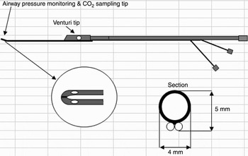
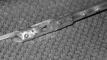
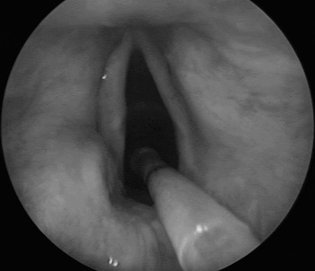
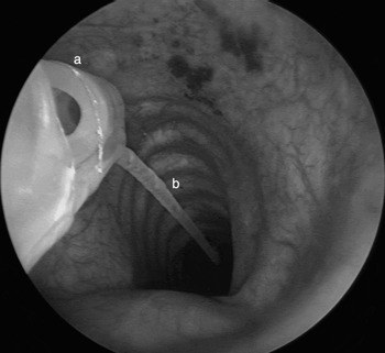
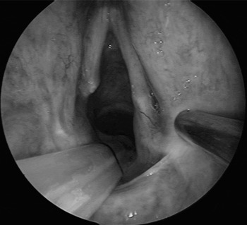
We assessed the development of subglottic jet ventilation at the Prince of Wales Hospital, Sydney, Australia, and here present our results for the newest version of this system. This new system consisted of a subglottic tube and companion ventilator (Figures 1 to 5). The tube was designed to have a minimal cross-sectional area and the facility for end-tidal carbon dioxide monitoring and subglottic airways pressure monitoring; it was also engineered to be non-flammable and safe for laser surgery. The main outcome measures assessed were anaesthetic safety, laser safety (Figure 2) and degree of microlaryngoscopic surgical access (Figures 3 to 5). Aspects of the tube which facilitated safe and reliable anaesthesia during laryngoscopy were identified. Comparison with other subglottic jet tubes was undertaken. Figures 3 to 5 illustrate the subglottic jet ventilation technique.
The second generation Prince of Wales Hospital tube.

Laser safety bench testing showed that, when struck by a CO2 laser with 100 per cent oxygen entrained, the Teflon Jockjet did not flare or burn.

Microlaryngoscopic view showing surgical access with the Jockjet tube in situ.

Microlaryngoscopic view showing subglottic jet ventilation with the Jockjet tube. a = Venturi port covering tube and Murphy's eye, ensuring a safe, diffused jet stream; b = subglottic airways pressure and CO2 monitoring line

Microlaryngoscopic view showing unilateral laser tenotomy result for bilateral vocal fold paralysis, with Jockjet tube in situ.

A focused review of the international literature was also undertaken to examine the development of subglottic jet ventilation for microlaryngeal and laryngeal laser surgery. The PubMed and Medline databases were searched for articles with combinations of keywords including: Anaesthesia, Larynx, Laser, Microlaryngoscopy, Subglottic Jet Ventilation. The final literature review was based upon the authors' experience together with conclusions from articles selected for inclusion.
Results
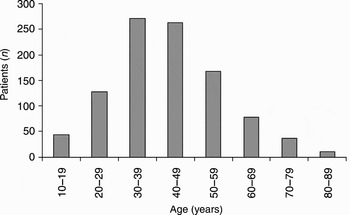
From June 2002 to March 2008 inclusive, 1000 consecutive patients underwent microlaryngeal surgery at our institutions. The patient group included 587 females and 413 males. Patients' ages ranged from 10 to 89 years of age (Figure 6).
Patient age range.

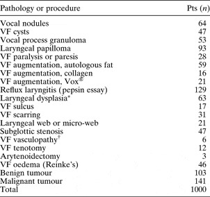
A range of benign and malignant pathologies was treated surgically (see Table I), via non-jet anaesthesia (16 patients) and by supraglottic (243) and subglottic (741) jetting techniques. The airway management techniques employed included microlaryngeal tube (16 patients), Hunsaker Mon-Jet (14), supraglottic jetting (243), Ben Jet (208), paediatric Ben Jet (187) and the second generation Prince of Wales Hospital jet is shown in Figure 1 (332), as outlined in Figure 1. The resection techniques employed included carbon dioxide laser (347 cases), ‘cold steel’ (611) and laryngeal microdebrider (42). In the 332 patients managed with subglottic jet ventilation via the Jockjet system (ULCO Medical, Sydney, Australia), a range of laryngeal procedures was undertaken, from cold steel to laser excision. Excellent microlaryngoscopic surgical access was achieved (Figures 3 to 5). No adverse anaesthetic outcomes were encountered in any of the 332 patients anaesthetised with subglottic jet ventilation, and surgical access was optimised.
Microlaryngeal pathology addressed and procedures undertaken

*Leukoplakia or erythroplakia. †Telangiectasia or venous lakes. Pts = patients; VF = vocal fold
In the remaining 668 patients managed without the Jockjet system, no serious anaesthetic complications were encountered. Three patients required continuous positive airway pressure post-operatively and stayed in the recovery room or high dependency unit overnight. No episodes of pneumonia, subcutaneous emphysema, pneumothorax, tension pneumothorax or pulmonary oedema were encountered.
Discussion
Airway management during microlaryngeal surgery has evolved from apnoeic techniques through to the controlled methods routinely employed today. Sanders described jet ventilation for diagnostic bronchoscopy, prior to its application for microlaryngoscopy.Reference Sanders1 Carden practised jet ventilation via needle puncture of the cricothyroid membrane (and encouraged his residents to jet the patient while he smoked a cigarette outside!).Reference Carden, Becker and Hamood2 El-Naggar et al. described jet ventilation for microlaryngeal procedures using a number five endotracheal tube.Reference El-Naggar, Keh, Stemmers and Collins3 Komesaroff and McKie described a device they named a ‘bronchoflater’.Reference Komesaroff and McKie4
Early experience with jet ventilation was beset with complications, such as pneumothorax.Reference O'Sullivan and Healy5–Reference Craft, Chambers, Ward and Goat7 Tracheobronchial injury due to the high pressure jet was not uncommon. Komesaroff and McKie's work highlighted the importance of tracheal pressure measurements, which were recorded via a fine polyethylene catheter attached to the outer wall of the bronchoscope. Benjamin and Gronow developed a technique of anaesthesia for microlaryngeal surgery using a subglottic jet ventilation catheter and a Venturi jet injector.Reference Benjamin and Gronow8 Tubes for subglottic jet ventilation were subsequently developed by Hunsaker, and at our own institution by Bridger and Maver.Reference Hunsaker9, Reference Bridger and Maver10
Over the last 20 years, subglottic jet ventilation tubes have improved. The current Prince of Wales Hospital jet ventilation tube is a fourth generation device; the previous three generations include the Hunsaker Mon-Jet, the Ben Jet and the first generation Prince of Wales Hospital tube.Reference Benjamin and Gronow8–Reference Brooker, Hunsaker and Zimmerman12
The Ben Jet tube provided good access via subglottic jet ventilation.Reference Rakover, Ben-Abraham, Avni, Nachtigal and Katz13 The stabilising flanges meant the tube was much less likely to be misaligned, and ensured a central, stable position in the trachea, which helped prevent tube flailing. However, the lack of carbon dioxide and pressure monitoring meant that ventilation with this type of tube carried an increased risk of hypercapnoea and pneumothorax.
The first generation Prince of Wales Hospital jet ventilation tube allowed true subglottic end-tracheal pressure monitoring, important for preventing high pressures and potential pneumothorax.Reference Bridger and Maver10 This tube allowed carbon dioxide measurement, to ensure adequate ventilation. However, as this tube was made of polyvinyl chloride (PVC), it was not safe for use with lasers.
The Hunsaker Mon-Jet tube allowed proximal but not subglottic jet monitoring. This tube was made of non-flammable fluoroplastic material, but was pliable and therefore at risk of being compressed or kinked by the laryngoscope, obstructing ventilation. Also, this tube's flexibility could lead to flailing in the trachea. Whilst the Mon-Jet allowed optimal laryngeal exposure during microlaryngeal surgery, we sought to develop a new tube with improvements based on our experience.Reference Orloff, Parhizkar and Ortiz14
Complications reported during the early use of subglottic jet ventilation included barotrauma and pneumothorax.Reference Werkhaven15 In order to improve anaesthetic safety and to avoid potential complications, the Jockjet system incorporated the facility for subglottic airway pressure and expired gas monitoring. The 4-mm diameter Teflon tube with 6-mm Venturi tip was designed to protect tracheal mucosa from the high velocity jet by producing a more diffused stream. In the authors' experience, no barotrauma-associated complications were encountered.
Teflon is rigid, enabling easy endotracheal intubation, and is flame-resistant, an essential feature for safe laryngeal laser surgery. Laryngeal procedures with carbon dioxide laser are increasingly routinely performed. Laser microlaryngoscopy techniques provide precise management for a wide range of upper airway conditions. The type of airway used is dictated by whether access to the hypopharynx, supraglottis, larynx or subglottis is required.Reference Patel and Rubin16 However, laser-related risks (including airway fire) are potentially fatal. Hunsaker assessed the laser safety of subglottic jet ventilation tubes, and demonstrated the danger of tube fires, even with low oxygen concentrations, when using Silastic®, rubber and PVC tubes in laser laryngeal surgery.Reference Hunsaker9 In our testing and experience, Teflon tubes are non-flammable (Figure 2).
Subglottic jetting may be used in all patients, including those with severe co-morbidities, obesity and difficult airway access.Reference Davies, Hillel, Maronian and Posner11 Subglottic jet ventilation may be safely employed in patients with difficult airways. The technique may be used for a wide variety of surgical pathology, from benign hyperfunction related laryngeal pathology to paralytic vocal fold conditions, papilloma, granuloma, dysplasia and malignancy.Reference Havas and Priestley17–Reference Miyawaki, Shono, Katori, Sakuragi and Higa19
• Jet ventilation techniques have enabled safe, controlled microlaryngoscopy and accurate treatment of laryngeal pathology
• This study assessed the development and use of the Jockjet subglottic jet ventilation system
In paediatric laryngeal microsurgery, the main indications for the use of high frequency jet ventilation are laryngomalacia, laryngeal papillomatosis and subglottic haemangioma. Paediatric and adult cases may be safely managed with subglottic jet ventilation.Reference El Hammar-Vergnes and Cros20–Reference Cook and Alexaner22 In children aged up to eight years, the jet ventilator adjustments must take into account the lower pulmonary compliance and the higher airway resistance. In order to avoid barotrauma, the jet ventilator must include an airway pressure monitoring system. The driving pressure and the frequency must be gradually increased while checking thoracic expansion. High frequency jet ventilation is reliable and safe in children.
A 2008 study explored airway management and ventilation during elective laryngeal surgery, focusing primarily on injector and jet ventilation (i.e. high pressure source ventilation).Reference Cook and Alexaner22 Complications during manual techniques led to critical care admissions and discharge delays. Several hospitals have reported major complications of high pressure source ventilation, including deaths. High frequency jet ventilation related complications have led to critical care admissions and discharge delay but no deaths. Complications were evenly spread between supraglottic, subglottic and transtracheal techniques. All deaths occurred in departments without high frequency jet ventilation. In the centres that perform more than 100 transtracheal jet ventilation cases per year no serious complications were reported.
Conclusion
This study examined how advances in jet ventilation tube design have facilitated safe anaesthesia and endolaryngeal surgery. Subglottic jet ventilation continues to improve. The facility to monitor end-tidal carbon dioxide and end-tracheal pressure is helpful for the delivery of safe anaesthesia; no complications were encountered in the present experience. With appropriate anaesthetic monitoring, subglottic jet ventilation may provide safe airway management and excellent surgical conditions during microlaryngeal and laryngeal laser surgery.